. 2022 Nov 5;13(11):928.
doi: 10.1038/s41419-022-05364-w.

Cadmium-induced apoptosis of Leydig cells is mediated by excessive mitochondrial fission and inhibition of mitophagy

Affiliations

Cadmium-induced apoptosis of Leydig cells is mediated by excessive mitochondrial fission and inhibition of mitophagy

Lingna Yi et al. Cell Death Dis. .

Abstract

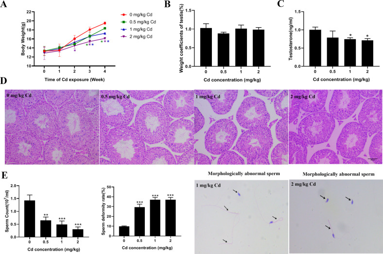
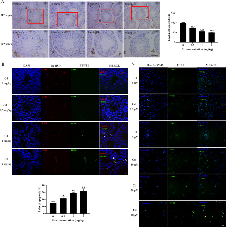
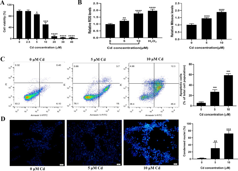
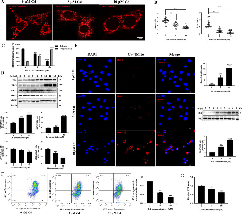
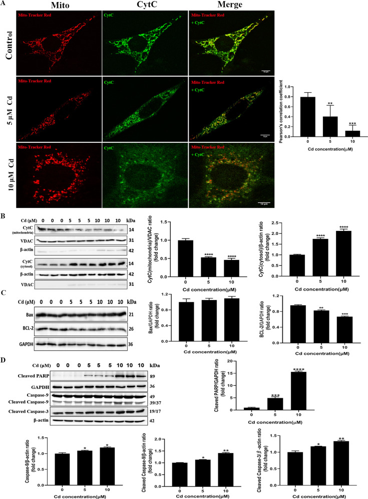
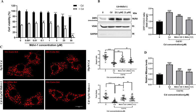
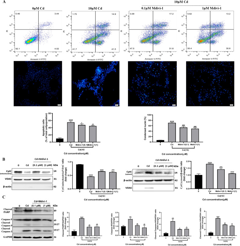
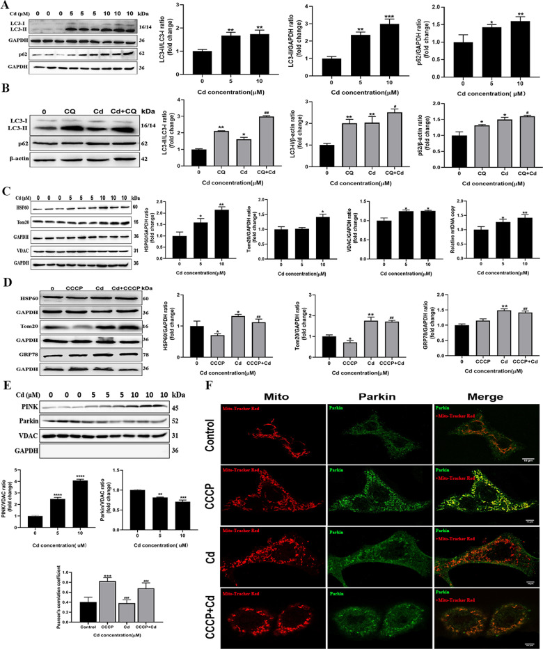
Cadmium is one of the environmental and occupational pollutants and its potential adverse effects on human health have given rise to substantial concern. Cadmium causes damage to the male reproductive system via induction of germ-cell apoptosis; however, the underlying mechanism of cadmium-induced reproductive toxicity in Leydig cells remains unclear. In this study, twenty mice were divided randomly into four groups and exposed to CdCl2 at concentrations of 0, 0.5, 1.0 and 2.0 mg/kg/day for four consecutive weeks. Testicular injury, abnormal spermatogenesis and apoptosis of Leydig cells were observed in mice. In order to investigate the mechanism of cadmium-induced apoptosis of Leydig cells, a model of mouse Leydig cell line (i.e. TM3 cells) was subjected to treatment with various concentrations of CdCl2. It was found that mitochondrial function was disrupted by cadmium, which also caused a significant elevation in levels of mitochondrial superoxide and cellular ROS. Furthermore, while cadmium increased the expression of mitochondrial fission proteins (DRP1 and FIS1), it reduced the expression of mitochondrial fusion proteins (OPA1 and MFN1). This led to excessive mitochondrial fission, the release of cytochrome c and apoptosis. Conversely, cadmium-induced accumulation of mitochondrial superoxide was decreased by the inhibition of mitochondrial fission through the use of Mdivi-1 (an inhibitor of DRP1). Mdivi-1 also partially prevented the release of cytochrome c from mitochondria to cytosol and attenuated cell apoptosis. Finally, given the accumulation of LC3II and SQSTM1/p62 and the obstruction of Parkin recruitment into damaged mitochondria in TM3 cells, the autophagosome-lysosome fusion was probably inhibited by cadmium. Overall, these findings suggest that cadmium induces apoptosis of mouse Leydig cells via the induction of excessive mitochondrial fission and inhibition of mitophagy.


Conflict of interest statement

The authors declare no competing interests.

Figures

Fig. 1
Fig. 1. The effect of cadmium exposure on testicular toxicity.
A The body weight of mice was recorded every day during the exposure period and weekly body weight was shown (n = 5 in each group). B Testis/body weight index of four groups. C Measurement of serum testosterone levels in four groups of mice. D Representative histology of the testis from each group, scale bar: 20 μm. E The number of sperm from cauda epididymides and deformity rate were examined, and abnormal sperm morphology was shown. Arrows indicate morphologically abnormal sperm. (*P < 0.05, **P < 0.01, ***P < 0.0001 vs. control).
Fig. 2
Fig. 2. Cadmium exposure decreased the number of Leydig cells and induced apoptosis in testes.
A Representative images of testis immunohistochemistry with anti-3β-HSD. The Leydig cells were stained in brown, scale bar: 20 μm. The number of 3β-HSD-positive cells was counted in 25 fields randomly selected on slides in each group of mice. B Testis immunohistochemistry combined TUNEL staining to analyze apoptosis of Leydig cells in vivo. White arrows indicate apoptotic Leydig cells. Broken white arrows indicate apoptotic spermatogenic cells. scale bar: 50 μm (C) Primary Leydig cells were exposed to different concentrations of cadmium and cell apoptosis was analyzed by Hoechst33342 and TUNEL staining, scale bar: 100 μm.
Fig. 3
Fig. 3. Cadmium exposure decreased cell viability and induced ROS and apoptosis of TM3 cells.
A TM3 cells were exposed to different concentrations of cadmium for 24 h. The cell viability was measured by CCK-8 assays. B Cellular ROS and Mitochondrial ROS production. C Apoptotic cells were detected by PI and Annexin V staining and represented as a percentage ratio. D Representative images of nuclear staining with Hoechst33342 in TM3 cells. Apoptotic cells were quantitated by nuclear condensation. Data represent the mean±SD for at least three independent experiments (**P < 0.01, ***P < 0.001, ****P < 0.0001 vs. control).
Fig. 4
Fig. 4. Cadmium induced mitochondrial fragmentation and dysfunction in TM3 cells.
A, B Mitochondria were stained with Mito-Tracker Red CMXRos after the cells were treated with cadmium. The mitochondrial morphology factors (aspect ratio and form factor) were evaluated. C The percentage of cells containing tubular or fragmented mitochondria was calculated. D The protein levels of FIS1, DRP1, OPA1 and MFN1 were measured by Western blot after treatment with cadmium. E The effects of cadmium on mitochondrial Ca2+ levels and expression of mitochondrial calcium uniporter (MCU) were analyzed by immunofluorescence and Western blot, respectively. F The effect of cadmium on mitochondrial ΔΨm was assayed by JC-1 staining. G Luciferase-based assays were used to determine cellular ATP production. The data represent the mean ± SD of three independent experiments (*P < 0.05, **P < 0.01, ***P < 0.001, ###P < 0.001, ****>P < 0.0001, ####P < 0.0001 vs. control).
Fig. 5
Fig. 5. Cadmium induced the release of cytochrome c from the mitochondria into the cytosol of TM3 cells and promoted apoptosis.
A, B The cellular distribution of cytochrome c was analyzed by immunofluorescence and Western blotting after the cells were treated with cadmium for 24 h. The cells were stained with mitochondrial probes (Mito-Tracker Red CMXRos), incubated with anti-CytC (Green) and then observed by laser confocal microscopy (100x). Pearson’s correlation coefficient was used to assess the colocalization between mitochondria and cytochrome c. VDAC and β-actin were used as internal references for mitochondrial and cytosolic fractions, respectively. C, D The levels of apoptosis-related proteins Bax, Bcl-2, cleaved PARP, Caspase-9, cleaved Caspase-9 and cleaved Caspase-3 were measured by Western blot. The data represent the mean ± SD of three independent experiments (**P < 0.01, ***P < 0.001, ****P < 0.0001 vs. control).
Fig. 6
Fig. 6. The DRP1 inhibitor Mdivi-1 protected mitochondrial damage caused by cadmium.
A TM3 cells were pretreated with a range of Mdivi-1 concentrations, followed by exposure to 10 μM Cd for 24 h and the cell viability was measured by CCK-8 assays. B The effect of Mdivi-1 on the mitochondrial location of DRP1 was determined by Western blot. VDAC and GAPDH were used as internal references for mitochondrial and cytosolic fractions, respectively. C The effect of Mdivi-1 on cadmium-induced mitochondrial deformation was analyzed by MitoTracker Red staining, and the aspect ratio and form factor were assessed. D Measurement of mitochondrial ROS levels. Data represent the mean±SD for at least three independent experiments (**P < 0.01, ***P < 0.001, ****P < 0.0001 vs. control; ####P < 0.0001 vs. Cd).
Fig. 7
Fig. 7. Mdivi-1 attenuated cadmium-induced apoptosis of TM3 cells.
A The effect of Mdivi-1 on cadmium-induced apoptosis was evaluated by PI/Annexin V staining and Hoechst33342 staining, respectively. B Cellular distribution of cytochrome c was analyzed by Western blot. VDAC and β-actin were used as internal references for mitochondrial and cytosolic fractions, respectively. C Mdivi-1 inhibited cadmium-induced expression of cleaved PARP, Caspase-9, cleaved Caspase-9 and cleaved Caspase-3. Data represent the mean±SD for three independent experiments (**P < 0.01, ***P < 0.001, ****P < 0.0001 vs. control; #P < 0.05, ##P < 0.01, ###P < 0.001, ####P < 0.0001 vs. Cd).
Fig. 8
Fig. 8. Cadmium-induced mitochondrial fragmentation was associated with inhibition of mitophagy.
A Expression levels of mitophagy markers LC3 and p62 were examined by Western blot after TM3 cells were treated with cadmium. B The cells were treated with or without chloroquine (CQ, 10 μM) in the presence or absence of cadmium for 24 h. The expression of LC3 and p62 was measured. C The effect of cadmium on mitochondrial mass was analyzed by immunoblotting for mitochondrial proteins (HSP60, TOM20 and VDAC) and measuring mitochondrial DNA (mtDNA) copy number. D The cells were exposed to 10 μM cadmium for 24 h with or without pretreatment with 10 μM CCCP for 2 h. The levels of mitochondrial proteins (HSP60 and Tom20) and an ER protein GRP78 were measured. E Mitochondria were isolated from the cadmium-treated cells and the expression of PINK and Parkin were measured by Western blot. VDAC and GAPDH were used as internal references for the mitochondria and cytosol, respectively (F) The cells were treated with cadmium and CCCP as above and then stained with mitochondrial probes (Mito-Tracker Red CMXRos) and anti-Parkin antibodies (Green). The colocalization between mitochondria and Parkin was analyzed by immunofluorescence. Data represent the mean±SD for at least three independent experiments (*P < 0.05, **P < 0.01, ***P < 0.001, ****P < 0.0001 vs. control; ##P < 0.01, ###P < 0.001, vs. CQ or CCCP).
Fig. 9
Fig. 9. Schematic illustration of cadmium-induced toxicity in TM3 cells.
Cadmium causes mitochondrial dysfunction and promotes excessive mitochondrial fission by controlling the expression of mitochondrial dynamics regulatory proteins (DRP1, FIS1, MFN1 and OPA1). Consequently, cytochrome c is released from the mitochondria to cytosol and the mitochondria-dependent intrinsic apoptosis of TM3 cells is induced. Moreover, cadmium probably disrupts calcium homeostasis and inhibits autophagic flux in the cells. The blocking of Parkin recruitment into damaged mitochondria by cadmium results in inhibition of mitophagy, production of excessive ROS and cell death.

References

    1. Skakkebaek NE, Rajpert-De Meyts E, Buck Louis GM, Toppari J, Andersson AM, Eisenberg ML, et al. Male reproductive disorders and fertility trends: influences of environment and genetic susceptibility. Physiol Rev. 2016;96:55–97. - PMC - PubMed
    1. Gabrielsen JS, Tanrikut C. Chronic exposures and male fertility: the impacts of environment, diet, and drug use on spermatogenesis. Andrology. 2016;4:648–61. - PubMed
    1. Ilacqua A, Izzo G, Emerenziani GP, Baldari C, Aversa A. Lifestyle and fertility: the influence of stress and quality of life on male fertility. Reprod Biol Endocrinol. 2018;16:115. - PMC - PubMed
    1. Cappelletti R, Ceppi M, Claudatus J, Gennaro V. Health status of male steel workers at an electric arc furnace (EAF) in Trentino, Italy. J Occup Med Toxicol. 2016;11:7. - PMC - PubMed
    1. Hong H, Xu Y, Xu J, Zhang J, Xi Y, Pi H, et al. Cadmium exposure impairs pancreatic beta-cell function and exaggerates diabetes by disrupting lipid metabolism. Environ Int. 2021;149:106406. - PubMed

Publication types