. 2022 Nov 5;8(1):446.
doi: 10.1038/s41420-022-01232-w.

SMG6 regulates DNA damage and cell survival in Hippo pathway kinase LATS2-inactivated malignant mesothelioma

Affiliations

SMG6 regulates DNA damage and cell survival in Hippo pathway kinase LATS2-inactivated malignant mesothelioma

Koya Suzuki et al. Cell Death Discov. .

Abstract

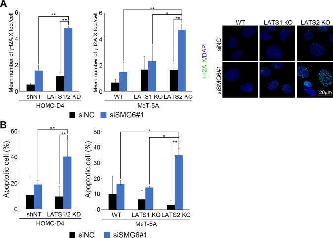
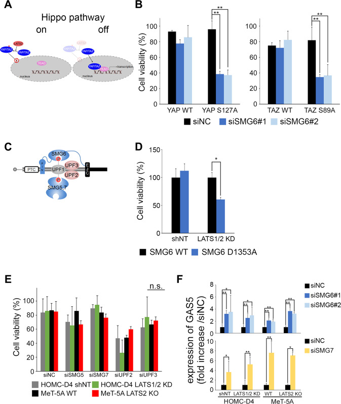
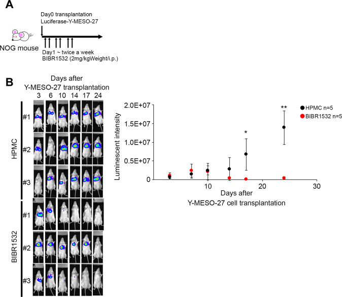
Many genes responsible for Malignant mesothelioma (MM) have been identified as tumor suppressor genes and it is difficult to target these genes directly at a molecular level. We searched for the gene which showed synthetic lethal phenotype with LATS2, one of the MM causative genes and one of the kinases in the Hippo pathway. Here we showed that knockdown of SMG6 results in synthetic lethality in LATS2-inactivated cells. We found that this synthetic lethality required the nuclear translocation of YAP1 and TAZ. Both are downstream factors of the Hippo pathway. We also demonstrated that this synthetic lethality did not require SMG6 in nonsense-mediated mRNA decay (NMD) but in regulating telomerase reverse transcriptase (TERT) activity. In addition, the RNA-dependent DNA polymerase (RdDP) activity of TERT was required for this synthetic lethal phenotype. We confirmed the inhibitory effects of LATS2 and SMG6 on cell proliferation in vivo. The result suggests an interaction between the Hippo and TERT signaling pathways. We also propose that SMG6 and TERT are novel molecular target candidates for LATS2-inactivated cancers such as MM.


Conflict of interest statement

The authors declare no competing interests.

Figures

Fig. 1
Fig. 1. LATS2-inactive cells showed cisplatin resistance.
Effect of cisplatin (A) and H2O2 (B) on LATS1/2 KD HOMC-D4 and LATS1 or LATS2 KO MeT-5A cells. The cells were treated with various concentrations of cisplatin or hydroperoxide for 24 h. After the medium change, the cells were cultured for an additional 48 h, and then cell viability was measured using CCK-8. All experiments were performed at least three independent times. Data are presented as means ± SD, and p values were calculated using the Tukey-Kramer method. * indicates p < 0.05 and ** indicates p < 0.01. n.s. means no significant difference.
Fig. 2
Fig. 2. Knockdown of SMG6 reduces the viability of LATS2-inactive cells.
A The mRNA expression levels of SMG6. B The protein expression levels of LATS1 and LATS2. * indicates a nonspecific band. C The knockdown efficiency of SMG6 mRNA. D Cell viability when SMG6 is knocked down. Cell viability was measured 72 h after siSMG6 treatment using CCK-8. All experiments were performed at least three independent times. Data are presented as means ± SD, and p values were calculated using the Tukey-Kramer method. ** indicates p < 0.01.
Fig. 3
Fig. 3. Knockdown of SMG6 induces DNA damage and apoptosis in LATS2-inactive mesothelial cells.
A γ-H2A.X immunofluorescence and DAPI staining in various cells infected with control or SMG6-specific siRNA (siSMG6#1). The number of γ-H2A.X foci in each cell was counted 72 h after siSMG6 treatment. γ-H2AX is green, and DAPI is blue. B The number of apoptotic cells was measured by flow cytometry using APO-Direct Kit 72 h after siSMG6 treatment. All experiments were performed at least three independent times. Data are presented as means ± SD; p-values were calculated using the Tukey-Kramer method. * indicates p < 0.05 and ** indicates p < 0.01.
Fig. 4
Fig. 4. YAP/TAZ activity was required to induce synthetic lethality but not related to the NMD pathway factors.
A Schematic drawing of Hippo pathway. B Non-phosphorylated forms of YAP/TAZ-overexpressing HOMC-D4 were transfected with siSMG6. Cell viability was measured after 144 h of siSMG6 treatment using CCK-8. C Schematic drawing of NMD complex. D (SMG6 WT and D1353A)-overexpressing plasmids were transfected into HOMC-D4 (non-target (shNT) and LATS1/2 KD cells. After 144 h of transfection, the cell viability was measured using CCK-8. E The viability of LATS2-inactive mesothelial cells treated with siSMG5, siSMG7, siUPF2, or siUPF3. Cell viability was measured using CCK-8. F The expression levels of GAS5 in siSMG6- (upper panel) or siSMG7- (lower panel) treated LATS2-inactive mesothelial cells. All experiments were performed at least three independent times. Data are presented as means ± SD, and p values were calculated using the Tukey-Kramer method. * indicates p < 0.05 and ** indicates p < 0.01. n.s. - no significant difference. ND means not detect.
Fig. 5
Fig. 5. Inactivation of LATS2 and inhibition of TERT show a synthetic lethal phenotype, requiring RdDP activity of TERT.
A Each cell was transfected with siTERT. Cell viability was measured after 144 h of siTERT treatment using CCK-8. B The knockdown efficiency of TERT. mRNA was collected after 72 h of siTERT treatment. C The IC50 of the TERT inhibitors (BIBR1532, trichostatin, doxorubicin, TMPyP4, suramin, and VX222) in MeT-5A (WT, LATS1 KO, and LATS2 KO). The diluted TERT inhibitor was applied to each cell, and cell viability was measured after 144 h of incubation using CCK-8. D hTERT D712A- and hTERT T249A-overexpressing plasmids were transfected into HOMC-D4 (non-target [shNT] and LATS1/2 KD) and MeT-5A (WT and LATS2 KO) cells. The cell viability was measured after 144 h of transfection using CCK-8. All experiments were performed at least three independent times. Data are presented as means ± SD, and p values were calculated using the Tukey-Kramer method. * indicates p < 0.05, ** indicates p < 0.01 and *** indicates p < 0.001. n.s. - no significant difference.
Fig. 6
Fig. 6. Synthetic lethality induced by inhibition of LATS2 and SMG6/TERT is also observed in a tumor-bearing mouse model.
A Timetable for transplantation, drug administration, and IVIS measurements. B The IVIS images were obtained after tumor engraft subcutaneous inoculation of luciferase induced-Y-MESO-27 cells. One day after Y-MESO-27 transplantation, HPMC (n = 5) or BIBR1532 (2 mg/kg, n = 5) were injected in peritoneal cavity twice a week. Tumor size was measured by IVIS twice weekly after tumor transplantation until day 30. The luminescent intensity indicated the levels of quantification of IVIS imaging. p values were calculated using student’s t-test.* indicates p < 0.05 and ** indicates p < 0.01.

Similar articles

Cited by

References

    1. Gomez DR, Rimner A, Simone CB, Cho BCJ, de Perrot M, Adjei AA, et al. The use of radiation therapy for the treatment of malignant pleural mesothelioma: expert opinion from the National Cancer Institute Thoracic Malignancy Steering Committee, International Association for the Study of Lung Cancer, and Mesothelioma Applied Research Foundation. J Thorac Oncol. 2019;14:1172–83. - PubMed
    1. Murthy SS, Testa JR. Asbestos, chromosomal deletions, and tumor suppressor gene alterations in human malignant mesothelioma. J Cell Physiol. 1999;180:150–7. - PubMed
    1. Rosenzweig KE, Giraud P. Radiation therapy for malignant pleural mesothelioma. Cancer Radiotherapie. 2017;21:73–76. - PubMed
    1. Baas P, Scherpereel A, Nowak AK, Fujimoto N, Peters S, Tsao AS, et al. First-line nivolumab plus ipilimumab in unresectable malignant pleural mesothelioma (CheckMate 743): a multicentre, randomised, open-label, phase 3 trial. Lancet. 2021;397:375–86. - PubMed
    1. Bueno R, Stawiski EW, Goldstein LD, Durinck S, de Rienzo A, Modrusan Z, et al. Comprehensive genomic analysis of malignant pleural mesothelioma identifies recurrent mutations, gene fusions and splicing alterations. Nat Genet. 2016;48:407–16. - PubMed