. 2022 Nov 5;13(1):6684.
doi: 10.1038/s41467-022-34607-9.

Autoinducer-2 and bile salts induce c-di-GMP synthesis to repress the T3SS via a T3SS chaperone

Affiliations

Autoinducer-2 and bile salts induce c-di-GMP synthesis to repress the T3SS via a T3SS chaperone

Shuyu Li et al. Nat Commun. .

Abstract

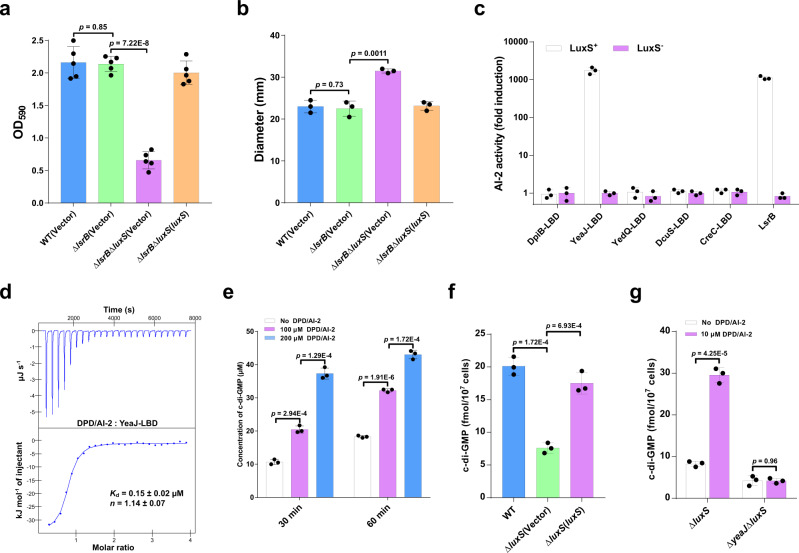
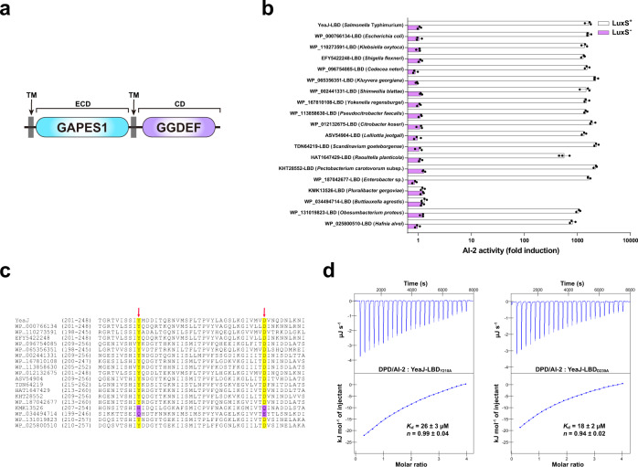
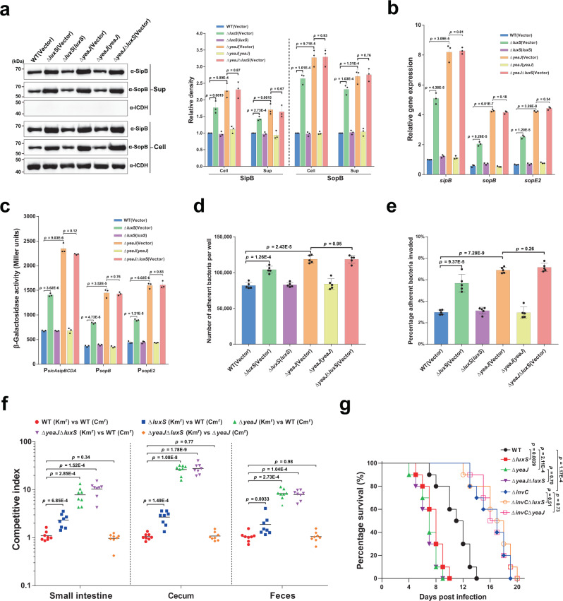
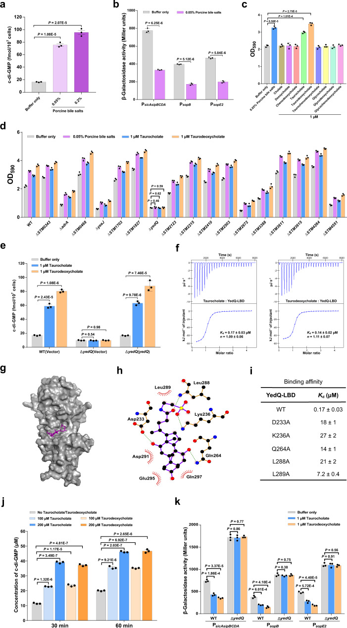
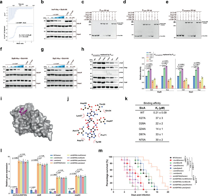
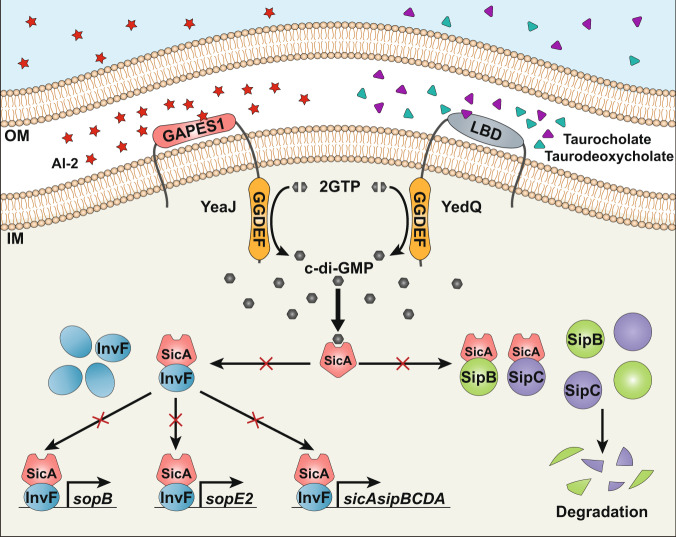
Cyclic di-GMP (c-di-GMP) transduces extracellular stimuli into intracellular responses, coordinating a plethora of important biological processes. Low levels of c-di-GMP are often associated with highly virulent behavior that depends on the type III secretion system (T3SS) effectors encoded, whereas elevated levels of c-di-GMP lead to the repression of T3SSs. However, extracellular signals that modulate c-di-GMP metabolism to control T3SSs and c-di-GMP effectors that relay environmental stimuli to changes in T3SS activity remain largely obscure. Here, we show that the quorum sensing signal autoinducer-2 (AI-2) induces c-di-GMP synthesis via a GAPES1 domain-containing diguanylate cyclase (DGC) YeaJ to repress T3SS-1 gene expression in Salmonella enterica serovar Typhimurium. YeaJ homologs capable of sensing AI-2 are present in many other species belonging to Enterobacterales. We also reveal that taurocholate and taurodeoxycholate bind to the sensory domain of the DGC YedQ to induce intracellular accumulation of c-di-GMP, thus repressing the expression of T3SS-1 genes. Further, we find that c-di-GMP negatively controls the function of T3SSs through binding to the widely conserved CesD/SycD/LcrH family of T3SS chaperones. Our results support a model in which bacteria sense changes in population density and host-derived cues to regulate c-di-GMP synthesis, thereby modulating the activity of T3SSs via a c-di-GMP-responsive T3SS chaperone.


Conflict of interest statement

The authors declare no competing interests.

Figures

Fig. 1
Fig. 1. AI-2 increases intracellular c-di-GMP levels by directly engaging the DGC YeaJ in S. Typhimurium.
a AI-2 regulates biofilm formation in S. Typhimurium independent of LsrB. Biofilms were stained with crystal violet and quantified using optical density measurement. Data were mean ± s.e.m. of five independent experiments. b AI-2 regulates the swimming motility of S. Typhimurium independent of LsrB. Data were mean ± s.e.m. of three independent experiments. c YeaJ-LBD is capable of retaining AI-2. Bioluminescence in V. harveyi MM32 (luxN, luxS) was induced by the addition of ligands released from purified proteins expressed in a luxS+ or luxS- E. coli strain. LsrB from S. Typhimurium was used as a positive control. AI-2 activity is reported as fold induction relative to the light production induced by a buffer control. Data were mean ± s.e.m. of three independent experiments. d ITC assays for the specific interaction between YeaJ-LBD and AI-2. The data shown are representative of three independent experiments with similar results. The Kd and complex stoichiometry (n) values were presented as mean ± s.d. of three independent experiments. e AI-2 stimulates the DGC activity of YeaJ in vitro. Data were mean ± s.e.m. of three independent experiments. f Liquid chromatography-tandem mass spectrometry (LC-MS/MS) measurements of cellular levels of c-di-GMP in S. Typhimurium strains. The bacterial cultures grown in LB broth at 37 °C with shaking to an OD600 of 1.3 were subjected to nucleotide extractions. Data represent mean ± s.d. from three biological replicates. g AI-2 increases cellular c-di-GMP concentration via YeaJ. The mutants ΔluxS and ΔyeaJΔluxS grown in LB medium to an OD600 of 1.3 were induced by 10 μM DPD/AI-2 or a buffer control for 30 min, and then bacterial cells were collected to extract nucleotides for LC-MS/MS analysis. Data shown are mean ± s.d. of three biological replicates. a, b, eg Statistical significance was evaluated using a two-tailed unpaired Student’s t-test and p < 0.05 was considered statistically significant. WT wildtype. Source data are provided as a Source Data file.
Fig. 2
Fig. 2. Widespread occurrence of GAPES1 domain-containing YeaJ homologs capable of sensing AI-2 in the order Enterobacterales.
a Schematic illustrating the predicted domain organization of YeaJ homologs. Protein sequences were analyzed using hmmscan program against the Pfam 34.0 database. TM transmembrane domain, ECD extracytoplasmic domain, CD cytoplasmic domain. b GAPES1 domains from YeaJ homologs in bacterial species belonging to the Enterobacteriaceae, Pectobacteriaceae, and Hafniaceae families are capable of retaining AI-2. GAPES1 domains from YeaJ homologs were expressed and purified as His6 fusion proteins in a luxS+ or luxS- E. coli strain. Bioluminescence in V. harveyi strain MM32 was measured following the addition of a buffer control or ligands released from the purified proteins upon denaturing by heating. YeaJ-LBD was used as a positive control. AI-2 activity is presented as mean ± s.e.m. of three independent experiments. The NCBI accession numbers for YeaJ homologs are provided and the bacterial species to which they belong are given in parentheses. c AI-2-binding GAPES1 domains of YeaJ homologs harbor two conserved residues corresponding to Y210 and D239 of YeaJ. GAPES1 domains of YeaJ and its homologs that have been tested for AI-2 binding activity were aligned using ClustalW embedded in MEGA7 software. Two positions corresponding to Y210 and D239 of YeaJ that may be involved in AI-2 binding are labeled with red arrows. The conserved residues in the two positions are highlighted in yellow and non-conserved residues are highlighted in purple. The NCBI accession number for each YeaJ homolog is included. d Isotherms representing binding of two mutants of YeaJ-LBD (Y210A or D239A) with DPD/AI-2. The binding affinity was determined using ITC. The data shown were one representative of three independent experiments with similar results. The Kd and complex stoichiometry (n) are presented as mean ± s.d. of three independent experiments. Source data are provided as a Source Data file.
Fig. 3
Fig. 3. AI-2 represses the T3SS-1 and negatively regulates the virulence of S. Typhimurium via YeaJ.
a AI-2 negatively regulates intracellular accumulation and secretion of SipB and SopB via YeaJ. Cell pellet (Cell) and concentrated supernatant (Sup) were probed for SipB and SopB by western blot analysis. Isocitrate dehydrogenase (ICDH) was probed as a loading control. The blots shown are representative of three independent experiments with similar results. The band intensities were quantified by scanning densitometry using ImageJ (NIH, USA), normalized to intracellular ICDH, and presented as values relative to that of the wildtype (mean ± s.d.; n = 3 independent experiments). b mRNA levels of T3SS-1 genes were determined by qRT-PCR analyses. Expression was normalized to 16 S rRNA and reported as fold change relative to that of the sipB gene of the wildtype. Data were mean ± s.e.m. of three independent experiments. c The promoter activities of T3SS-1 genes measured using β-galactosidase activity assays (mean ± s.e.m.; n = 3 independent experiments). d, e, Adherence to (d) and invasion of (e) Caco-2 cells by S. Typhimurium strains. Data were mean ± s.e.m. of five independent experiments. f Six-week-old female BALB/c mice were infected orally with a 1:1 mixture of two S. Typhimurium strains carrying kanamycin-resistant (Kmr) pKT100 and chloramphenicol-resistant (Cmr) pBBR1MCS1, respectively. A competitive index (CI) was calculated as the ratio of the test strain carrying pKT100 versus (vs) the control strain carrying pBBR1MCS1 recovered from mice. Horizontal lines represent the geometric mean CI value for each group (n = 8 mice per group). g Deletion of luxS or yeaJ leads to enhanced virulence of S. Typhimurium in mice. Six-week-old female BALB/c mice were infected orally with each S. Typhimurium strain and survival was monitored daily. Data were illustrated as a percentage of mice survival (n = 10 mice per group). Statistical significance was evaluated by two-tailed unpaired Student’s t-test (ae), two-tailed Mann–Whitney U-test (f), or Log-rank (Mantel–Cox) test (g). P values <0.05 indicate significant differences. Source data are provided as a Source Data file.
Fig. 4
Fig. 4. Bile components taurocholate and taurodeoxycholate repress the expression of T3SS-1 genes via stimulating the c-di-GMP synthase activity of YedQ.
a Bile salts stimulate an increase in intracellular c-di-GMP concentrations. Data were shown as mean ± s.d. of three biological replicates. b Bile salts inhibit the promoter activities of T3SS-1 genes. The promoter activity was determined by quantifying β-galactosidase activity. Data were mean ± s.e.m. of three independent experiments. c Bile salts and the individual bile components taurocholate and taurodeoxycholate stimulate biofilm formation in S. Typhimurium. Data were mean ± s.e.m. of three independent experiments. d Bile-induced enhancement of biofilm formation in S. Typhimurium requires YedQ. Data were presented as mean ± s.e.m. of three independent experiments. e An increase in intracellular c-di-GMP concentrations in response to taurocholate and taurodeoxycholate requires YedQ. Data were mean ± s.d. of three biological replicates. f Taurocholate and taurodeoxycholate bind to the LBD of YedQ with high affinity. ITC data shown are one representative of three independent experiments with similar results. Kd and complex stoichiometry (n) are presented as mean ± s.d. of three independent experiments. g Surface representation of the structural model of YedQ-LBD in complex with taurocholate, prepared using PyMOL. Taurocholate is shown as purple sticks. h Schematic of the predicted contacts between taurocholate and YedQ-LBD from the taurocholate-binding conformation. Potential hydrogen bonds are indicated as green dashed lines. i Binding of taurocholate to YedQ-LBD and its mutants. The binding affinity was measured by ITC. The Kd values are presented as mean ± s.d. of three independent experiments. j Taurocholate and taurodeoxycholate enhance the DGC activity of YedQ in vitro. Data represent mean ± s.e.m. of three independent experiments. K The promoter activities of T3SS-1 genes were inhibited by taurocholate and taurodeoxycholate in the wild-type strain, but not in ΔyedQ. Data were mean ± s.e.m. of three independent experiments. ae, j, K P values were determined using the two-tailed unpaired Student’s t-test. A p value less than 0.05 was considered to be statistically significant. Source data are provided as a Source Data file.
Fig. 5
Fig. 5. Regulation of the expression and secretion of T3SS-1 effectors by c-di-GMP signaling is dependent on the binding of c-di-GMP to SicA.
a ITC analysis of c-di-GMP binding to SicA. Data shown are one representative of three independent experiments with similar results, with Kd and complex stoichiometry (n) presented as mean ± s.d. b Co-IP of InvF-His6 with SicA-HA is impaired by c-di-GMP, but not by cGMP or c-di-AMP. The immunoprecipitated proteins (HA-IP) and the total cell lysates (Input) were assessed by western blot analysis. ce EMSAs for InvF/SicA binding to promoters of sopB (c), sopE2 (d), and the sicAsipBCDA operon (e) in the presence and absence of nucleotides. f, g c-di-GMP decreased the Co-IP of SipB-His6 (f) and SipC-His6 (g) with SicA-HA in a dose-dependent fashion. h Western blot analysis of intracellular accumulation and secretion of SipB and SipC by strains with PsicAsipBCDA replaced by PinvF. Band intensities were presented as values relative to that of the wildtype with promoter replacement (mean ± s.d.; n = 3 independent experiments). i Surface representation of the homology model of SicA in complex with c-di-GMP. c-di-GMP is shown as purple sticks. j Schematic of the predicted contacts between c-di-GMP and SicA. Potential hydrogen bonds are indicated as green dashed lines. k Binding of c-di-GMP to SicA and its mutants, as measured by ITC (Kd = mean ± s.d.; n = 3 independent experiments). l qRT-PCR analyses of the mRNA levels of T3SS-1 genes. Expression was presented as values relative to that of sicA of the wildtype (mean ± s.e.m.; n = 3 independent experiments). m Survival curves of 6-week-old female BALB/c mice infected orally with S. Typhimurium strains. Data were illustrated as a percentage of mice survival (n = 10 mice per group). bh Gels or blots shown are one representative of three independent experiments with similar results. Statistical significance was calculated using the Student’s t-test (h, l) or Log-rank (Mantel–Cox) test (m). h, l, m Differences were considered statistically significant at p < 0.05. Source data are provided as a Source Data file.
Fig. 6
Fig. 6. Model of how AI-2 and bile stimulate the synthesis of c-di-GMP to repress the T3SS through targeting the CesD/SycD/LcrH family of chaperones.
The QS signal AI-2 and host-derived cues, including bile components taurocholate and taurodeoxycholate induce an increase in intracellular c-di-GMP concentrations via the DGCs YeaJ and YedQ, respectively. When the intracellular c-di-GMP level is elevated, higher amounts of the T3SS chaperone SicA bound by c-di-GMP will result in less binding of SicA to InvF, SipB, and SipC, thus reducing transcription of the T3SS-1 genes such as sopB, sopE2, sicA, sipB, and sipC while impairing the presecretory stabilization and efficient secretion of SipB and SipC. OM outer membrane, IM inner membrane.

Similar articles

Cited by

References

    1. Jenal U, Reinders A, Lori C. Cyclic di-GMP: second messenger extraordinaire. Nat. Rev. Microbiol. 2017;15:271–284. - PubMed
    1. Herbst S, et al. Transmembrane redox control and proteolysis of PdeC, a novel type of c-di-GMP phosphodiesterase. EMBO J. 2018;37:e97825. - PMC - PubMed
    1. Hengge R. High-specificity local and global c-di-GMP signaling. Trends Microbiol. 2021;29:993–1003. - PubMed
    1. Srivastava D, Waters CM. A tangled web: regulatory connections between quorum sensing and cyclic Di-GMP. J. Bacteriol. 2012;194:4485–4493. - PMC - PubMed
    1. Almblad, et al. Bacterial cyclic diguanylate signaling networks sense temperature. Nat. Commun. 2021;12:1986. - PMC - PubMed

Publication types

MeSH terms

LinkOut - more resources