. 2022 Nov 5;15(1):163.
doi: 10.1186/s13045-022-01376-3.

Safe and effective off-the-shelf immunotherapy based on CAR.CD123-NK cells for the treatment of acute myeloid leukaemia

Affiliations

Safe and effective off-the-shelf immunotherapy based on CAR.CD123-NK cells for the treatment of acute myeloid leukaemia

Simona Caruso et al. J Hematol Oncol. .

Abstract

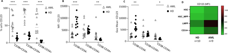
Background: Paediatric acute myeloid leukaemia (AML) is characterized by poor outcomes in patients with relapsed/refractory disease, despite the improvements in intensive standard therapy. The leukaemic cells of paediatric AML patients show high expression of the CD123 antigen, and this finding provides the biological basis to target CD123 with the chimeric antigen receptor (CAR). However, CAR.CD123 therapy in AML is hampered by on-target off-tumour toxicity and a long "vein-to-vein" time.

Methods: We developed an off-the-shelf product based on allogeneic natural killer (NK) cells derived from the peripheral blood of healthy donors and engineered them to express a second-generation CAR targeting CD123 (CAR.CD123).

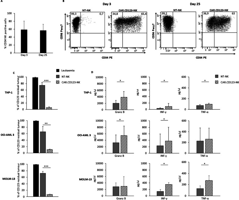
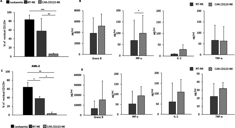
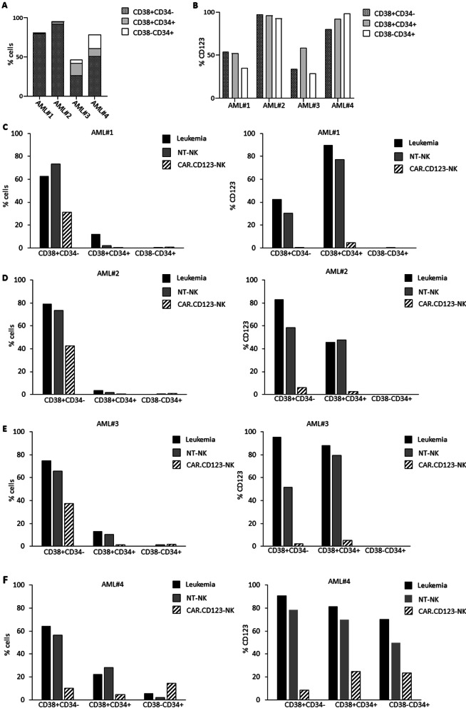
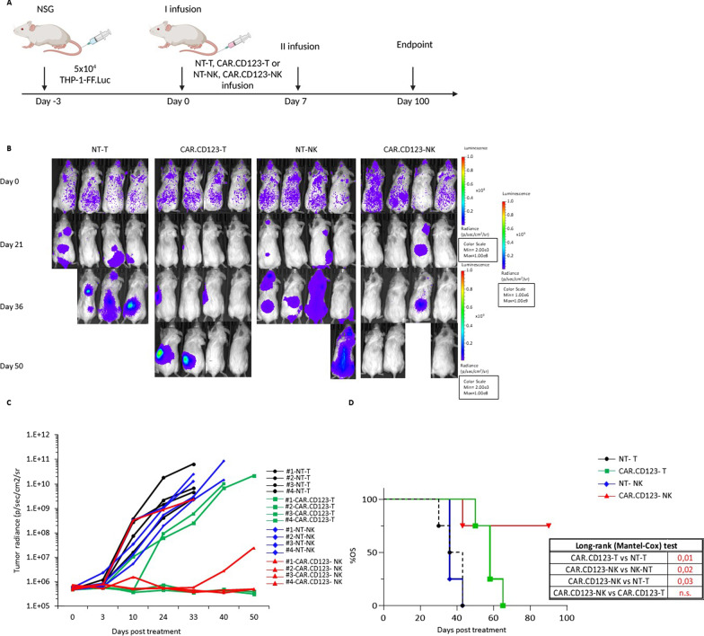
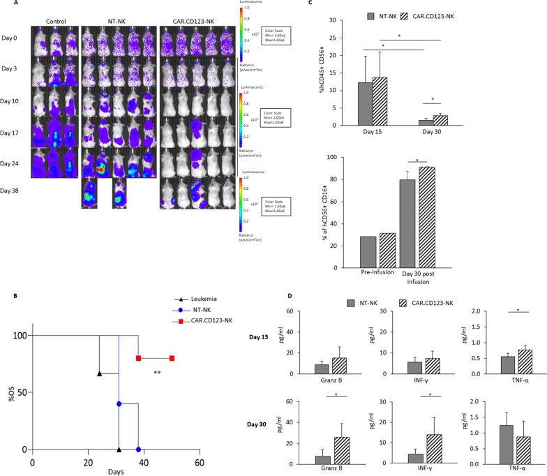
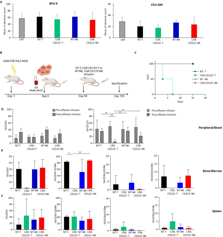
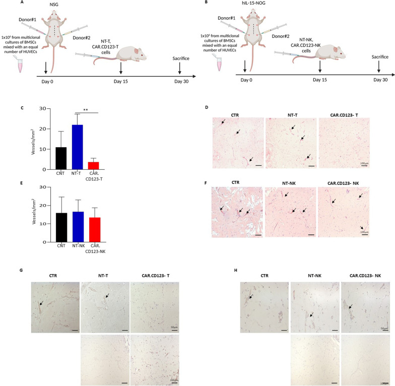
Results: CAR.CD123-NK cells showed significant anti-leukaemia activity not only in vitro against CD123+ AML cell lines and CD123+ primary blasts but also in two animal models of human AML-bearing immune-deficient mice. Data on anti-leukaemia activity were also corroborated by the quantification of inflammatory cytokines, namely granzyme B (Granz B), interferon gamma (IFN-γ) and tumour necrosis factor alpha (TNF-α), both in vitro and in the plasma of mice treated with CAR.CD123-NK cells. To evaluate and compare the on-target off-tumour effects of CAR.CD123-T and NK cells, we engrafted human haematopoietic cells (hHCs) in an immune-deficient mouse model. All mice infused with CAR.CD123-T cells died by Day 5, developing toxicity against primary human bone marrow (BM) cells with a decreased number of total hCD45+ cells and, in particular, of hCD34+CD38- stem cells. In contrast, treatment with CAR.CD123-NK cells was not associated with toxicity, and all mice were alive at the end of the experiments. Finally, in a mouse model engrafted with human endothelial tissues, we demonstrated that CAR.CD123-NK cells were characterized by negligible endothelial toxicity when compared to CAR.CD123-T cells.

Conclusions: Our data indicate the feasibility of an innovative off-the-shelf therapeutic strategy based on CAR.CD123-NK cells, characterized by remarkable efficacy and an improved safety profile compared to CAR.CD123-T cells. These findings open a novel intriguing scenario not only for the treatment of refractory/resistant AML patients but also to further investigate the use of CAR-NK cells in other cancers characterized by highly difficult targeting with the most conventional T effector cells.


Conflict of interest statement

The authors have no conflicting financial interests to disclose.

Figures

Fig. 1
Fig. 1
CD123 is highly expressed on primary blasts collected from paediatric AML patients. A Percentage of CD123 expression levels in primary BM cells obtained from AML patients (triangles) and HD (circles) were assessed by flow cytometry analysis, across 3 main haematopoietic cell subpopulations, namely the mature CD38+CD34 cells, the intermediate CD38+ CD34+ cells, and the most immature CD38 CD34+ stem cells. The gating strategy is shown in Additional file 1: Fig. S1. B Median fluorescence intensity (MFI) for CD123 on AML primary blasts or BM-derived cells from HDs. MFI was adjusted for cell size by dividing MFI by the forward scatter (FSC). C Geometric mean fluorescence intensities (GeoMean) of CD123 on AML primary blasts or BM-derived cells from HDs. Data are shown as average ± SD. *p < 0.05, **p < 0.01, ***p ≤ 0.001. D The heatmap shows the CD123 MFI detected on HSC precursors from BM of AML patients and HDs
Fig. 2
Fig. 2
CAR.CD123-NK cells exert a significant cytotoxicity against CD123+ AML cells. A–B CAR.CD123 is stably expressed on NK cells over extensive in vitro culture (evaluated up to 25 days). A The CAR.CD123 expression on NK cells from 9 different HDs was monitored at Day 3 and at Day 25 after transduction and is represented as average ± SD (B) Representative FACS analysis of CAR.CD123 expression in NK cells at Day 3 and Day 25 after transduction is shown. CAR expression was assessed by the use of anti-CD34 for the CAR detection in combination with anti-CD56 for NK detection. C Long-term 6-day cocultures were performed in 9 independent experiments, in which CD123+ leukaemia cell lines (namely, THP-1, OCI-AML3, and MOLM-13) (black bars) were cocultured with either NT-NK (grey bars) or CAR.CD123-NK cells (line black bars) derived from 9 independent HDs at the E:T ratio of 1:1. Data are shown as average ± SD. D Granz B, IFN-γ and TNF-α were measured by ELISA assay in 24 h culture supernatant of NT-NK (grey bars) or CAR.CD123-NK cells (line black bars) in response to CD123.+ leukaemia cell lines (namely, THP-1, OCI-AML3, and MOLM-13). Cytokine analysis was performed in 4 independent experiments, and data are shown as average ± SD. *p < 0.05, **p < 0.01, ***p < 0.001
Fig. 3
Fig. 3
CAR.CD123-NK cells efficiently recognize and kill primary AML blasts. A Leukaemic cells obtained from BM samples of 3 AML patients (AML#1, AML#3, AML#4) at diagnosis (black bars representing leukaemic cells alone) were cocultured for 6 days with NT-NK (grey bar), or CAR.CD123-NK (line black bar) derived from a single HD. Coculture was performed at an E:T ratio of 1:1. Data of residual CD123+ leukaemic cells is shown as average ± SD. B Granz B, IFN-γ, IL-2 and TNF-α were measured by ELISA assay in 24-h culture supernatant of NT-NK (grey bars) or CAR.CD123-NK cells (line black bars) in response to CD123+ leukaemic cells. C Leukaemic CD123+ cells from BM sample of one single AML patient (AML #2; black bar represent leukaemic cells alone) were cocultured with NT-NK (grey bars) or CAR.CD19-NK (line black bars) derived from 3 independent HDs. Coculture was performed at the E:T ratio of 1:1. Data of residual total CD123+ leukaemic cells is shown as average ± SD. *p < 0.05. D Granz B, IFN-γ, IL-2 and TNF-α were measured by ELISA assay in 24 h culture supernatant of NT-NK (grey bars) or CAR.CD123-NK cells (line black bars) in response to CD123.+ leukaemic cells from AML#2 patient. *p < 0.05
Fig. 4
Fig. 4
CAR.CD123-NK cells show a strong anti-leukaemia activity against the most relevant AML haematopoietic maturation subsets. A Percentage of CD38+CD34 (dotted bar), CD38+CD34+ (light grey bar) and CD38CD34+(white bar) cells obtained from BM samples of AML #1, AML #2, AML#3 and AML#4 were analysed by flow cytometry at thawing. B Percentage of CD123+ cells in the different haematopoietic maturation subsets, namely CD38+CD34 (dotted bar), CD38+CD34+ (light grey bar) and CD38CD34+(white bar) analysed at thawing of AML #1, AML #2, AML#3 and AML#4 BM samples (analysis performed before the in vitro culture of the cells). C AML #1, (D) AML #2, (E) AML#3 and (F) AML#4 BM cells have been independently analysed for the residual percentage of total CD38+CD34, CD38+CD34+ and CD38CD34+ cells (on the left) and for the residual percentage of CD123+ cells (on the right panel) after 6-day culture of leukaemic cells alone (black bars), leukaemic cells with NT-NK cells (grey bars) or with CAR.CD123-NK cells (black line bars) at the E:Tratio of 1:1
Fig. 5
Fig. 5
CAR.CD123-NK cells exert a significant anti-leukaemia activity in an AML xenograft NGS model. A Illustration of experimental setting in which NSG mice were systemically infused with THP-1-FF-Luc.GFP cells and, at the time of leukaemia engraftment, treated with NT-T, CAR.CD123-T, NT-NK and CAR.CD123-NK cells. B Time course of in vivo bioluminescence imaging of the treated NSG mice from Day 0 (Day of effector cells infusion) to Day 50. C Graph shows bioluminescence analysis of each leukaemia-bearing mouse treated with NT-T (black line with circles), CAR.CD123-T (green line with squares), NT-NK (blue line with pentagon) and CAR.CD123-NK (red line with triangles) cells. D One hundred days probability of overall survival (OS) of leukaemia-bearing mice treated with 2 consecutive adoptive transfers of NT-T (black line with circles), CAR.CD123-T (green line with squares), NT-NK (blue line with pentagon) and CAR.CD123-NK (red line with triangles) cells. On the right bottom, the statistical analysis of 100-Day OS of leukaemia-bearing mice treated with 2 consecutive adoptive transfers of NT-T, CAR.CD123-T, NT-NK, and CAR.CD123-NK cells
Fig. 6
Fig. 6
CAR.CD123-NK cells maintain efficacy and safety profile in an AML xenograft animal model based on hIL15-NOG mice. A hIL15-NOG mice were systemically engrafted with THP-1-FF-Luc.GFP cells and, at the time of leukaemia engraftment, treated or not with NT-NK and CAR.CD123-NK cells. Time course of in vivo bioluminescence imaging of the treated NOG mice. B Fifty-day probability of OS for NSG mice systemically infused with THP-1-FF-Luc.GFP cells after adoptive transfer of NT-NK (blue bars with circles) or CAR.123-NK cells (red bars with squares). The untreated control is represented by a black line with triangles. C Flow cytometry analysis of human NK cells in PB of mice (n = 5 mice receiving NK-NT; n = 5 mice receiving CAR.CD123-NK) performed at Day 15 and Day 30 after effector cell infusion. Plots show the percentage of hCD45+ hCD56+ hCD3 cells gated on the total cells present in PB of the mice (left panel) and as the percentage of hCD45+ hCD16+ cells of the total hCD45+ hCD56+ CD3. cells present in PB of the mice (right panel). Data are shown as average ± SD. *p < 0.05. D Cytokine concentration was measured in the plasma of mice bearing THP-1-FF-Luc.GFP cells and treated with NT-NK (grey bars) and CAR.CD123-NK (black line bars) cells at Day 15 and at Day 30 after effector cell infusion. Granz B, IFN-γ and TNF-α were measured by ELISA assay, and data are shown as average ± SD. *p < 0.05, **p < 0.01
Fig. 7
Fig. 7
In vitro and in vivo on-target off-tumour effect of both CAR.CD123-T and CAR.CD123-NK cells against hematopoietic precursors. A Colony-forming unit (CFU) assay was performed with CD34+ cells derived from BM samples of 3 HDs after a short-term 4-h coculture with media alone (control condition, CNT; black line bars), un-transduced T cells (NT-T; black bars), CAR.CD123-T (green bars), un-transduced NK cells (NT-NK; blue bars) or CAR.CD123-NK cells (red bars). BFU-E and CFU-GM colonies were counted after 14 days. Results are shown as average of absolute count ± SD. B Schematic representation of humanized mouse model of hGM-CSF/hIL3 NOG (hu-NOG) mice engrafted with CD34+ cells derived from cord blood (CB) and infused with effector cells. C 15-Day probability of OS for humanized mouse model engrafted with adoptive transfer of NT-T (black line with circle), CAR.CD123-T (dotted green line with squares), NT-NK (blue line with rhombus) and CAR.CD123-NK (red line with triangles) cells. D Flow cytometry analysis of hCD45 cells in PB of hu-NOG mice before (grey bars) and (squared pattern bars) after 4 days from the effector cells infusion. Graphs show the percentage of hCD45+ cells on the total mononuclear cells present in PB (left panel), and the percentage of hCD123+ cells on the total hCD45+ cells present in the PB samples (right panel). Data are shown as average of ± SD. EF Flow cytometry analysis of human hematopoietic cell quantification in tibia BM (E) and spleen (F) of hu-NOG mice receiving NT-T (black bars), CAR.CD123-T (green bars), NK-NT (blue bars) or CAR.CD123-NK (red bars). Graphs report the percentage of hCD45+ cells on the total mononuclear cells present in the investigated tissue (first panel), the percentage of hCD34CD38+ cells on the total hCD45+ cells (second panel), the percentage of hCD34+CD38+ cells on the total hCD45+ cells (third panel), and the percentage of hCD34+CD38 cells on the total hCD45.+ cells (fourth panel). Data are shown as average ± SD. *p < 0.05, **p < 0,001
Fig. 8
Fig. 8
CAR.CD123-NK cells do not target the human endothelial, preserving the vessels integrity in the murine model. AB Schematic representation of angiogenesis assay in NSG mice engrafted at Day 15 with NT-T or CAR.CD123-T and hIL-15NOG mice engrafted at Day 15 with NT-NK or CAR.CD123-NK cells. Mice were killed at 30 days post-Matrigel injection. C The number of vessels per mm2 was reduced in CAR.CD123-T cells-bearing NSG mice (red column) as compared to control (mice that were not infused with effector cells, black column) or to mice receiving NT-T cells (blue column). Data are shown as average ± SD; **p < 0,001. D Haematoxylin and eosin-stained sections of Matrigel plugs showing the formation of an extensive network of capillary-like blood vessels in control and in NT-T infused mice, in contrast to CAR.CD123-T infused mice. E hIL-15NOG mice carrying Matrigel plugs were injected with NK-NT (blue column) or CAR.CD123-NK cells (red column), and no differences were observed in the vessels number compared to the control condition (CNT, black column). Data are shown as average of ± SD. F Haematoxylin and eosin-stained sections of Matrigel plugs derived from hIL15NOG mice infused with CAR.CD123-NK show no differences in the newly formed capillary network. G Representative images of immunohistochemical staining for CD123 (top panels) and CD45 (bottom panels) in samples derived from mice infused with NT-T or CAR.CD123-T cells. H Representative images of immunohistochemical staining for CD123 (top panels) and CD45 (bottom panels) in samples derived from mice infused with NT-NK or CAR.CD123-NK cells

References

    1. Kantarjian H, et al. Acute myeloid leukemia: current progress and future directions. Blood Cancer J. 2021;11:41. - PMC - PubMed
    1. Hernandez-Lopez A, Tellez-Gonzalez MA, Mondragon-Teran P, Meneses-Acosta A. Chimeric antigen receptor-T cells: a pharmaceutical scope. Front Pharmacol. 2021;12:720692. - PMC - PubMed
    1. Haubner S, et al. Coexpression profile of leukemic stem cell markers for combinatorial targeted therapy in AML. Leukemia. 2019;33:64–74. - PMC - PubMed
    1. Fiorenza S, Turtle CJ. CAR-T cell therapy for acute myeloid leukemia: preclinical rationale, current clinical progress, and barriers to success. BioDrugs. 2021;35:281–302. - PubMed
    1. Bras AE, et al. CD123 expression levels in 846 acute leukemia patients based on standardized immunophenotyping. Cytom B Clin Cytom. 2019;96:134–142. - PMC - PubMed

Publication types

Substances