. 2023 Jan;34(1):99-107.
doi: 10.1111/jce.15734. Epub 2022 Nov 16.

Characteristics of pulsed electric field cardiac ablation porcine treatment zones with a focal catheter

Affiliations

Characteristics of pulsed electric field cardiac ablation porcine treatment zones with a focal catheter

Atul Verma et al. J Cardiovasc Electrophysiol. 2023 Jan.

Abstract

Objectives: Pulsed electric field (PEF) therapies employ punctuated energy delivery to kill cells in a volume of tissue through mechanisms that are not dependent on thermal processes. A key component to successful cardiac ablation procedures is ensuring the generation of transmural, contiguous ablation zones, which requires in-depth knowledge regarding treatment sizes for a given therapeutic application.

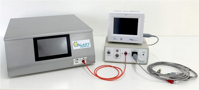
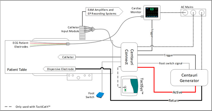
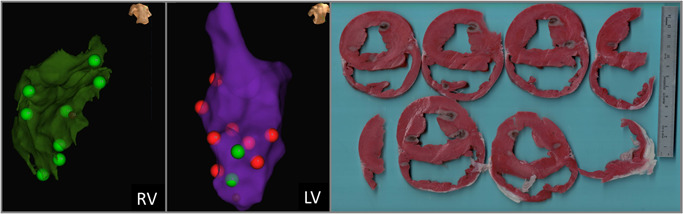
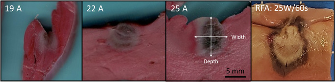
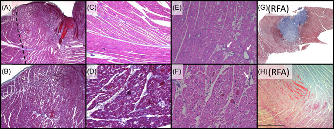
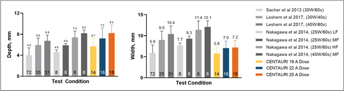
Methods: In this study, a series of acute treatments were delivered to porcine ventricles, where triphenyl tetrazolium chloride (TTC) vitality stain was used to identify treatment effect sizes for the three focal monopolar CENTAURI PEF cardiac ablation energy settings.

Results: Treatment depths were 5.7, 7.2, and 8.2 mm for the 19, 22, and 25 A energy settings, respectively. Gross pathology indicated umbral zones of hemorrhage surrounded by pale avital TTC-negative-negative tissue, which contrasted significantly from radiofrequency ablation (RF) controls. Histologically, treatment zones are identified by regions of contraction band necrosis and cardiomyocytolysis, which contrasted with RF control lesions composed primarily of coagulation necrosis.

Conclusions: Together, these data indicate the ability for focal monopolar PEF treatments to generate deep treatment zones in cardiac ablation without incurring the gross or histological coagulative characteristics of RF thermal lesions.

Keywords: Aliya; CENTAURI; dose; electroporation; lesion; size.


Conflict of interest statement

Dr. Verma reports grants from Bayer, Biosense Webster, Abbott, and Medtronic; advisory for Biosense Webster, Medtronic, Boston Scientific, Adagio Medical, Medlumics, and Ablacon; research with Galaxy Medical. Drs. Neal, Vachani, and Evans are researchers with Galaxy Medical. Dr. Deneke reports advisory for Galaxy Medical and Farapulse; Dr. Nakagawa reports research for Biosense Webster, Galaxy Medical.

Figures

Figure 1
Figure 1
The Galaxy Medical CENTAURI System consists of the generator, the catheter connection box, and all associated cabling for the delivery of focal monopolar biphasic PEF energy at three distinct energy settings. PEF, pulsed electric field
Figure 2
Figure 2
The PEF system set‐up and integration with standard electroanatomical mapping system, EP recording system, and the TactiCath SE cardiac ablation catheter. PEF, pulsed electric field
Figure 3
Figure 3
Matching EnSite Precision treatment sites with gross pathology treatment effects. The maps and anatomical landmarks were used to delineate respective treatments. Here, green lesions tags denote where the 19 Amp energy setting was used, while red lesion tags denote the 25 Amp energy setting.
Figure 4
Figure 4
Representative acute (>2 h) gross pathology results from the three energy settings. TTC stained vital tissue red, while avital tissue is pale. An umbral region of acute hemorrhage is identifiable, which was observed to resolve 3 days after PEF delivery (data not shown). A panel of a representative acute RFA ventricle lesion (25 W, 60 s, 25 g of contact force, with 17 ml/min irrigation) is included to demonstrate the clear difference between monopolar PEF energy and thermal ablation modalities, where a large core of tan and brown tissue is found, surrounded by a penumbra of hemorrhage. TTC, triphenyl tetrazolium chloride
Figure 5
Figure 5
Characteristic histology for the acute monopolar PEF system tested relative to RFA lesions with (A–D, G, H) Masson's Trichrome and (E, F) Hematoxylin and Eosin. (A) Low power magnification (×2) of a left ventricle lesion, demonstrating a focal ablation site characterized by acute myocardial necrosis in the treated area (right of dashed line). (B) Higher magnification (×4) of Panel A, demonstrating the transition zone between necrotic and viable myocytes. (C) Adjacent control (untreated) tissue (×10), which contrasts with (D) the treatment (×20) showing acute contraction band necrosis. (E: ×10, F: ×20) lesion region showing myocyte contraction, hypereosinophilia, and cardiomyocytolysis “moth eaten” appearance of necrotic muscle, with interspersed intact blood vessels (arrows). (G) Low power magnification of a left ventricle RFA lesion characterized by a large zone of coagulation necrosis (blue) surrounded by a penumbra of hemorrhagic contraction band necrosis (dark purple). (F) Higher magnification of the boundary zone, further contrasting the difference in lesion histopathology between Galaxy PEF treatments and RF thermal ablation. (A): Low mag Tri lesion boundary. (B): Mid‐mag Tri lesion boundary. (C): Control Mid‐high zoom. (D): High zoom lesion CBN. (E): Midhigh‐mag HE lesion (myocytolysis). (F): High mag HE lesion. (G): low mag RF. (H): mid‐mag RF. PEF, pulsed electric field; RF, radiofrequency
Figure 6
Figure 6
Comparing focal monopolar PEF ventricle ablation depths (left) and widths (right) with RF., , RF contact forces indicated by low force (LF, 3–8 g), medium force (MF, 18–27 g), and high force (HF, 40–62 g). Trial numbers for each test condition are indicated inside the bars. PEF, pulsed electric field; RF, radiofrequency

References

    1. Stewart MT, Haines DE, Miklavčič D, et al. Safety and chronic lesion characterization of pulsed field ablation in a Porcine model. J Cardiovasc Electrophysiol. 2021;32:958‐969. 10.1111/jce.14980 - DOI - PMC - PubMed
    1. Rubinsky B, Onik G, Mikus P. Irreversible electroporation: a new ablation modality—clinical implications. Technol Cancer Res Treat. 2007;6(no. 1):37‐48. 10.1177/153303460700600106 - DOI - PubMed
    1. Maor E, Ivorra A, Leor J, Rubinsky B. The effect of irreversible electroporation on blood vessels. Technol Cancer Res Treat. 2007;6(no. 4):307‐312. 10.1177/153303460700600407 - DOI - PubMed
    1. du Pré BC, van Driel VJ, van Wessel H, et al. Minimal coronary artery damage by myocardial electroporation ablation. EP Europace. 2013;15(no. 1):144‐149. 10.1093/europace/eus171 - DOI - PubMed
    1. Song Y, Zheng J. The esophagus survives non‐thermal irreversible electroporation ablation and gradually rehabilitates. In Review, preprint. 2020:1‐15. 10.21203/rs.3.rs-18960/v1 - DOI

Publication types