. 2022 Dec:86:104312.
doi: 10.1016/j.ebiom.2022.104312. Epub 2022 Nov 4.

Leukemia inhibitory factor is a therapeutic target for renal interstitial fibrosis

Affiliations

Leukemia inhibitory factor is a therapeutic target for renal interstitial fibrosis

Shihui Xu et al. EBioMedicine. 2022 Dec.

Abstract

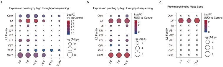
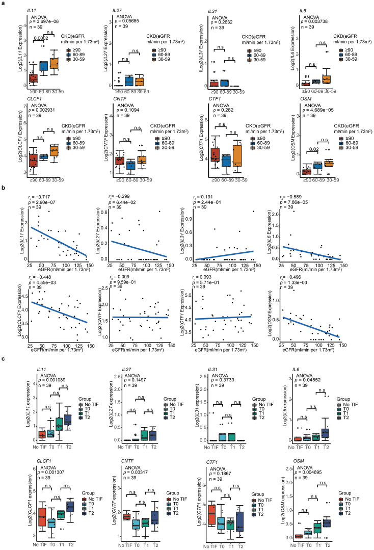
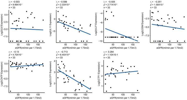
Background: The role of the IL6 family members in organ fibrosis, including renal interstitial fibrosis (TIF), has been widely explored. However, few studies have ever simultaneously examined them in the same cohort of patients. Besides, the role of leukemia inhibitory factor (LIF) in TIF remains unclear.

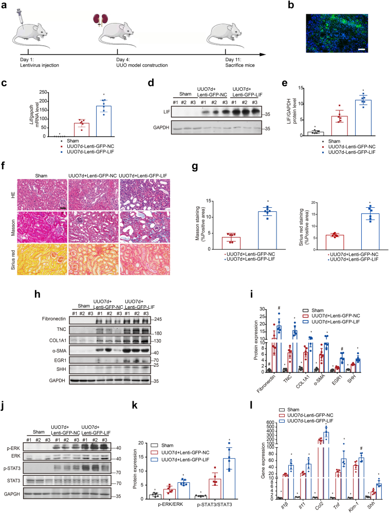
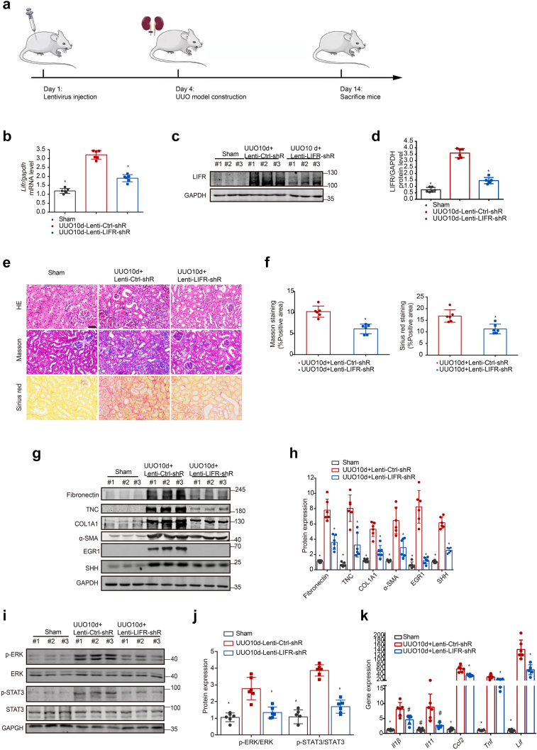
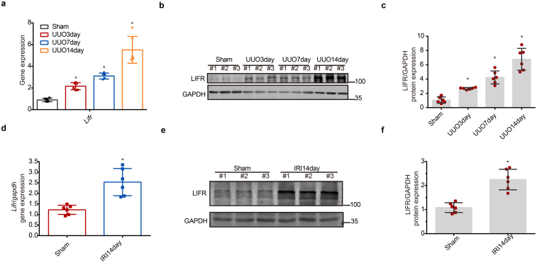
Methods: RNA-seq data of kidney biopsies from chronic kidney disease (CKD) patients, in both public databases and our assays, were used to analyze transcript levels of IL6 family members. Two TIF mouse models, the unilateral ureteral obstruction (UUO) and the ischemia reperfusion injury (IRI), were employed to validate the finding. To assess the role of LIF in vivo, short hairpin RNA, lenti-GFP-LIF was used to knockdown LIF receptor (LIFR), overexpress LIF, respectively. LIF-neutralizing antibody was used in therapeutic studies. Whether urinary LIF could be used as a promising predictor for CKD progression was investigated in a prospective observation patient cohort.

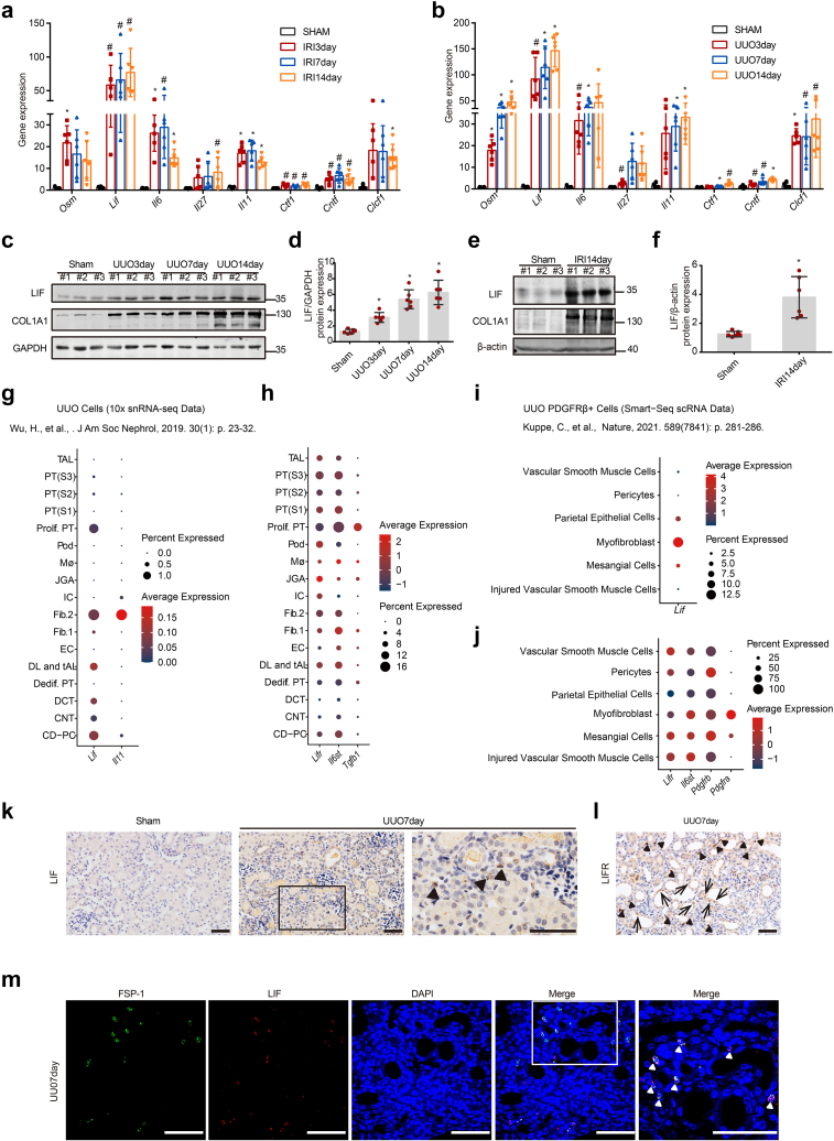
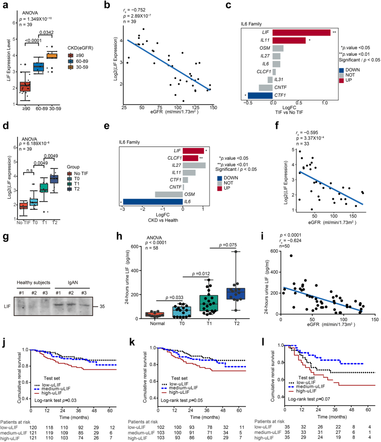
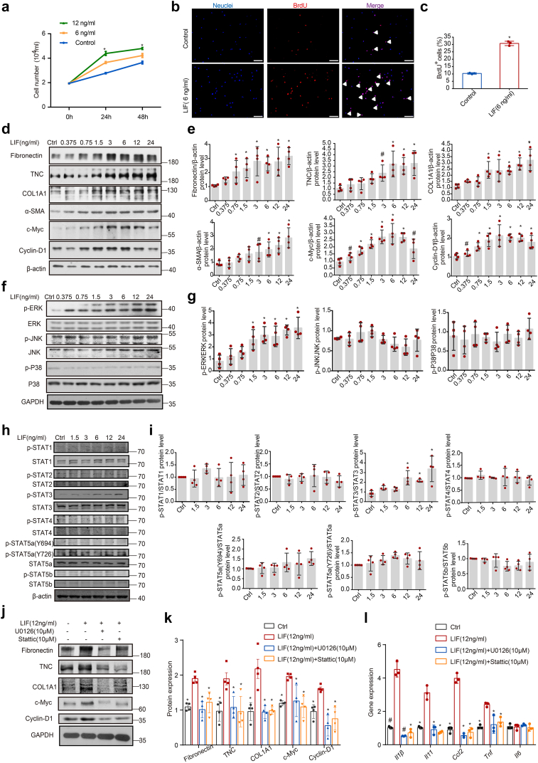
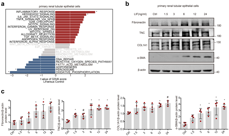
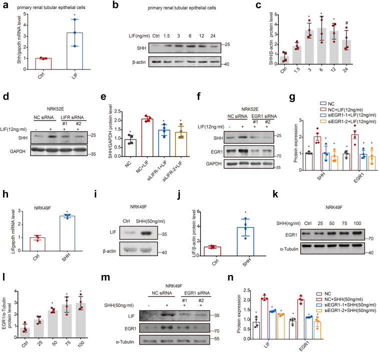
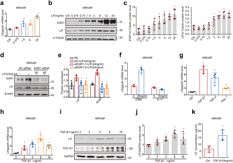
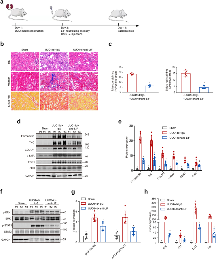
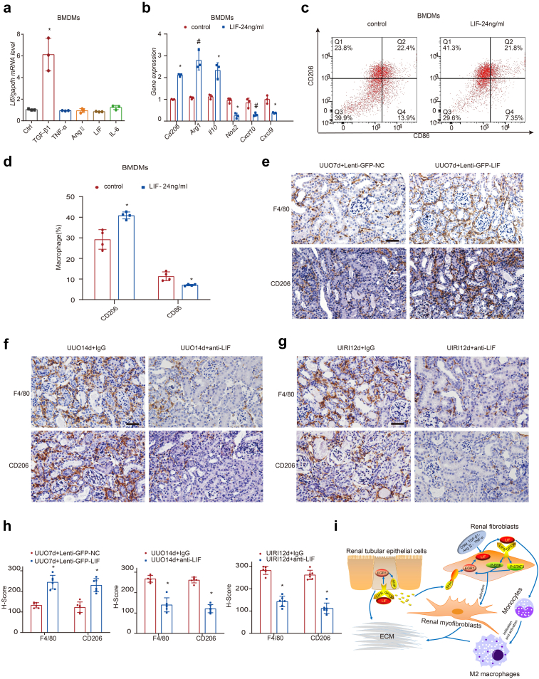
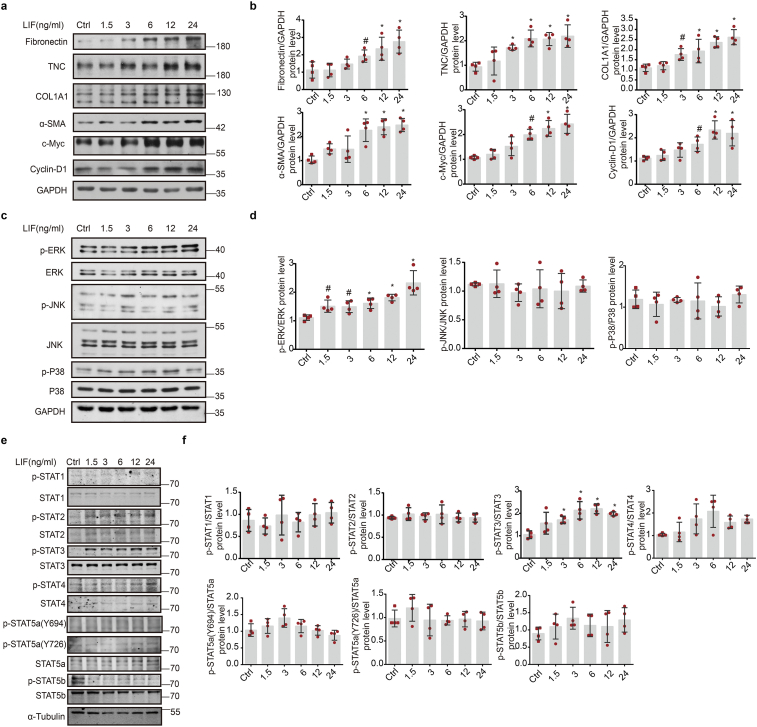
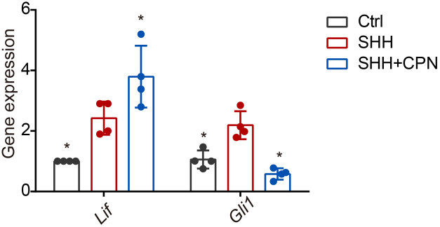
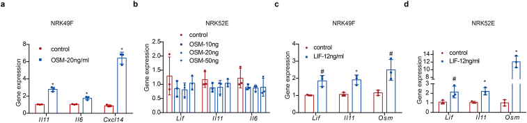
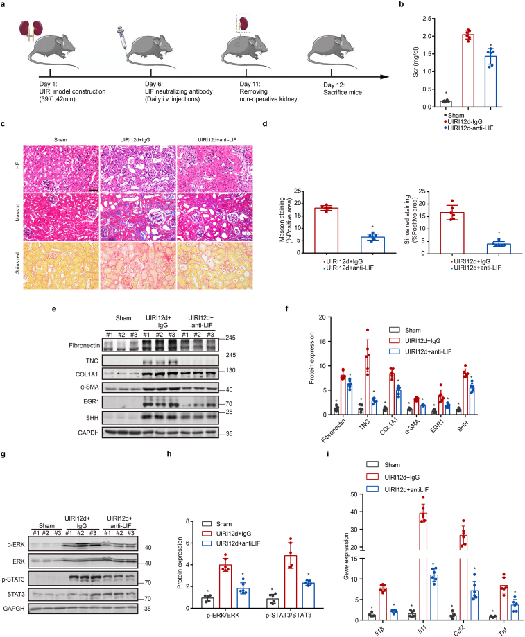
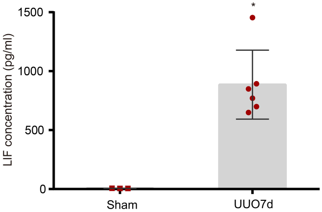
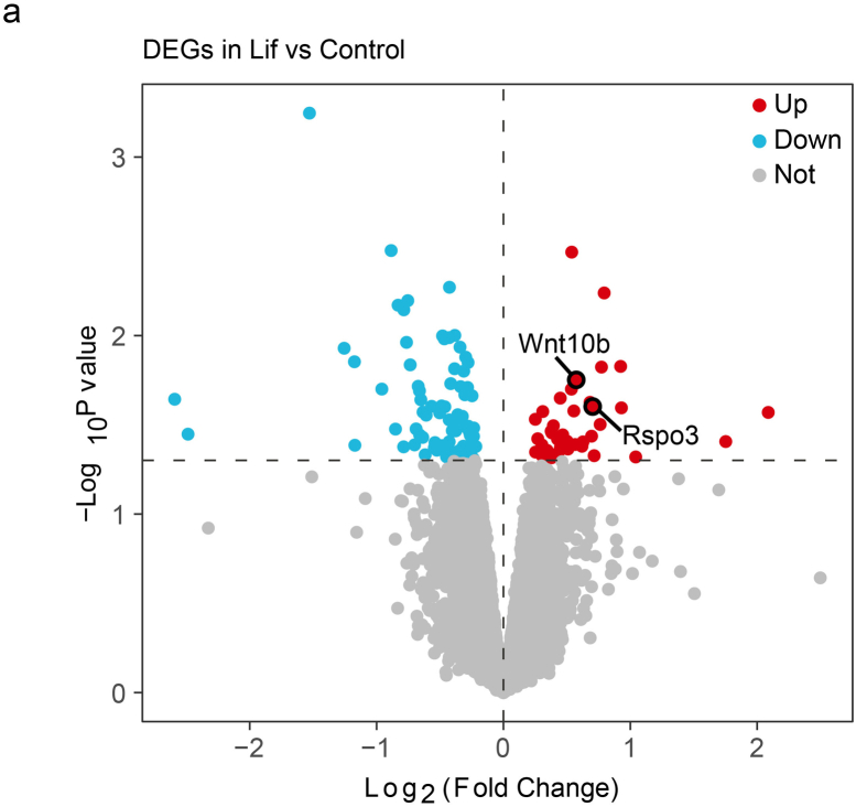
Findings: Among IL6 family members, LIF is the most upregulated one in both human and mouse renal fibrotic lesions. The mRNA level of LIF negatively correlated with eGFR with the strongest correlation and the smallest P value. Baseline urinary concentrations of LIF in CKD patients predict the risk of CKD progression to end-stage kidney disease by Kaplan-Meier analysis. In mouse TIF models, knockdown of LIFR alleviated TIF; conversely, overexpressing LIF exacerbated TIF. Most encouragingly, visible efficacy against TIF was observed by administering LIF-neutralizing antibodies to mice. Mechanistically, LIF-LIFR-EGR1 axis and Sonic Hedgehog signaling formed a vicious cycle between fibroblasts and proximal tubular cells to augment LIF expression and promote the pro-fibrotic response via ERK and STAT3 activation.

Interpretation: This study discovered that LIF is a noninvasive biomarker for the progression of CKD and a potential therapeutic target of TIF.

Fundings: Stated in the Acknowledgements section of the manuscript.

Keywords: Chronic kidney disease; Fibroblast activation; IL6 family; LIF; Renal fibrosis.


Conflict of interest statement

Declaration of interests All the authors declared no conflict of interest.

Figures

Fig. 1
Fig. 1
LIF was upregulated in fibroblasts in mouse models of TIF induced by UUO and IRI. (a) The mRNA level of IL6 cytokines family at days 3, 7 and 14 after IRI (n = 6), normalized with gapdh. (b) The mRNA level of IL6 cytokines family at days 3, 7 and 14 after UUO (n = 6), normalized with gapdh. (c and d) Western blot analysis (c) and densitometric quantification (d) of LIF protein at days 3, 7 and 14 after UUO (n = 6). (e and f) Western blot analysis (e) and densitometric quantification (f) of LIF protein at day 14 after IRI (n = 6). (g) Dotplot illustrating expression of Lif and Il11 in single nucleus RNA sequencing (snRNA-seq) dataset of fibrotic kidney from mice 14 days after UUO. (h) Dotplot illustrating expression of Lifr, Il6st and Tgfb1 in single nucleus RNA sequencing (snRNA-seq) dataset of fibrotic kidney from mice 14 days after UUO. (i) Dotplot illustrating expression of Lif in a published Smart-Seq dataset of PDGFRβ+ cells from mice 10 days after UUO. (j) Dotplot illustrating expression of Lifr, Il6st, pdgfrb and pdgfra in a published Smart-Seq dataset of PDGFRβ+ cells from mice 10 days after UUO. (k) Representative LIF immunostaining in renal sections from sham mice (left panel) and UUO mice (middle and right panel). Magnification, ×400 (left and middle panel), ×1000 (right panel). Arrows indicate positive LIF expression. The boxed region was shown at higher magnification (right panels). Scale bar, 50 μm. (l) Representative LIFR immunostaining in renal sections from UUO 7 day mice. LIFR was detected in mesenchymal cells (Use triangle to indicate) and some tubular epithelial cells (Use arrows to indicate). Magnification, ×400. (m) Representative fluorescent images of coimmunostaining LIF (red) with FSP-1 (green) in renal sections from UUO mice. Arrows in magnified image indicate colocalization of LIF and FSP-1. The boxed region was showed at higher magnification. Scale bar, 50 μm. Data were expressed as means ± SD. #P < 0.05, ∗P < 0.01 versus sham mice. P values were determined by Student's t-test in (f) and one-way ANOVA (Least-Significant Difference test or Dunnett's T3 test) in (a), (b) and (d). Data in (g) and (h) referenced frome Wu, H., et al., 2019. Data in (i) and (j) referenced frome Kuppe, C., et al., 2021.
Fig. 2
Fig. 2
LIF expression in renal biopsies and urine from CKD patients. (a) The expression of LIF along with the decline of eGFR (n = 39). (b) The association between the log2 of LIF expression and eGFR (n = 39). (c) Differential expression of IL6 family cytokines between subjects with (n = 33) and without (n = 6) TIF. (d) The expression of LIF in different stages of Oxford-T grades of IgAN (n = 39). (e) Differential expression of IL6 family cytokines between CKD (n = 28) and healthy subjects (n = 9) from the ERCB Nephrotic Syndrome Data set. (f) The association between the log2 of LIF expression and eGFR in subjects (n = 33) from the ERCB Nephrotic Syndrome Data set. (g) Western blot analysis of LIF protein in 24 h-urine. (h) The amount of LIF protein in 24 h-urine was increased along with the stage of Oxford-T grades of IgAN (n = 50) and healthy controls (n = 8). The amount of LIF protein in 24 h-urine was measure by ELISA. (i) The amount of LIF protein in 24 h-urine negatively correlated with eGFR of IgAN patients (n = 50). (j–l) The level of uLIF at baseline predicts the risk of CKD progression to ESRD. Tertiles of uLIF-7 levels had a graded relationship with the risk of CKD progression to ESRD in total cohort (j), in subgroups of patients with eGFR <60 ml/min/1.73 m2 (k) and patients with proteinuria >1.0 g/d (l). Data in (a), (d) and (h) were represented as median ± interquartile range (IQR) and P values were determined by one-way ANOVA (Least-Significant Difference test). The Spearman correlation analysis was used in (b), (f) and (i). The R package limma (3.46.0) was used in (c) and (e) for gene differential expression analysis between two groups. Data in (j), (k) and (l) were generated using the Kaplan–Meier method and compared using the log-rank test.
Fig. 3
Fig. 3
LIF promoted proliferation, activation of rat fibroblasts through ERK1/2 and STAT3 pathway. (a) NRK49F cells were incubated with LIF (6 ng/ml or 12 ng/ml) for 24 h or 48 h, and then cell numbers were counted. #P < 0.05, ∗P < 0.01 versus controls (n = 3). (b and c) Representative micrographs of BrdU incorporation and quantitative determination of the percentage of BrdU-positive cells. Arrows indicate BrdU-positive cells. Scale bar, 100 μm. #P < 0.05, ∗P < 0.01 versus controls (n = 3). (d and e) Western blot analyses (d) and quantitative data (e) showed that LIF upregulated the expression of proliferation and activation-related proteins in fibroblasts in a dose-dependent manner. NRK49F cells were incubated with indicated concentrations of LIF for 48 h. Cell lysates were subjected to Western blot analysis for c-Myc, Cyclin-D1, α-SMA, TNC, Fibronectin, and COL1A1. β-actin was used to verify equivalent loading. #P < 0.05, ∗P < 0.01 versus controls (n = 4). (f and g) Representative Western blot (f) and quantitative data (g) showed that LIF induced phosphorylation of ERK1/2 in a dose-dependent manner. NRK49F cells were treated with indicated concentration of LIF for 30 min. #P < 0.05, ∗P < 0.01 versus controls (n = 4). (h and i) NRK49F cells were treated with gradient concentration of LIF for 2 h. Representative Western blot (h) and quantitative data (i) showed that LIF induced phosphorylation of STAT3 in a dose-dependent manner. #P < 0.05, ∗P < 0.01 versus controls (n = 4). (j and k) Representative Western blot (j) and quantitative data (k) showed that blockade of Mek1/ERK1/2 or STAT3 signaling abolished LIF-induced fibroblast proliferation and activation. NRK49F cells were pretreated with specific MEK1 and MEK2 inhibitor U0126 (10 μM) or STAT3 inhibitor stattic (10 μM) for 30 min followed by incubation with LIF (6 ng/ml) or vehicle for 24 h. Cell lysates were subjected to Western blot analyses for c-Myc, Cyclin-D1, TNC, Fibronectin, and COL1A1. #P < 0.05, ∗P < 0.01 versus LIF alone (n = 4). (l) Real-time PCR analysis showed blockade of Mek1/ERK1/2 or STAT3 signaling reduced LIF-induced expression of pro-inflammatory cytokines in fibroblasts. NRK49F cells were pretreated with specific MEK1 and MEK2 inhibitor U0126 (10 μM) or STAT3 inhibitor stattic (10 μM) for 30 min, followed by incubation with LIF or vehicle for 24 h. Relative mRNA levels are reported after normalization with GAPDH. #P < 0.05, ∗P < 0.01 versus LIF alone (n = 3). Data were expressed as means ± SD. P values were determined by Student's t-test in (c) or one-way ANOVA (Least-Significant Difference test or Dunnett's T3 test) in (a), (e), (g), (i), (k) and (l).
Fig. 4
Fig. 4
LIF promotes pro-fibrotic response in renal tubular cells. (a) Top enriched hallmark pathways identified by GSVA. The R package limma (3.46.0) was used for pathways differential expression analysis between two groups. (b and c) Representative Western blot (b) and quantitative data (c) showed LIF upregulated the expression of Fibronectin, TNC, COL1A1 and α-SMA in primary renal tubular epithelial cells in a dose-dependent manner. #P < 0.05, ∗P < 0.01 versus controls (n = 4). One-way ANOVAs (Least-Significant Difference test or Dunnett's T3 test) in (c) was used for determining the P values while comparing between each pair of groups, respectively.
Fig. 5
Fig. 5
LIF and SHH mediated the crosstalk between fibroblasts and renal tubular cells. (a) Quantitative real-time PCR showed the mRNA level of Shh induced by LIF (6 ng/ml, 24 h) in primary renal tubular epithelial cells, normalized with gapdh. ∗P < 0.01 versus control (n = 3). (b and c) Representative Western blot (b) and quantitative data (c) showed LIF upregulated the expression of SHH in primary renal tubular epithelial cells in a dose-dependent manner. #P < 0.05, ∗P < 0.01 versus controls (n = 4). (d and e) Representative Western blot (d) and quantitative data (e) showed silencing LIFR abolished LIF-induced SHH expression in NRK52E cells. NRK52E cells were transfected with scramble siRNA or siLIFR respectively, and then treated with 12 ng/ml LIF for 72 h. #P < 0.05, ∗P < 0.01 versus scramble siRNA + LIF (n = 4). (f and g) Representative Western blot (f) and quantitative data (g) showed silencing EGR1 abolished LIF-induced SHH expression in NRK52E cells. NRK52E cells were transfected with scramble siRNA or siEGR1 respectively, and then treated with 12 ng/ml LIF for 72 h. (h) Quantitative real-time PCR showed that SHH (50 ng/ml, 24 h) upregulated the mRNA level of Lif in NRK49F cells. ∗P < 0.01 versus control (n = 4). (i and j) Representative Western blot (i) and quantitative data (j) showed that SHH (50 ng/ml, 24 h) increased the protein level of LIF in NRK49F cells. ∗P < 0.01 versus control (n = 4). (k and l) Western blot analyses (k) and quantitative data (l) showed the upregulation of EGR1 by SHH in NRK49F cells. ∗P < 0.01 versus controls (n = 4). (m and n) Western blot (m) and quantitative data (n) showed SHH-induced LIF expression was attenuated by EGR1 siRNA. ∗P < 0.01 versus LIF alone (n = 4). #P < 0.05, ∗P < 0.01 versus scramble siRNA + LIF (n = 4). Data were expressed as means ± SD. Student's t-test in (a), (h) and (j) or one-way ANOVAs (Least-Significant Difference test or Dunnett's T3 test) in (c), (e), (g), (l) and (n) was used for determining the P values while comparing between each pair of groups, respectively.
Fig. 6
Fig. 6
Upstream regulators of LIF expression. (a–e) LIF upregulated the expression of itself in NRK49F cells through ERK-EGR1 axis. NRK49F cells were treated with LIF for 24 h. (a) Real-time PCR showed that LIF upregulated itself in NRK49F cells. Relative Lif mRNA levels were shown as fold induction over controls after normalization with gapdh, respectively. ∗P < 0.01 versus controls (n = 3). (b and c) Western blot analyses (b) and quantitative data (c) showed that LIF induced the expression of itself and EGR1 in a dose-dependent manner. #P < 0.05, ∗P < 0.01 versus controls (n = 4). (d and e) Western blot analyses (d) and quantitative data (e) showed that knocking down EGR1 diminished LIF-induced LIF production. #P < 0.05, ∗P < 0.01 versus NC+LIF (n = 4). (f) Upstream regulators of Lif expression. NRK49F cells were treated with OSM (20 ng/ml) and IL11 (10 ng/ml). (g) Upstream regulators of Lif expression. NRK49F cells were treated with TGF-β1 (8 ng/ml), TNF-α (8 ng/ml) and Ang II (10 nM/ml) for 24 h. Real-time PCR measured the mRNA level of Lif. Relative Lif mRNA levels were showed as fold induction over controls after normalization with gapdh, respectively. ∗P < 0.01 versus controls (n = 4). (h) The mRNA level of Lif were induced by TGF-β1 induced LIF expression in a dose-dependent manner in NRK49F cells. ∗P < 0.01 versus controls (n = 4). (i and j) Western blotting Representative Western blot (i) and quantitative data (j) showed that the protein level of LIF were induced by TGF-β1 in a dose-dependent manner in NRK49F cells. #P < 0.05, ∗P < 0.01 versus controls (n = 4). (k) NRK49F cells were treated with TGF-β1 (8 ng/ml) for 24 h, and the LIF concentration in the culture medium was determined by ELISA. ∗P < 0.01 versus controls (n = 4). Data were expressed as means ± SD. Student's t-test in (f), (g) and (k) or one-way ANOVAs (Least-Significant Difference test or Dunnett's T3 test) in (a), (c), (e), (h) and (j) was used for determining the P values while comparing between each pair of groups, respectively.
Fig. 7
Fig. 7
Ectopic expression of LIF exacerbated TIF induced by UUO. (a) Schematic diagram showed the experimental design. (b) The frozen section of positive GFP-LIF staining at day 7 after UUO. Scale bar, 50 μm. (c) Real-time PCR showed the mRNA levels of Lif at day 7 after UUO. (d and e) The protein levels of LIF were assessed by Western blot at day 7 after UUO. Representative Western blot (d) and quantitative data (e) are showed. (f and g) Representative images of H&E, Masson trichrome and Sirius red staining of renal cortex at day 7 after UUO in three groups (f) and quantitative analysis of fibrotic area (g). Scale bar, 50 μm. (h and i) Western blotting showed the protein levels of TNC, Fibronectin, COL1A1, α-SMA, EGR1, SHH at day 7 after UUO. Representative Western blot (h) and quantitative data (i) are showed. (j and k) The protein levels of p-ERK, ERK, p-STAT3 and STAT3 were assessed by Western blot analyses at day 7 after UUO. Representative Western blot (j) and quantitative data (k) are showed. (l) Real-time PCR showed the mRNA levels of Il1β, Il11, Ccl2, Tnf, Kim-1 and Shh at day 7 after UUO. Data were expressed as means ± SD. Student's t-test in (g) or one-way ANOVAs (Least-Significant Difference test or Dunnett's T3 test) in (c), (e), (i), (k) and (l) was used for determining the P values while comparing between each pair of groups, respectively, #P < 0.05, ∗P < 0.01 versus UUO 7day-lenti-GFP-NC (n = 6).
Fig. 8
Fig. 8
Knocking down LIFR ameliorated renal fibrotic lesions induced by UUO. (a) Schematic diagram of the experimental design. (b) Real-time PCR showed the mRNA levels of Lifr at day 10 after UUO. (c and d) Representative Western blot (c) and quantitative data (d) showed the protein level of LIFR at day 10 after UUO. (e and f) Representative images of H&E, Masson trichrome and Sirius red staining of renal cortex at day 10 after UUO in three groups (e) and quantitative analyses of fibrotic area (f). Scale bar, 50 μm. (g and h) Representative Western blot (g) and quantitative data (h) showed the protein levels of TNC, Fibronectin, COL1A1, α-SMA, EGR1, SHH at day 10 after UUO. (i and j) Representative Western blot (i) and quantitative data (j) showed the protein levels of p-ERK, ERK, p-STAT3 and STAT3 at day 10 after UUO. (k) Real-time PCR showed the mRNA levels of Il1β, Il11, Ccl2, Tnf and Lif at day 10 after UUO. Data were expressed as means ± SD. Student's t-test in (f) or one-way ANOVAs (Least-Significant Difference test or Dunnett's T3 test) in (b), (d), (h), (j) and (k) was used for determining the P values while comparing between each pair of groups, respectively, #P < 0.05, ∗P < 0.01 versus UUO10day+Lenti-Ctrl-shR (n = 6).
Fig. 9
Fig. 9
Neutralizing LIF antibody attenuated renal fibrosis induced by UUO. (a) Schematic diagram of the experimental design. (b and c) Representative images of H&E, Masson trichrome and Sirius red staining of renal cortex at day 14 after UUO in three groups (b) and quantitative analyses of fibrotic area (c). Scale bar, 50 μm. (d and e) Representative Western blot (d) and quantitative data (e) showed the protein levels of TNC, Fibronectin, COL1A1, α-SMA, EGR1and SHH at day 14 after UUO. (f and g) Representative Western blot (f) and quantitative data (g) showed the protein levels of p-ERK, ERK, p-STAT3 and STAT3 at day 14 after UUO. (h) Real-time PCR showed the mRNA levels of Il1β, Il11, Ccl2 and Tnf at day 14 after UUO. Data were expressed as means ± SD. Student's t-test in (c) or one-way ANOVAs (Least-Significant Difference test or Dunnett's T3 test) in (e), (g) and (h) was used for determining the P values while comparing between each pair of groups, respectively, #P < 0.05, ∗P < 0.01 versus UUO14day-IgG (n = 6).
Fig. 10
Fig. 10
LIF promoted macrophage infiltration and induced M2 macrophages polarization. (a) BMDMs were treated with TGF-β1 (8 ng/ml), TNF-α (8 ng/ml), Ang II (10 nM/ml), LIF (24 ng/ml) and IL6 (10 ng/ml) for 24 h. #P < 0.05, ∗P < 0.01 versus controls (n = 3). (b) BMDMs were treated with LIF for 48 h. Real-time PCR measured the mRNA level of Cd206, Arg1, II10, Nos2, Cxcl10 and Cxcl9. Relative mRNA levels were showed as fold induction over controls after normalization with gapdh, respectively. #P < 0.05, ∗P < 0.01 versus controls (n = 3). (c and d) Flow cytometry analysis of BMDMs polarization with or without LIF stimulation (c) and quantitative data (d). #P < 0.05, ∗P < 0.01 versus controls (n = 4). (e) Representative F4/80 and CD206 IHC image at day 7 after UUO in two groups. (f and g) Representative F4/80 and CD206 IHC image at day 14 after UUO (f) and at day 12 after UIRI (g). (h) H-score of F4/80 and CD206 staining at day 7, 14 after UUO and day 12 after UIRI. #P < 0.05, ∗P < 0.01 versus UUO7d-Lenti-GFP-NC, UUO14d-IgG and UIRI-12d-IgG, respectively (n = 6). (i) Schematic presentation the role of LIF in macrophages and in the crosstalk between renal fibroblasts and renal tubular epithelial cells and the mechanism to pro-fibrotic response during renal fibrosis. Data were expressed as means ± SD. Student's t-test was used for determining the P values while comparing between each pair of groups, respectively. #P < 0.05, ∗P < 0.01.
Supplementary Fig. S1
Supplementary Fig. S2
Supplementary Fig. S3
Supplementary Fig. S4
Supplementary Fig. S5
Supplementary Fig. S6
Supplementary Fig. S7
Supplementary Fig. S8
Supplementary Fig. S9
Supplementary Fig. S10

Comment in

Similar articles

Cited by

References

    1. Chen T.K., Knicely D.H., Grams M.E. Chronic kidney disease diagnosis and management: a review. JAMA. 2019;322(13):1294–1304. doi: 10.1001/jama.2019.14745. - DOI - PMC - PubMed
    1. Kakitapalli Y., Ampolu J., Madasu S.D., Sai K.M. Detailed review of chronic kidney disease. Kidney Dis (Basel) 2020;6(2):85–91. doi: 10.1159/000504622. - DOI - PMC - PubMed
    1. Liu Y. Cellular and molecular mechanisms of renal fibrosis. Nat Rev Nephrol. 2011;7(12):684–696. doi: 10.1038/nrneph.2011.149. - DOI - PMC - PubMed
    1. Djudjaj S., Boor P. Cellular and molecular mechanisms of kidney fibrosis. Mol Aspects Med. 2019;65:16–36. doi: 10.1016/j.mam.2018.06.002. - DOI - PubMed
    1. Lebleu V.S., Taduri G., O'Connell J., et al. Origin and function of myofibroblasts in kidney fibrosis. Nat Med. 2013;19(8):1047–1053. doi: 10.1038/nm.3218. - DOI - PMC - PubMed