. 2022 Nov;53(7):694-710.
doi: 10.1016/j.arcmed.2022.10.007. Epub 2022 Nov 3.

Bioinformatic Analysis of B- and T-cell Epitopes from SARS-CoV-2 Structural Proteins and their Potential Cross-reactivity with Emerging Variants and other Human Coronaviruses

Affiliations

Bioinformatic Analysis of B- and T-cell Epitopes from SARS-CoV-2 Structural Proteins and their Potential Cross-reactivity with Emerging Variants and other Human Coronaviruses

Diana Laura Pacheco-Olvera et al. Arch Med Res. 2022 Nov.

Abstract

Background: The mutations in SARS-CoV-2 variants of concern (VOC) facilitate the virus' escape from the neutralizing antibodies induced by vaccines. However, the protection from hospitalization and death is not significantly diminished. Both vaccine boosters and infection improve immune responses and provide protection, suggesting that conserved and/or cross-reactive epitopes could be involved. While several important T- and B-cell epitopes have been identified, mainly in the S protein, the M and N proteins and their potential cross-reactive epitopes with other coronaviruses remain largely unexplored.

Aims: To identify and map new potential B- and T-cell epitopes within the SARS-CoV-2 S, M and N proteins, as well as cross-reactive epitopes with human coronaviruses.

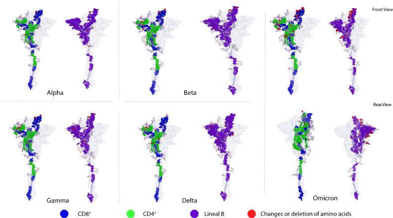
Methods: Different bioinformatics tools were used to: i) Identify new and compile previously-reported B-and T-cell epitopes from SARS-CoV-2 S, M and N proteins; ii) Determine the mutations in S protein from VOC that affect B- and T-cell epitopes, and; iii) Identify cross-reactive epitopes with coronaviruses relevant to human health.

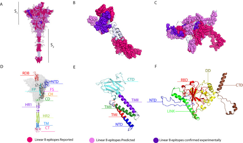
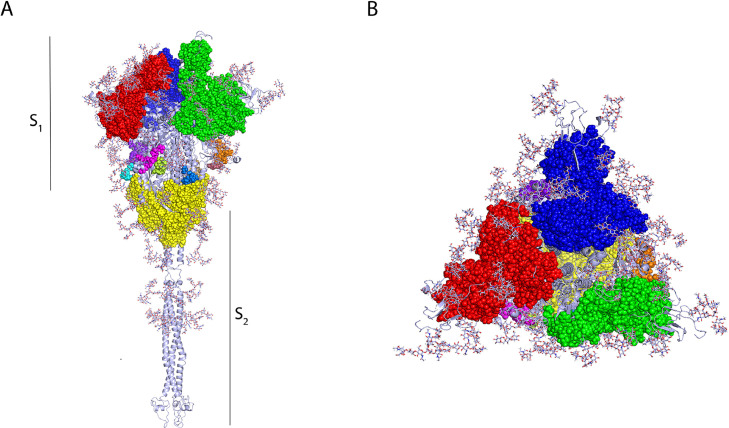
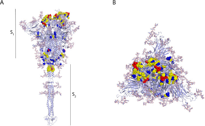
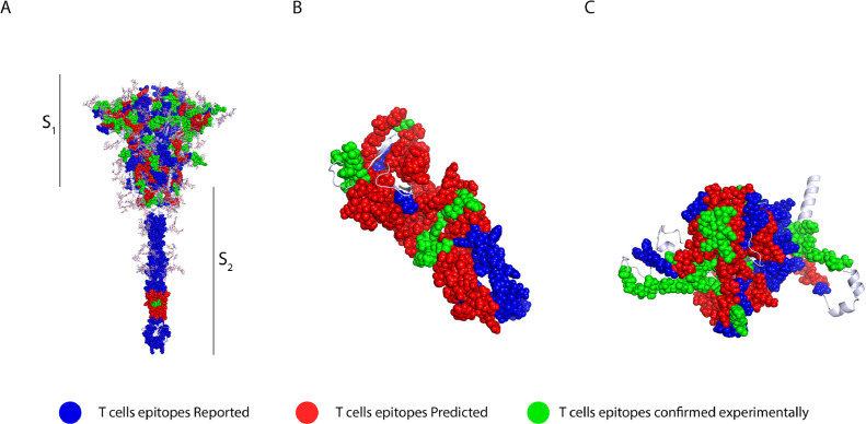
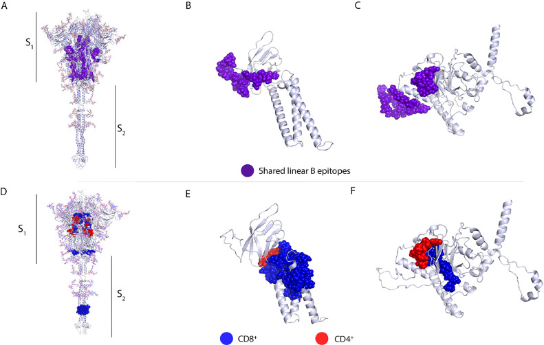
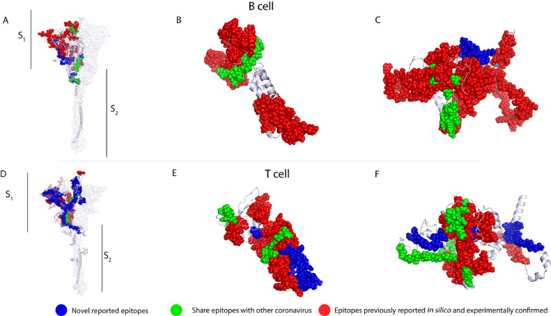
Results: New, potential B- and T-cell epitopes from S, M and N proteins as well as cross-reactive epitopes with other coronaviruses were found and mapped within the proteins' structures.

Conclusion: Numerous potential B- and T-cell epitopes were found in S, M and N proteins, some of which are conserved between coronaviruses. VOCs present mutations within important epitopes in the S protein; however, a significant number of other epitopes remain unchanged. The epitopes identified here may contribute to augmenting the protective response to SARS-CoV-2 and its variants induced by infection and/or vaccination, and may also be used for the rational design of novel broad-spectrum coronavirus vaccines.

Keywords: B- and T-cell epitopes; COVID-19 vaccines.Introduction; Cross-reactivity; Membrane protein; Nucleocapsid protein; SARS-CoV-2; Spike protein.


Conflict of interest statement

Conflicts of Interest All authors declare no conflicts of interest.

Figures

Figure 1
Figure 1
Linear B-cell epitopes in spike, membrane and nucleocapsid proteins. 3D graphical representations of A. Spike, B. Membrane and C Nucleocapsid proteins of SARS-CoV2. Linear B-cell epitopes reported by in silico studies found in the literature are shown in magenta color, epitopes predicted in this study are shown in pink, and predicted epitopes that have been experimentally confirmed to be immunogenic are shown in purple. The following domains and regions are also shown: D. S protein, in red the receptor binding region (RBD, 336-518), in dark blue the N-terminal domain (NTD, 16-291), in magenta the furin cleavage site (FS), in cyan the fusion peptide (FP, 817-834) (S1/S2), in orange the central helix (CH, 987-1034), in green the connecting domain (CD, 1080-1135), in purple and in green the heptad repeat domains 1 (HR1, 1047-1163) and 2 (HR2, 1163-1210) respectively, in light blue the transmembrane domain (TM, 1214-1234), and in pink the cytoplasmic tail (CT, 1235-1273); E. M protein, in cyan color the connecting domain (CTD, 101-222), the three transmembrane domains in red (TMI, 20-40), green (TMII, 51-71) and purple (TMIII, 80-100) respectively and in dark blue the N-terminal domain (NTD, 1-19 ) F. N protein, in red RNA binding domain (RBD, 50-174), in dark blue the N-terminal domain (NTD, 1-50), in yellow the dimerization domain (DD, 246-365), in brown the connection domain (CTD, 366-419) and in green central linker (LINK, 174-246). X-ray crystallography S protein ID: 6VSB_1_1, M protein ID: QHD43419 and N protein ID: QHD43423.
Figure 2
Figure 2
Conformational B-cell epitopes of the SARS-CoV-2 spike glycoprotein displayed in trimer representation, A. Side and B. Top view. Predictions of conformational epitopes were obtained with the Ellipro tool. The major epitopes between chains A and B are shown in red, chains B and C are shown in green, and chains A and C are shown in yellow.
Figure 3
Figure 3
Conformational B-cell epitopes of the SARS-CoV-2 spike glycoprotein displayed in monomer representation, A. Side and B. Top view. Predictions of conformational epitopes were obtained with the DiscoTope tool. The selected residues are mainly distributed in 4 domains. In particular, one residue (N282) belongs to the NTD, and the RBD contains 28 of the predicted residues, 27 of them located in the RBM. Within the S2 region, three residues (P793, I794 and P809) are located in the fusion peptide (FP) and three other residues (N914, Y917 and E918) are located within HR1. The predicted residues are shown in Figure 3B, where predicted residues with high specificity (91–100%) are shown in red, residues with moderate specificity (86–90%) are shown in yellow and residues with low specificity (75–85%) are shown in blue.
Figure 4
Figure 4
T-cell epitopes in spike, membrane and nucleocapsid proteins. 3D graphical representations of A. Spike, B. Membrane and C. Nucleocapsid proteins of SARS-CoV2. T-cell epitopes reported by in silico studies found in the literature are shown in blue, epitopes predicted in this study are shown in green, and predicted epitopes that have been experimentally confirmed to be immunogenic are shown in red. The epitopes described in the literature are found throughout the spike protein structure; the predicted epitopes are present throughout the structure, except for a notable gap in the transmembrane domain (TD). In the M protein, EPITS are evenly distributed throughout the protein, whereas ECEs are mainly located in the N-terminal and C-terminal domains. On the other hand, the 3D model of the N protein shows that the distribution of epitopes is throughout the entire protein. In contrast, the ECEs were mapped mainly in the RNA-binding domain, the binding site and the dimerization domain.
Figure 5
Figure 5
Amino acid changes in SARS-CoV-2 variants being monitored (Alpha, Beta and Gamma) and variants of concern (Delta and Omicron). 3D graphical representations of the spike protein of SARS-CoV2 where changes or deletions in specific amino acids are shown in red for Alpha, Beta, Gamma, Delta and Omicron variants. CD8+ T-cell epitopes are shown in blue, CD4+ T-cell epitopes are shown in green, and B-cell epitopes are shown in purple. All epitopes compiled in this work (ER, EP and ED) are shown. RBD, NTD, and S1 incision sites are the main regions of the protein that present amino acid deletions and substitutions. Front and rear views of the Omicron variant are included to highlight the high number of amino acid changes along the protein sequence. X-ray crystallography S protein ID: 6VSB_1_1.
Figure 6
Figure 6
Conserved linear B-cell and T-cell epitopes in spike, membrane and nucleocapsid proteins of human coronaviruses. 3D graphical representations of the A, and D. Spike, B, and E. Membrane and C, and F. Nucleocapsid proteins of SARS-CoV2. Epitopes that have above 50% shared identity with epitopes from seasonal coronaviruses (HKU1, NL63, 229E and OC43), SARS-CoV and MERS are highlighted in different colors. Linear B-cell epitopes are shown in purple A–C. CD8+ T-cell epitopes are represented in blue, CD4+ T-cell epitopes are shown in red. All epitopes (ERIS, EPITS and EEC) were included in the analysis. X-ray crystallography S protein ID: 6VSB_1_1, M protein ID: QHD43419 and N protein ID: QHD43423.
Figure 7
Figure 7
The locations of proposed epitopes set for a possible broad-spectrum T and B-cell vaccine candidate, as well as potential cross-reactive epitopes, including, newly reported epitopes in blue, shared epitopes with other coronavirus are shown in green and epitopes previously reported in silico and experimentally confirmed are shown in red. 3D graphical representations of spike A and D. membrane B and E. and nucleocapsid C and F proteins of SARS-CoV-2.

Similar articles

Cited by

References

    1. Yang H, Rao Z. Structural biology of SARS-CoV-2 and implications for therapeutic development. Nat Rev Microbiol. 2021;19:685–700. doi: 10.1038/s41579-021-00630-8. - DOI - PMC - PubMed
    1. ArcGIS Dashboards 2020. Available from: https://www.arcgis.com/apps/dashboards/bda7594740fd40299423467b48e9ecf6. (Accessed November 22, 2021).
    1. Dong E, Du H, Gardner L. An interactive web-based dashboard to track COVID-19 in real time. Lancet Infect Dis. 2020;20:533–534. doi: 10.1016/S1473-3099(20)30120-1. - DOI - PMC - PubMed
    1. Ssentongo P, Ssentongo AE, Voleti N, et al. SARS-CoV-2 vaccine effectiveness against infection, symptomatic and severe COVID-19: a systematic review and meta-analysis. BMC Infect Dis. 2022;22:439. doi: 10.1186/s12879-022-07418-y. - DOI - PMC - PubMed
    1. Farmacovigilancia de vacunas para COVID-19 - Catálogo. Farmacovigilancia de vacunas para COVID-19 2022. Available from: https://covid-19pharmacovigilance.paho.org/(Accessed August 10, 2022).

Publication types

Substances

Supplementary concepts