

Sternal lymphadenopathy in dogs with malignancy in different localizations: A CT retrospective study of 60 cases
- PMID: 36337196
- PMCID: PMC9634218
- DOI: 10.3389/fvets.2022.1019196
Sternal lymphadenopathy in dogs with malignancy in different localizations: A CT retrospective study of 60 cases
Abstract
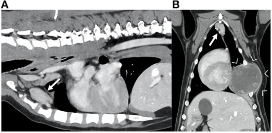
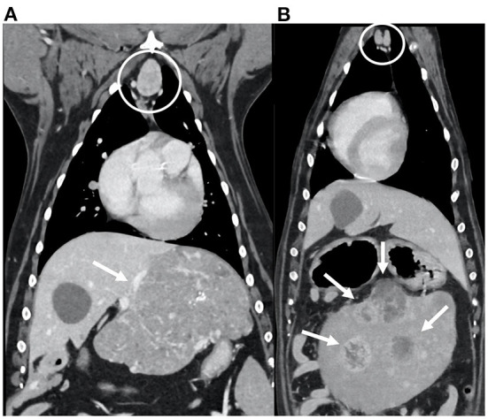
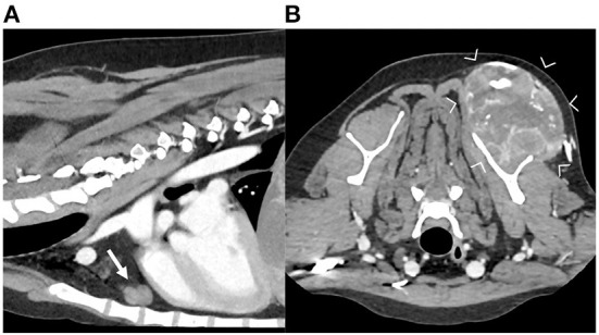
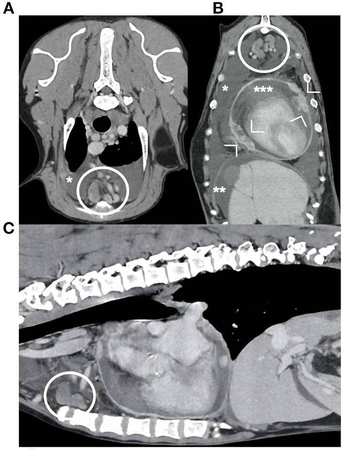
Sternal lymph nodes (SLNs) drain a multitude of regions in dogs, including the pectoral and shoulder region, the thoracic wall and mammary glands, the mediastinum, thymus, diaphragm, and the ventral abdominal wall and peritoneal cavity. Neoplastic conditions of these regions can lead to sternal lymphadenopathy. The aim of this study was to assess the most frequent localizations of the primary neoplasia and the most frequent tumor types in dogs with sternal lymphadenopathy. Computed tomographic (CT) characteristics of SLNs in dogs with confirmed neoplasia were also described. For this single-center retrospective descriptive study, dogs with sternal lymphadenopathy and cytological or histological diagnosis of neoplasia were included. Sixty dogs fulfilled the inclusion criteria: 30 (50%) with thoracic neoplasia, 19 (32%) with abdominal neoplasia, 6 (10%) with neoplasia of the front limbs and 5 (8%) with generalized neoplasia. Based on the cytological/histological diagnosis of the primary neoplasia, 31/60 (52%) dogs presented with a sarcoma, 15/60 (25%) with carcinoma, and 14/60 (23%) with round cell tumor. The presence of heterogeneous contrast enhancement was more frequent in dogs with sarcoma, while the concomitant presence of other abnormal lymph nodes was more frequent in dogs with round cell neoplasia. Tumors of different types and in different location can result in sternal lymphadenopathy in dogs. The most frequent in this study were thoracic and abdominal neoplasia, followed by neoplasia of the shoulder region. Sarcoma was the most common tumor type detected in this study, and the main CT characteristic of the SLNs in case of sarcoma was heterogeneous contrast enhancement.
Keywords: computed tomography; neoplasia; sternal lymph node; thorax; tumor.
Copyright © 2022 Cordella, Saunders and Stock.
Conflict of interest statement
The authors declare that the research was conducted in the absence of any commercial or financial relationships that could be construed as a potential conflict of interest.
Figures
References
-
- Barone R. (Editor). Système lymphatique du Chien. In: Anatomie comparée des mammiferes domestiques Tome 5: Angiologie—Chapitre IV: Système Lymphatique−2eme edn. Baronne Edition (2021). p. 821.
-
- Bezuidenhout AJ. The lymphatic system. In: Evans HE, editor. Miller's Anatomy of the Dog, 3rd edn. (Philadelphia, PA: WB Saunders) (1993), 731–734.
-
- Baum H. Lymph nodes of the thorax and associated organs. In: The Lymphatic System of the Dog. (2022), 60–72. Available online at: https://openpress.usask.ca/k9lymphaticsystem/ (accessed May 23, 2022).
-
- Thrall DE. Canine and feline mediastinum. In: Thrall ED, editor. Textbook of veterinary diagnostic radiology, 7th edn. (St. Louis, MO: Elsevier) (2018), 649–69. 10.1016/B978-0-323-48247-9.00045-0 - DOI
LinkOut - more resources
Full Text Sources