. 2022 Oct 19:13:1006962.
doi: 10.3389/fmicb.2022.1006962. eCollection 2022.

A serralysin-like protein of Candidatus Liberibacter asiaticus modulates components of the bacterial extracellular matrix

Affiliations

A serralysin-like protein of Candidatus Liberibacter asiaticus modulates components of the bacterial extracellular matrix

Lucila Garcia et al. Front Microbiol. .

Abstract

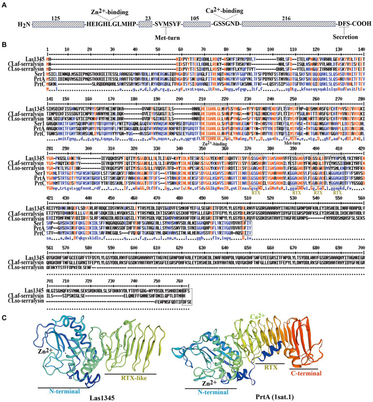
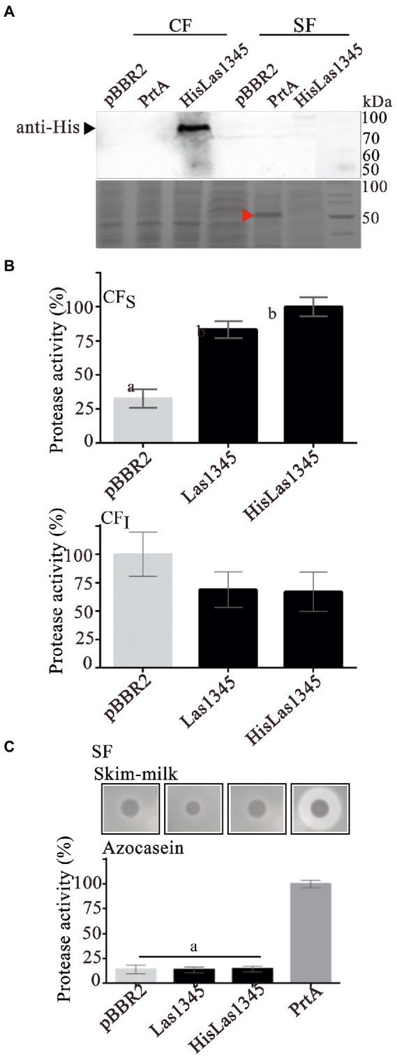
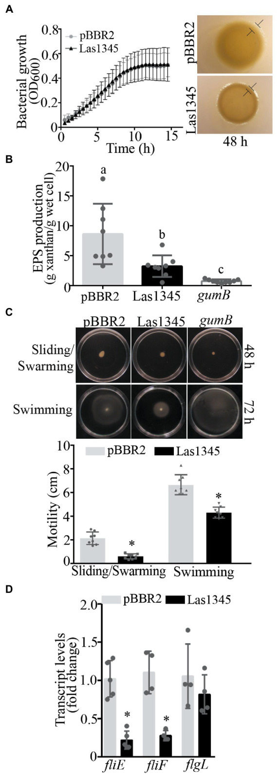
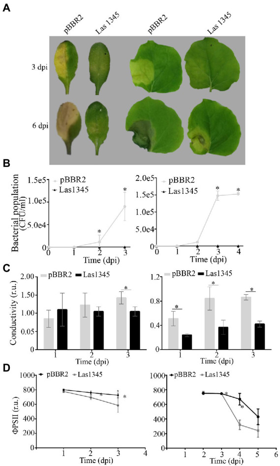
Huanglongbing (HLB), the current major threat for Citrus species, is caused by intracellular alphaproteobacteria of the genus Candidatus Liberibacter (CaL), with CaL asiaticus (CLas) being the most prevalent species. This bacterium inhabits phloem cells and is transmitted by the psyllid Diaphorina citri. A gene encoding a putative serralysin-like metalloprotease (CLIBASIA_01345) was identified in the CLas genome. The expression levels of this gene were found to be higher in citrus leaves than in psyllids, suggesting a function for this protease in adaptation to the plant environment. Here, we study the putative role of CLas-serralysin (Las1345) as virulence factor. We first assayed whether Las1345 could be secreted by two different surrogate bacteria, Rhizobium leguminosarum bv. viciae A34 (A34) and Serratia marcescens. The protein was detected only in the cellular fraction of A34 and S. marcescens expressing Las1345, and increased protease activity of those bacteria by 2.55 and 4.25-fold, respectively. In contrast, Las1345 expressed in Nicotiana benthamiana leaves did not show protease activity nor alterations in the cell membrane, suggesting that Las1345 do not function as a protease in the plant cell. Las1345 expression negatively regulated cell motility, exopolysaccharide production, and biofilm formation in Xanthomonas campestris pv. campestris (Xcc). This bacterial phenotype was correlated with reduced growth and survival on leaf surfaces as well as reduced disease symptoms in N. benthamiana and Arabidopsis. These results support a model where Las1345 could modify extracellular components to adapt bacterial shape and appendages to the phloem environment, thus contributing to virulence.

Keywords: Huanglongbing; biofilm; protease; surrogate bacteria; virulence factor.


Conflict of interest statement

The authors declare that the research was conducted in the absence of any commercial or financial relationships that could be construed as a potential conflict of interest.

Figures

Figure 1
Figure 1
Multiple sequence alignment and representative tertiary structure of Las1345. (A) Schematic representation of Las1345 (CLIBASIA_01345). Conserved motifs associated with serralysin family protein are shown. HExxHxUGUxH, zinc-binding motif; SxMxY, a Met-turn motif; GGxGxDxUx, calcium-binding motif (glycine-rich repeats) where U represents an aliphatic amino acid and X can be any residue; Dxxx, T1SS secretion signal. (B) Sequence alignment of the serralysin-like proteins encoded by Candidatus Liberibacter (CaL) asiaticus (CLas) strain psy62 (Las1345, WP_015452346); CaL africanum strain PTSAPSY (CLaf_Serralysin, AKK19938.1); CaL solanacearum strain ZC1 (CLsoB_Serralysin, WP_013461860); Serratia marcescens (PrtA, CAA39139.1); S. liquefaciens strain FK01 (Ser1, BAK39731) and Dickeya chrisantemy strain B374 (PrtC; PDB 1K7I_A). Alignment was obtained by ClustalW and Multaline. Low consensus alignment (50%) is shown in blue letters while high consensus alignment (90%) is shown in red. Protein domains were identified by Pfam. Protein domains, showing the metalloprotease sequence (HExxHxxGxxHP) motif, the met-turn (SxMSYF/W) motif, the four RTX repeats (the classic, GGxGxD and the divergent, GSxGxD) and the ABC exporter motif (Dxxx) are indicated in boxes. (C) Tertiary structure of Las1345 domains based on a homology model derived from PrtA structure (PBD ID 1sat.1; Baumann, 1994; QMEANG 0.65 ± 0.5). The N terminal Zn-binding domain and the RTX β-rolls are colored in rainbow. Zinc and calcium ions are represented by gray and green spheres, respectively.
Figure 2
Figure 2
Las1345 has intracellular protease activity in Rhizobium leguminosarum. (A) Immunodetection of Las1345 (~75 kDa) in cellular (CF) and supernatant cell free fraction (SF) from Rhizobium leguminosarum A34 (A34) cells expressing Las1345, HisLas1345 or transformed with the empty vector (pBBR2), grown for 16 h at 30°C. Rhizobium adhering protein A1 (RapA1, ~36 kDa) and Glucose-6-phosphate dehydrogenase (G6PD, ~55 kDa) were used as extracellular and intracellular controls, respectively. (B) Protease activity in SF from A34 cultures (C), Protease activity in soluble and insoluble CF (CFS and CFI) from A34 cultures. The relative activity was expressed as the percentage of activity detected with respect to the maximum protease activity in the assay. Values are expressed as means ± standard deviations from six independent biological replicates. Different letters indicate significant differences at p < 0.05 (one-way analysis of variance, Tukey’s test).
Figure 3
Figure 3
Las1345 protease activity in Serratia marcescens. (A) Immunodetection of Las1345 (~75 kDa) in cellular (CF) and supernatant cell free fraction (SF) prtA cells from S. marcescens expressing Las1345, HisLas1345 or transformed with an empty vector (pBBR2), grown for 16 h at 30°C. PrtA (~50 kDa) was visualized by Coomassie staining (red arrow) and used as control of extracellular secretion (SF). (B) Protease activity in soluble and insoluble CF (CFS and CFI) from prtA/pBBR2, prtA/Las1345, prtA/FIGURE 3 (Continued)HisLas1345, and prtA/PrtA cells. The relative activity was expressed as the percentage of activity detected with respect to the maximum protease activity in the assay (C) Protease activity in SF from prtA/pBBR2, prtA/Las1345, prtA/HisLas1345, and prtA/PrtA cells was measured by detecting degradation of milk proteins (LB agar-skim milk plate), seen as a halo around the colony and by the azocaseinase assay. Values are expressed as means ± standard deviations from six independent biological replicates. Different letters indicate significant differences at p < 0.05 (one-way analysis of variance, Tukey’s test).
Figure 4
Figure 4
Las1345 expression in plant cells does not affect cell integrity. (A) Quantification of cell death in N. benthamiana leaves overexpressing GFP, Las1345-GFP, or Las1345 by measurement of conductivity at 3 days post agroinfiltration (dpi). Values are expressed as mean ± standard deviation of six samples. Each sample was obtained from three leaf disks of 1 cm2 collected randomly from different agroinfiltrated leaves. Data was analyzed by two-way analysis of variance and Tukey’s test at p < 0.05. Each(Continued)FIGURE 4 (Continued)assay was repeated three times. Scale bar, 10 μM. (B) N. benthamiana leaves expressing Las1345-GFP (green, white arrow) or GFP were imaged by confocal microscopy at 2 days post infiltration (dpi). FM4-64 (FM) and DAPI were used to distinguish membrane (red, orange arrow) and nucleus (blue, red arrow), respectively. Co-localization of GFP and FM is shown in yellow. (C) Protease activity using azocasein as substrate in total plant protein extracts (50 μg) from N. benthamiana leaves overexpressing GFP, Las1345GFP or Las1345 at 2 dpi. Activity is expressed as percentage relative to the highest activity value obtained among samples. Values are expressed as mean ± standard deviation of three samples. Each sample contains six disks from different agroinfiltrated leaves. Data was analyzed by two-way analysis of variance and Tukey’s test at p < 0.05.
Figure 5
Figure 5
Las1345 expression reduce xanthan production and cell motility. (A), Xanthomonas campestris pv. campestris (Xcc) growth in NYGB medium. Values are expressed as means ± standard deviation of five biological replicates. This assay was repeated five times. Macrocolony phenotype of Las1345-expressing Xcc and(Continued)FIGURE 5 (Continued)the control (Xcc/pBBR2) grown on 1% (w/v) agar NYGB medium at 28°C during 48 h. Differences at boundary zone are indicated with T-markers. (B) Quantification of xanthan secreted in Las1345-expressing Xcc cultures grown for 20 h at 28°C compared with control Xcc/pBBR2 and a xanthan deficient mutant (gumB). Values are expressed as means ± standard deviation of eight biological replicates. Different letters indicate significant differences at p < 0.05 (one-way analysis of variance, Tukey’s test). This assay was repeated three times. (C) Xcc/Las1345, Xcc/pBBR2 and gumB cultures were normalized by OD600 and inoculated on 0.5% (w/v) agar NYGB medium (sliding/swarming) and on 0.25% (w/v) agar NYGB medium (swimming). Pictures were taken after 48 or 72 h of incubatio at 28°C. Motility was measured as colony diameter (cm). Values are expressed as means ± standard deviation of 10 biological replicates. (D) Quantitative reverse transcription (qPCR) analysis of flagellum assembly-related genes (fliE, fliF, and flgL). Fold change of RNA levels, normalized to 16 s and relative to Xcc/pBBR2 is shown. Values are expressed as means ± standard deviation (SD) of five samples (replicates). Asterisks indicates significant differences between Xcc/Las1345 and Xcc/pBBR2 at *p < 0.05 (Students t-test).
Figure 6
Figure 6
Biofilm architecture of Xanthomonas campestris pv. campestris (Xcc) is disrupted when Las1345 is expressed. GFP-labeled Xcc cells (Xcc-GFP) expressing Las1345 (Xcc-GFP/Las1345) were grown in 8-well chambers with a 1-mm thick borosilicate glass containing Y minimal medium and visualized at different stages of biofilm formation under confocal laser scanning microscopy for 4 days after inoculation at 28°C. Biofilm structure is shown as 2D-images of a single layer in the XY plane at different distances from the bottom well along the Z-axis (0–11898.21 nm) at day 1, 2 and 4. Green channel shows Xcc-GFP/Las1345 (Las1345) or Xcc-GFP/pBBR2 (pBBR2) cells. At day 1, a detail (right corner) of the biofilm structure in Xcc-GFP/Las1345 and control cells is shown. At Day 2, Biomass and Roughness coefficient using COMSTAT 2.0 were calculated. Data is shown as means ± standard deviation of three replicates. Asterisks indicate significant differences at p < 0.05 (Student’s t-test). At day 4, biofilm structure is showed by GFP (green channel) and Congo red (CR) emission (red channel) to indicate cells and exopolysaccharides production in the extracellular matrix, respectively. A detail of the biofilm structures is shown for both strains. C, GFP cells; EPS, exopolysaccharide; M, extracellular matrix. Scale bars, 20 μm.
Figure 7
Figure 7
Biofilm modifications induced by Las1345 expression alters Xanthomonas campestris pv. campestris (Xcc) virulence. (A) Characterization of symptom development induced by Las1345-expresing Xcc in host plants. Infection of Arabidopsis thaliana and Nicotiana benthamiana with Xcc/Las1345 or Xcc/pBBR2 (107 CFU/ml) using pressure infiltration. Photos of disease symptoms were taken at different days post inoculation (dpi). (B) Bacterial population of Xcc transformants in N. benthamiana and A. thaliana leaves. Values are expressed as means ± standard deviation from five samples. Each sample was obtained from three leaf disks of 1 cm2 from different inoculated leaves. (C) Quantification of cell death in leaves treated as described in (A) by measuring electrolyte leakage (conductivity) at different dpi. Values are expressed as means ± standard deviation from six samples. Each sample contains three leaf disks of 1 cm2 from 10 inoculated leaves. (D) Photosystem II quantum efficiency (ϕPSII) were measured on inoculated leaf. Values are expressed as means ± standard deviation from 15 leaves. Asterisk indicates significant differences p < 0.05 (Student’s t-test).

Similar articles

Cited by

References

    1. Achor D., Welkerm S., Ben-Mahmoud S., Wangm C., Folimonovam S. Y., Duttm M., et al. . (2020). Dynamics of 'Candidatus Liberibacter asiaticus' movement and sieve-pore plugging in citrus sink cells. Plant Physiol. 182, 882–891. doi: 10.1104/pp.19.01391, PMID: - DOI - PMC - PubMed
    1. Alves M. N., Cifuentes-Arenas J. C., Raiol-Junior L. L., Ferro J. A., Peña L. (2021). Early population dynamics of 'Candidatus Liberibacter asiaticus' in susceptible and resistant genotypes after inoculation with infected Diaphorina citri feeding on young shoots. Front. Microbiol. 12:683923. doi: 10.3389/fmicb.2021.683923, PMID: - DOI - PMC - PubMed
    1. An A.-Q., Potnis N., Dow J. M., Vorhölter F.-J., He Y.-Q., Becker A., et al. . (2020). Mechanistic insights into host adaptation, virulence and epidemiology of the phytopathogen Xanthomonas. FEMS Microbiol. Rev. 44, 1–32. doi: 10.1093/femsre/fuz024 - DOI - PMC - PubMed
    1. Andrade M. O., Pang Z., Achor D. S., Wang H., Yao T., Singer B. H., et al. . (2020). The flagella of 'Candidatus Liberibacter asiaticus' and its movement in planta. Mol. Plant Pathol. 21, 109–123. doi: 10.1111/mpp.12884, PMID: - DOI - PMC - PubMed
    1. Andrade M. O., Wang N. (2019). The tad pilus apparatus of 'Candidatus Liberibacter asiaticus' and its regulation by Vis NR. Mol. Plant-Microbe Interact. 32, 1175–1187. doi: 10.1094/MPMI-02-19-0052-R, PMID: - DOI - PubMed

LinkOut - more resources