. 2022 Oct 20:12:955719.
doi: 10.3389/fonc.2022.955719. eCollection 2022.

ERCC1 is a potential biomarker for predicting prognosis, immunotherapy, chemotherapy efficacy, and expression validation in HER2 over-expressing breast cancer

Affiliations

ERCC1 is a potential biomarker for predicting prognosis, immunotherapy, chemotherapy efficacy, and expression validation in HER2 over-expressing breast cancer

Yilun Li et al. Front Oncol. .

Abstract

Objective: To investigate the relationship between Excision repair cross-complementation 1 (ERCC1) expression, clinicopathological features, and breast cancer prognosis in patients treated with trastuzumab. Further, we aim to explore the immune status of ERCC1 in breast cancer.

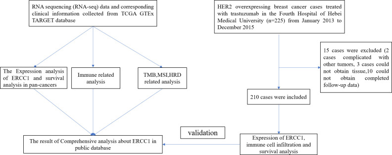
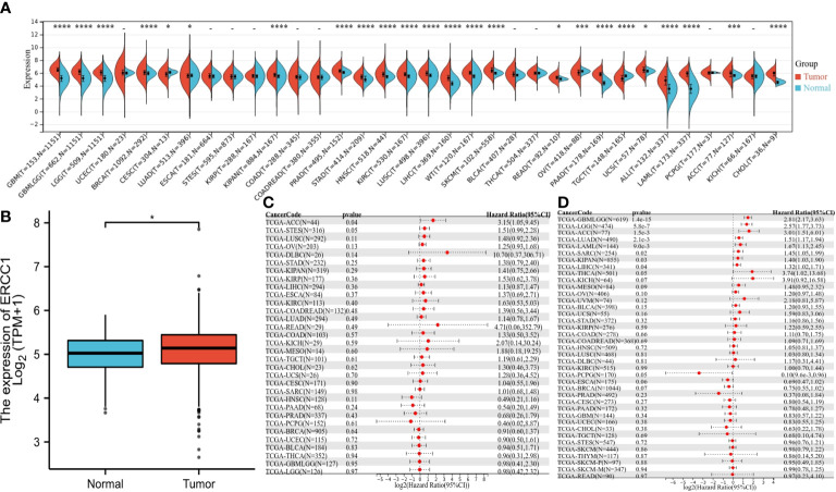
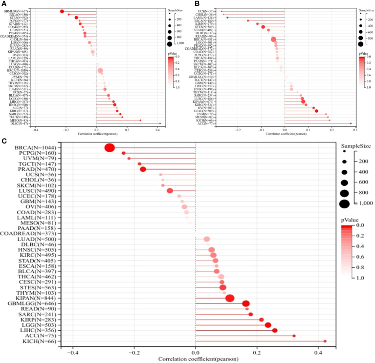
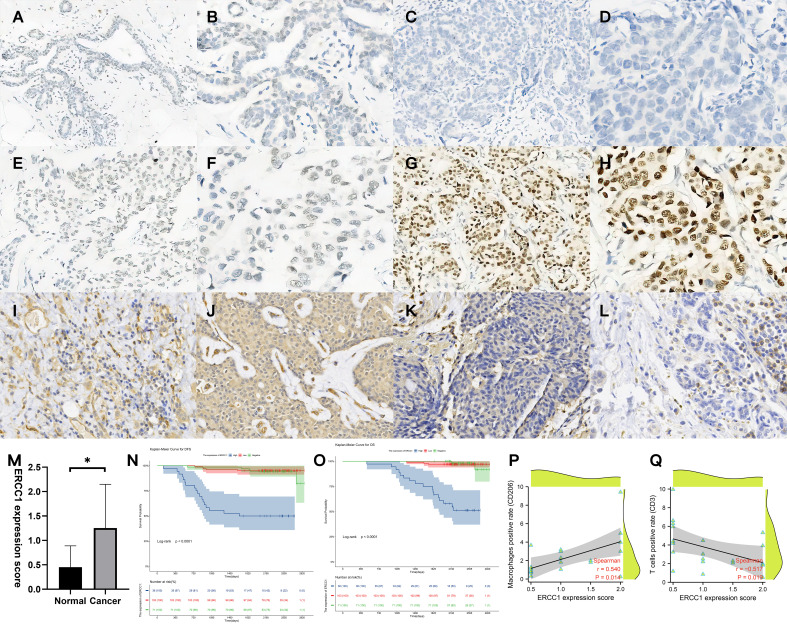
Methods: The data were retrieved from publicly available databases like the Cancer Genome Atlas, Therapeutically Applicable Research to Generate Effective Treatments, and the Genotype-Tissue Expression. The data was used to perform differential expression analyses between tumor and normal tissues in pan-cancers, immune-related analysis, homologous recombination deficiency (HRD), tumor mutation burden, and microsatellite instability. A total of 210 patients with HER2 over-expressing breast cancer from the Fourth Hospital of Hebei Medical University between January 2013 to December 2015 were enrolled in the study. Ten adjacent normal tissues were used to study the expression pattern of ERCC1 in normal tissues. Immunohistochemistry was performed to study ERCC1 expression and immune cell infiltration in different status of ERCC1 expression. Further, the correlation between ERCC1 expression, immune cell infiltration clinicopathological features, and the prognosis of patients with breast cancer was analyzed.

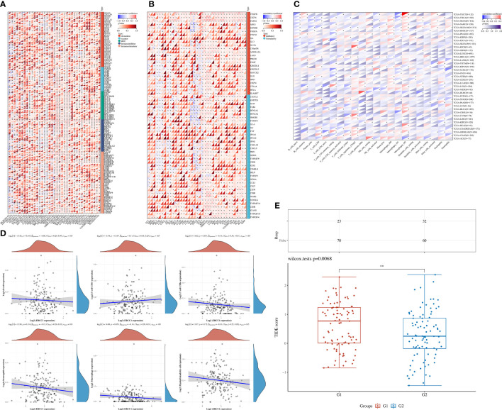
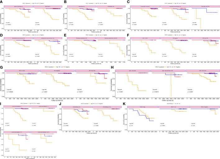
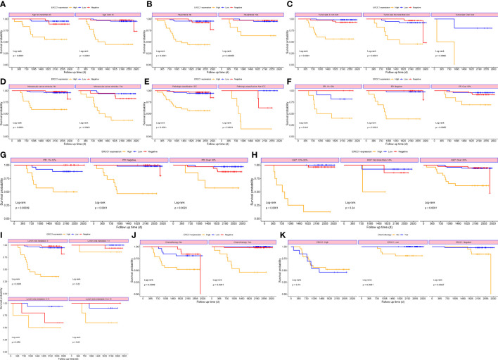
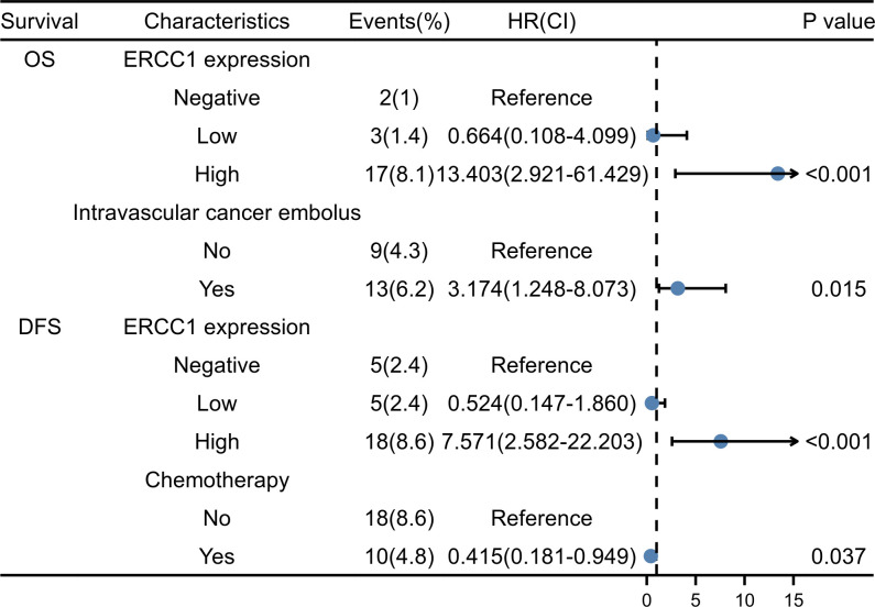
Results: The immune analysis revealed a significant correlation between CD8+ T cell, CD4+ T cell, T helper cell, macrophages, mast cells, and ERCC1 expression. Spearman analysis show that ERCC1 expression is related to macrophages and T cells. A close correlation was observed between increased ERCC1 expression and high tumor immune dysfunction and exclusion (TIDE) score as well as HRD. The results revealed a significant correlation among ERCC1, chemotherapy and estrogen receptor (ER; P < 0.05) expression. Univariate survival analysis revealed a significant correlation (P < 0.05) between that ERCC1 and ER expression, blood vessel invasion, and disease-free survival (DFS). ERCC1 and ER expression, tumor size, blood vessel invasion, pathological type, and lymph node metastases significantly correlated (P < 0.05) with overall survival in patients. Multivariate regression analysis revealed that ERCC1 expression and chemotherapy were independent factors that influence DFS. ERCC1 expression and vascular tumor thrombus were independent influencing factors that influence OS.

Conclusion: A correlation was observed between high ERCC1 expression and poor patient prognosis. High ERCC1 expression also influences the efficacy of immunotherapy and chemotherapy.

Keywords: breast cancer; clinicopathological feature; excision repair cross-complementary gene 1; human epidermal growth factor receptor-2; prognosis.


Conflict of interest statement

The authors declare that the research was conducted in the absence of any commercial or financial relationships that could be construed as a potential conflict of interest.

Figures

Figure 1
Figure 1
The flowchart of the complete data analysis and validation of ERCC1 expression.
Figure 2
Figure 2
The expression of ERCC1 and survival analysis in pan-cancers, (A) ERCC1 expression in pan-cancers. (B) ERCC1 expression in breast cancer. (C) Univariate survival analysis in pan-cancers [disease-free survival (DFS]). (D) Univariate survival analysis in pan-cancers [overall survival (OS)]. *P < 0.05; ***P < 0.005; ****P < 0.001.
Figure 3
Figure 3
The effect of ERCC1 on immune status. (A) Correlation between ERCC1 and 150 immune pathways marker-related genes in pan-cancers. (B) Correlation between ERCC1 and immune checkpoints in pan-cancers. (C) The correlation between ERCC1 and tumor-associated immune cells was evaluated using Cell-type Identification by Estimating Relative Subsets of RNA Transcripts in pan-cancers. (D) Correlation between ERCC1 and tumor-associated immune cells in HER2 over-expressing breast cancer. (E) Tumor immune dysfunction and exclusion score in HER2 over-expressing breast cancer. G1: High expression of ERCC1. G2: Low expression of ERCC1 (Divided by average expression). *P < 0.05; **P < 0.01; ***P < 0.005; ****P < 0.001.
Figure 4
Figure 4
Homologous recombination deficiency (HRD), Microsatellite instability (MSI), and Tumor mutation burden (TMB) analysis in pan-cancers. (A) Correlation between ERCC1 and TMB in pan-cancers. (B) Correlation between ERCC1 and MSI in pan-cancers. (C) Correlation between ERCC1 and HRD in pan-cancers.
Figure 5
Figure 5
Validation of ERCC1 expression, survival analysis, and immune cell infiltration in patients. (A, B) Immunohistochemical (IHC) staining of ERCC1 in normal tissues, (A) Magnification, 10×10, (B) Magnification, 20×10. (C, D) IHC staining of ERCC1 in HER2 over-expressing breast cancer tissues (Expression: negative). (C) Magnification, 10×10, (D) Magnification, 20×10. (E, F) IHC staining of ERCC1 in HER2 over-expressing breast cancer tissues (Expression: low level). (E) Magnification, 10×10, (F) Magnification, 20×10. (G, H) IHC staining of ERCC1 in HER2 over-expressing breast cancer tissues (Expression: high level). (G) Magnification, 10×10, (H) Magnification, 20×10. (I, J) IHC localization of CD206 indicates macrophage infiltration in 20 patients with different levels of ERCC1 expression. (I) ERCC1 expression level: low, magnification, 20×10. (J) ERCC1 expression level: high, magnification, 20×10. (K, L) IHC localization of CD3 indicates T cell infiltration in 20 patients with different levels of ERCC1 expression. (K) ERCC1 expression level: low, magnification, 20×10. (L) ERCC1 expression level: high, magnification, 20×10. (M) Expression of ERCC1 in normal and cancer tissue. (N) Survival analysis grouped by ERCC1 expression (DFS), (O) Survival analysis grouped by ERCC1 expression (OS) Blue: high level, Red: low level, Green: negative. (P) Spearman’s rank correlation analysis determined the correlation between ERCC1 expression score and positive cell percentage of T cells (CD3). (Q) Spearman’s rank correlation analysis determined the correlation between ERCC1 expression score and positive cell percentage of macrophages (CD206). *P < 0.05.
Figure 6
Figure 6
Survival curves stratified based on clinical factors (OS) (A) Age (B) menopause (C) Tumor size (D) Intravascular cancer embolus (E) Pathological classification (F) ER (G) PR (H) Ki-67 (I) Lymph node metastasis (J) Chemotherapy (K) ERCC1 expression.
Figure 7
Figure 7
Survival curves stratified based on clinical factors (DFS) (A) Age (B) menopause (C) Tumor size (D) Intravascular cancer embolus (E) Pathologic classification (F) ER (G) PR (H) Ki-67 (I) Lymph node metastasis (J) Chemotherapy (K) ERCC1 expression.
Figure 8
Figure 8
The forest plot of multivariate COX survival analysis.

Similar articles

Cited by

References

    1. Siegel RL, Miller KD, Fuchs HE, Jemal A. Cancer statistic. CA Cancer J Clin (2021) 71(1):7–33. doi: 10.3322/caac.21654 - DOI - PubMed
    1. Li Y, Ma L. Nomograms predict survival of patients with lymph node-positive, luminal a breast cancer. BMC Cancer (2021) 21(1):965. doi: 10.1186/s12885-021-08642-6 - DOI - PMC - PubMed
    1. Duffy MJ, Walsh S, McDermott EW, Crown J. Biomarkers in breast cancer: Where are we and where are we going? Adv Clin Chem (2015) 71:1–23. doi: 10.1016/bs.acc.2015.05.001 - DOI - PubMed
    1. Di Fiore PP, Pierce JH, Kraus MH, Segatto O, King CR, Aaronson SA. erbB-2 is a potent oncogene when overexpressed in NIH/3T3 cells. Science (1987) 237(4811):178–82. doi: 10.1126/science.2885917 - DOI - PubMed
    1. Li H, Zhou L, Ma J, Zhu Y, Fan J, Li N, et al. . Distribution and susceptibility of ERCC1/XPF gene polymorphisms in han and uygur women with breast cancer in xinjiang, China. Cancer Med (2020) 9(24):9571–80. doi: 10.1002/cam4.3547 - DOI - PMC - PubMed