. 2021 Jul-Aug:47:206-222.
doi: 10.1016/j.mattod.2021.03.012. Epub 2021 May 20.

Stimuli-responsive transdermal microneedle patches

Affiliations

Stimuli-responsive transdermal microneedle patches

Pooyan Makvandi et al. Mater Today (Kidlington). 2021 Jul-Aug.

Abstract

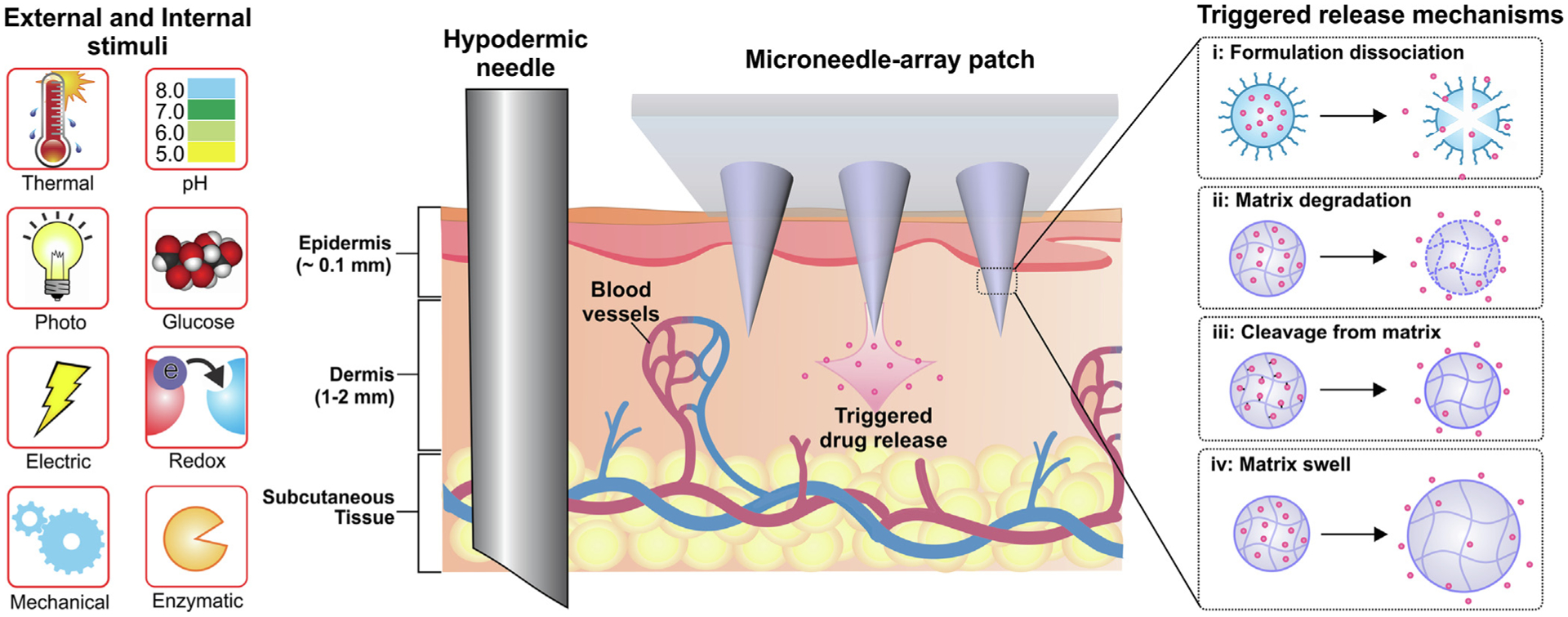
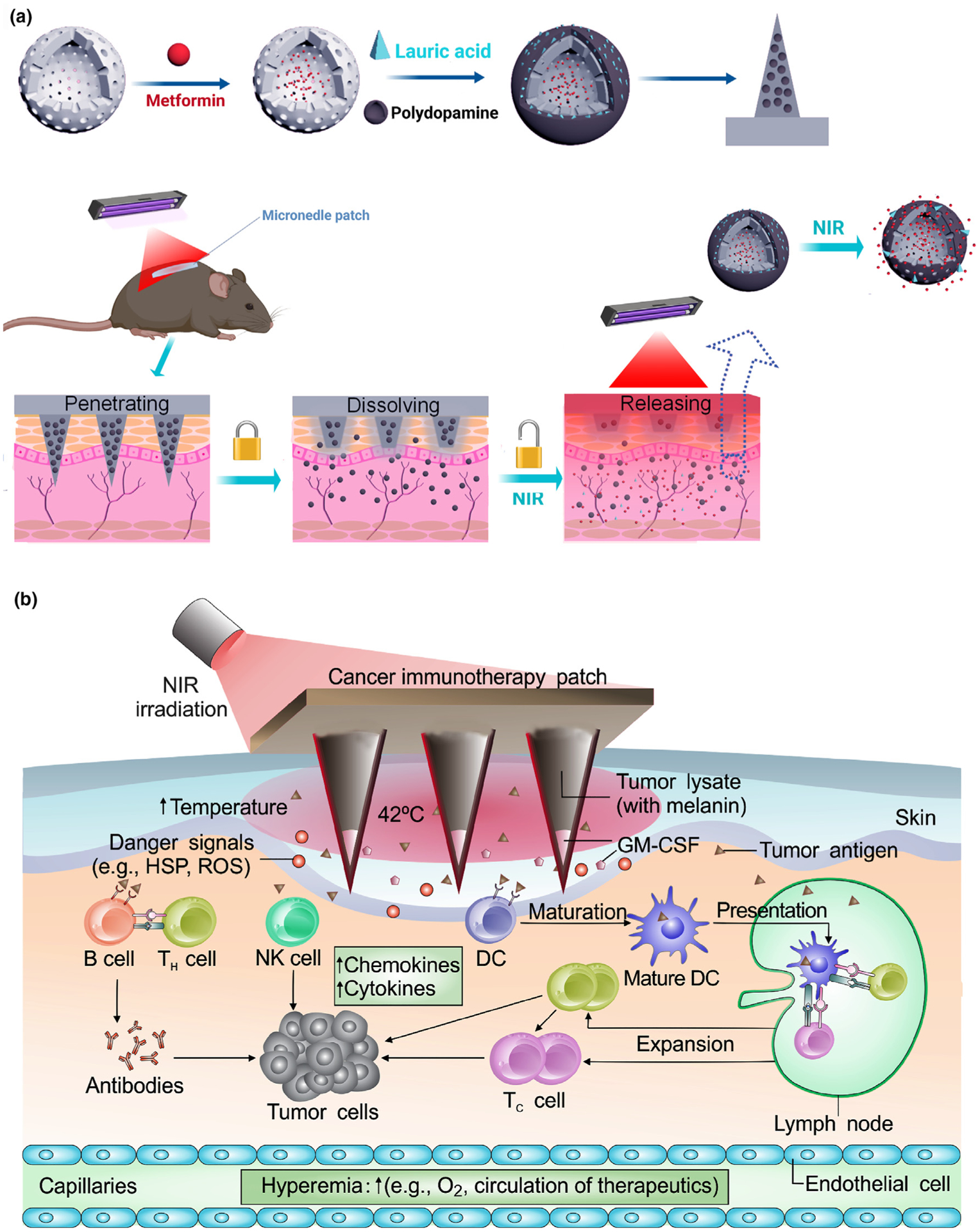
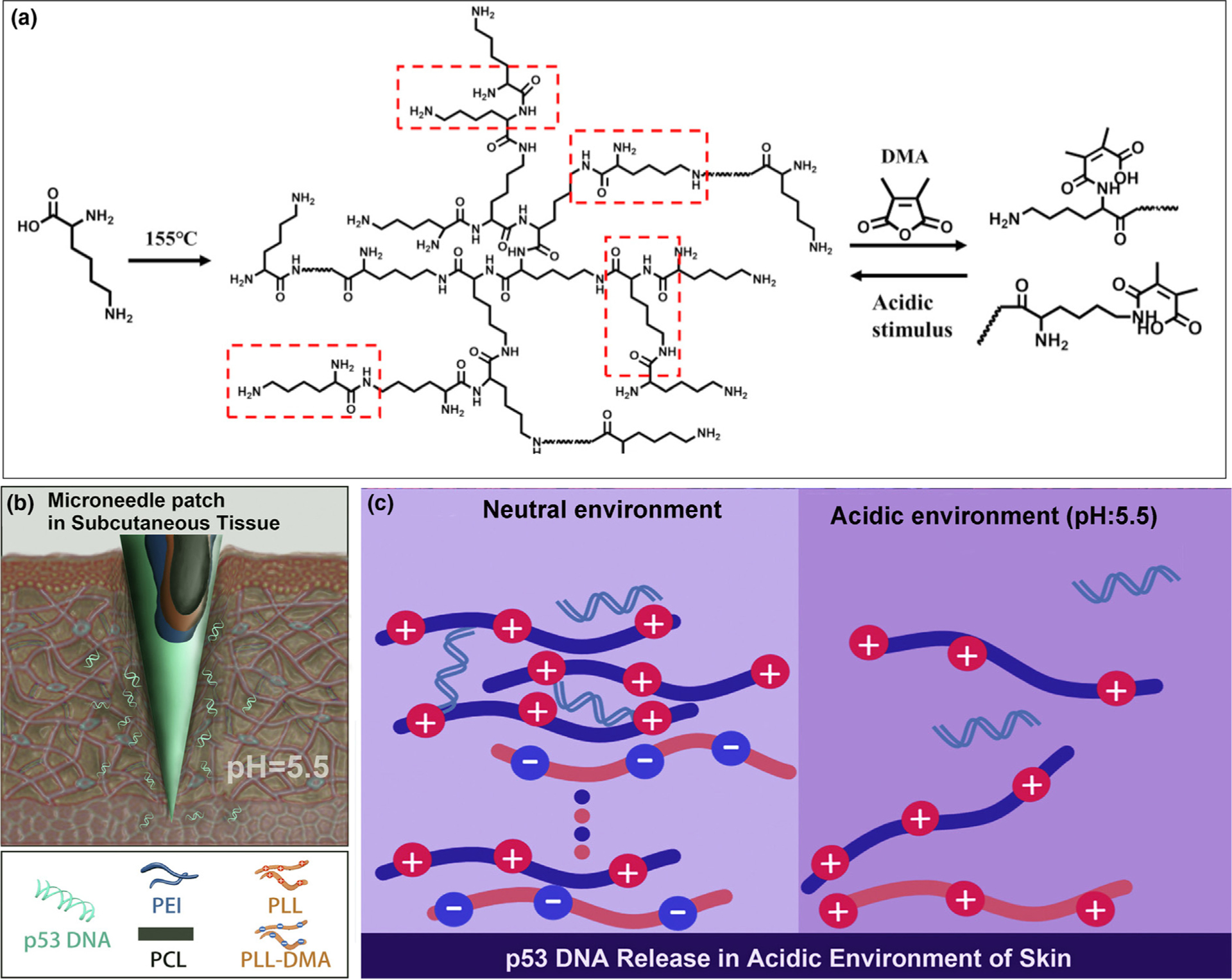
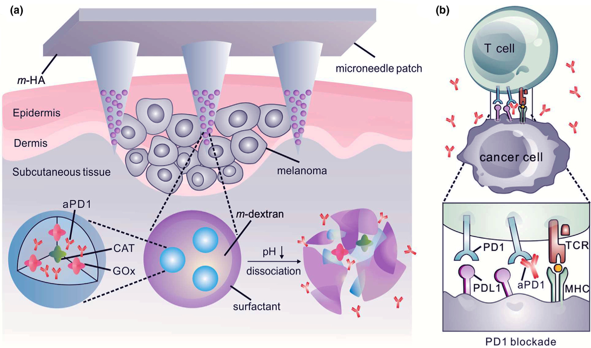
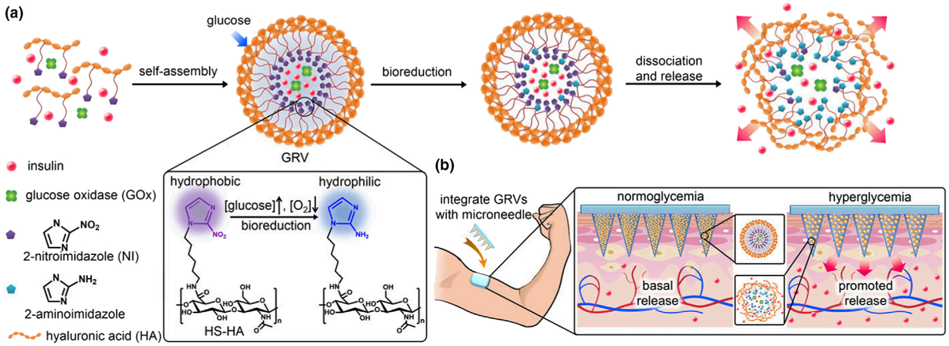
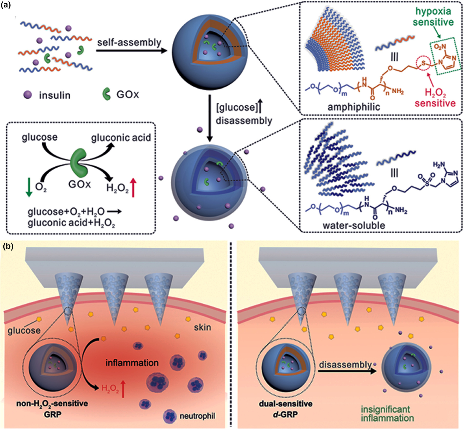
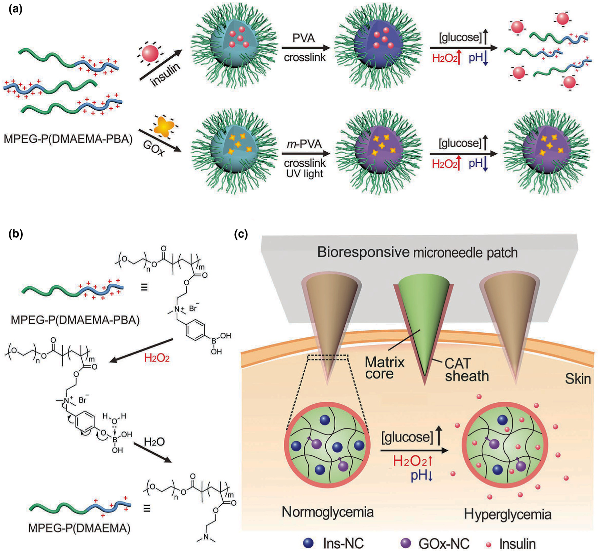
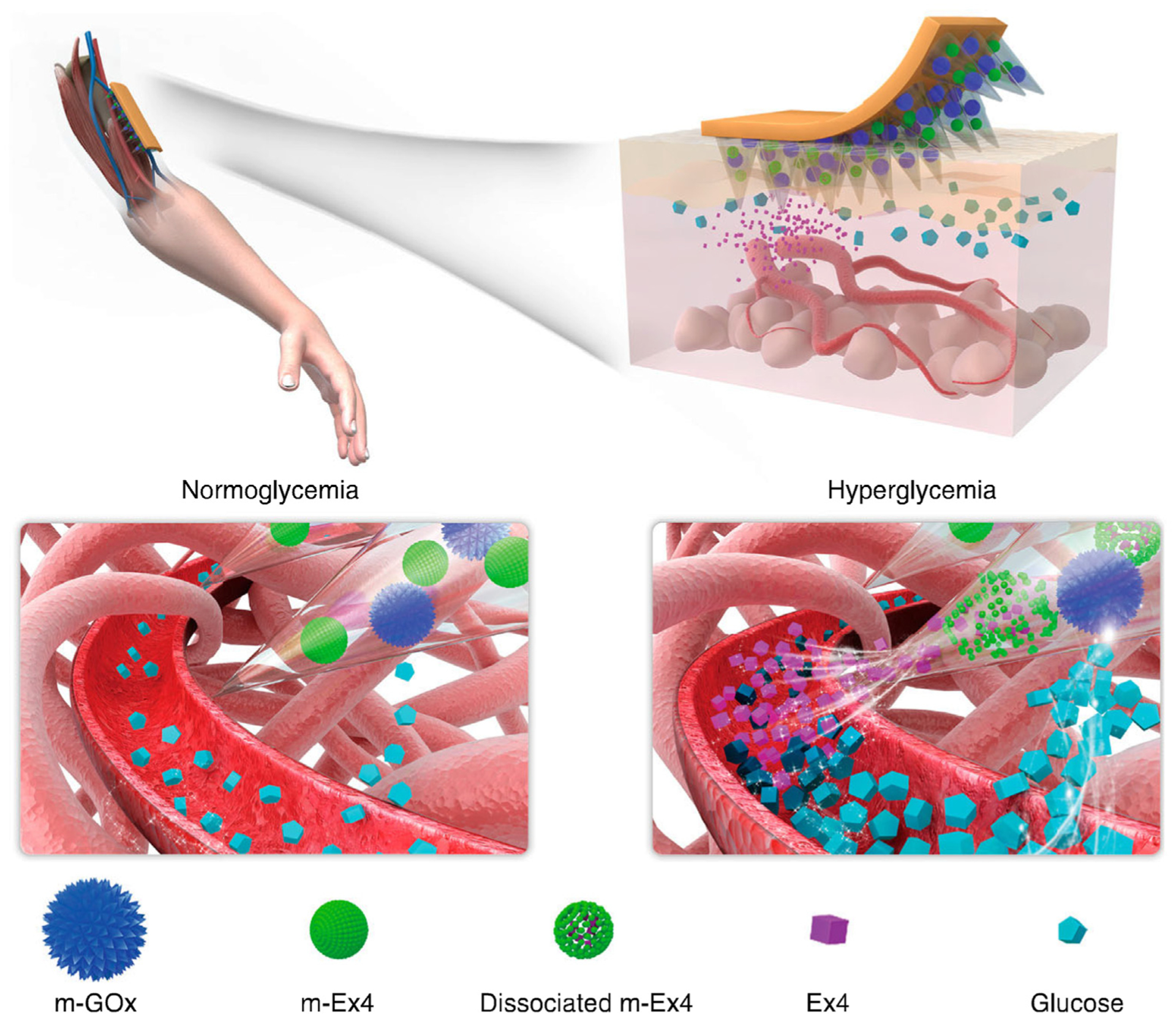
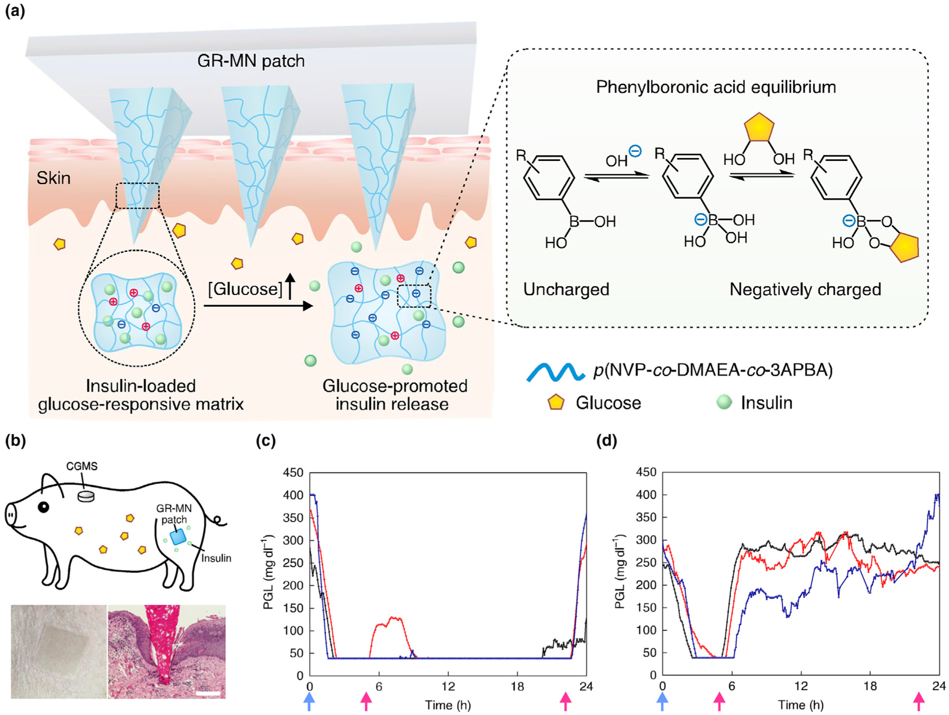
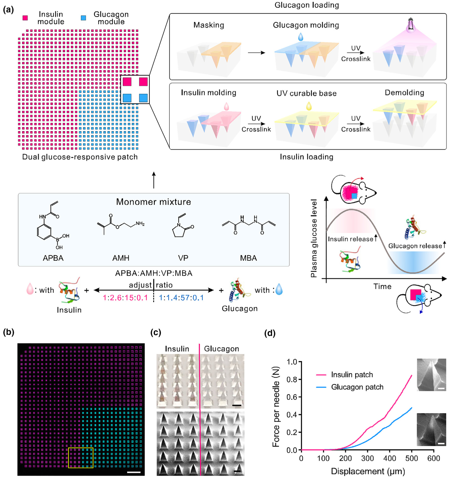
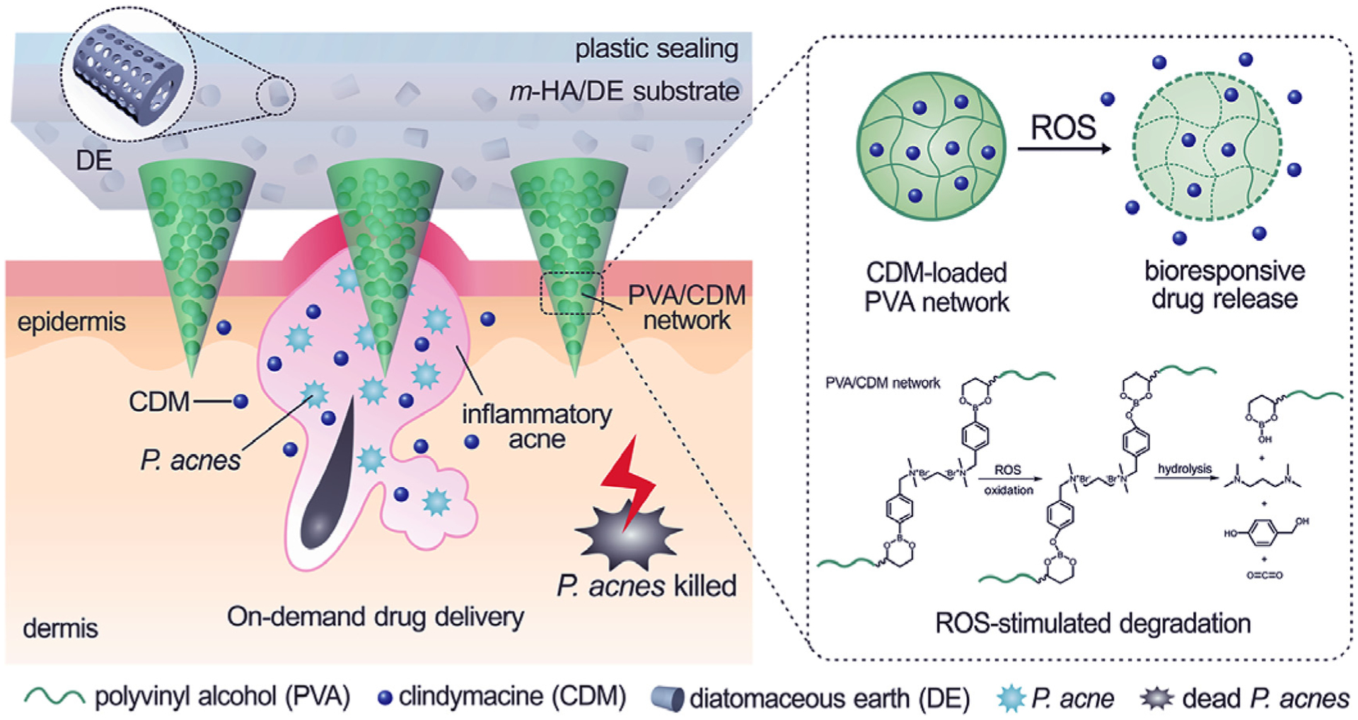
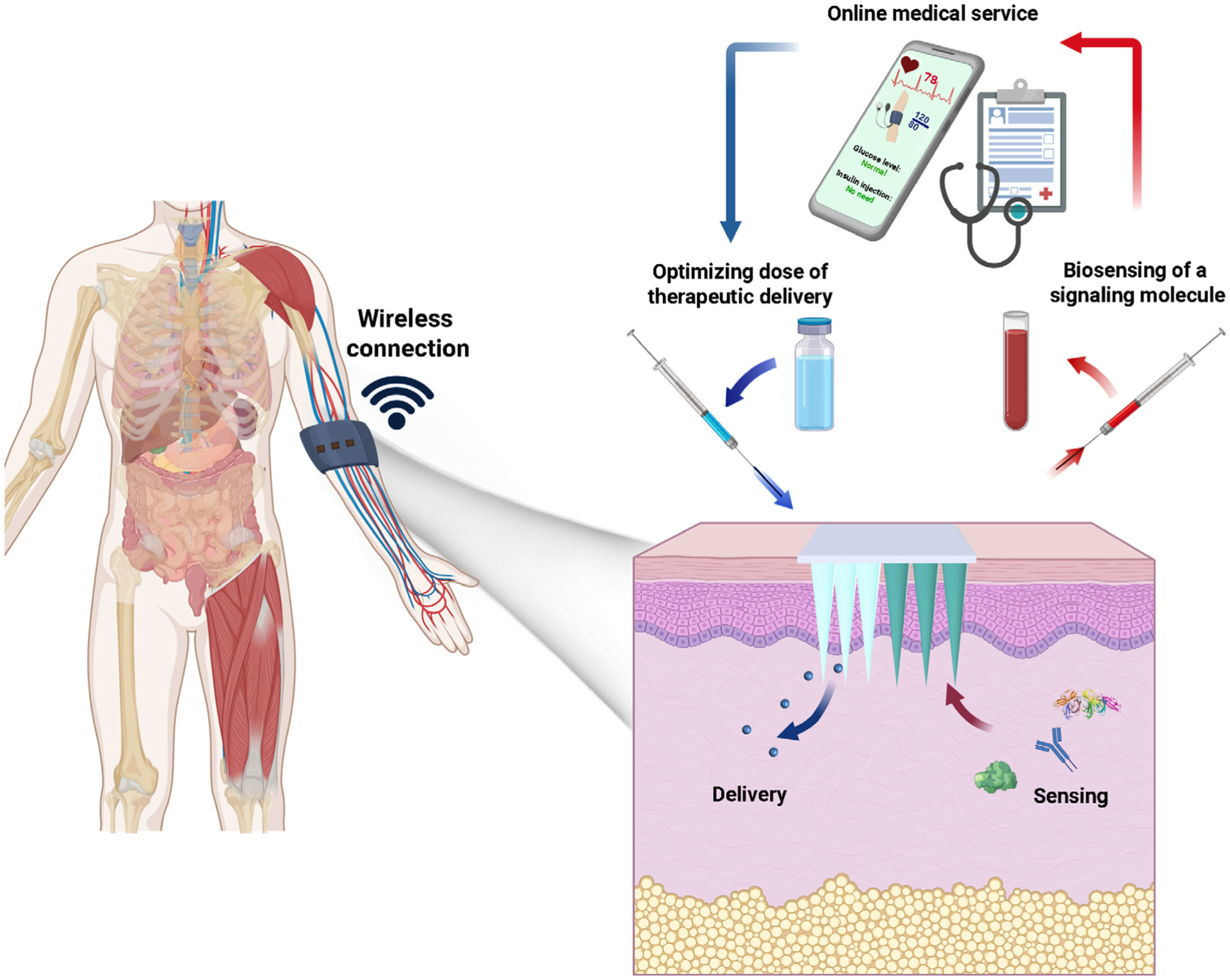
Microneedle (MN) patches consisting of miniature needles have emerged as a promising tool to perforate the stratum corneum and translocate biomolecules into the dermis in a minimally invasive manner. Stimuli-responsive MN patches represent emerging drug delivery systems that release cargos on-demand as a response to internal or external triggers. In this review, a variety of stimuli-responsive MN patches for controlled drug release are introduced, covering the mechanisms of action toward different indications. Future opportunities and challenges with respect to clinical translation are also discussed.


Conflict of interest statement

Competing interests Z.G. is a scientific co-founder of Zenomics Inc. and Zcapsule Inc. R.L. is a scientific advisor to Zenomic Inc. and Zcapsule Inc. For a list of entities with which R.L. is involved, compensated or uncompensated, see https://tinyurl.com/RLCOINBME. The remaining authors declare no competing interests.

Figures

FIGURE 1
FIGURE 1
The different types of internal and external stimuli and trigger release mechanisms for stimuli-responsive transdermal MN patch systems.
FIGURE 2
FIGURE 2
(a) Schematic of the preparation of metformin-loaded hollow mesoporous SiO2 (met/HMSN) and decoration of polydopamine (PDA) and lauric acid (LA) on the nanoparticles. Near-infrared (NIR)-responsive release of the loaded metformin on diabetic rats by the transdermal delivery is illustrated. Reproduced with modification from [23]. (b) Illustration of melanin-mediated cancer immunotherapy through a transdermal MN vaccine patch. The presence of melanin, the natural-occurring pigment in the whole tumor lysate, leads to the local release of heat via controllable near-infrared light emission. HSP: heat shock protein; ROS: reactive oxygen species; GM-CSF: granulocyte–macrophage colony-stimulating factor; NK cell: natural killer cell; DC: dendritic cell. Reprinted with permission from [24].
FIGURE 3
FIGURE 3
pH-responsive multilayer gene delivery microneedle. (a) Polylysine (PLL) and dimethylmaleic anhydride-modified polylysine (PLL-DMA) synthesis. Hyperbranched PLL, a positively-charged amino-rich polymer, is synthesized from lysin under high temperature. When exposed to DMA, the majority of amino groups form bonds with DMAt, producing PLL-DMA that is rich in –COOH functional groups. In acidic conditions, the amide groups are unstable, and PLL-DMA returns to hyperbranched PLL. Charge alteration here is responsible for pH sensitivity of this system. (b) The polyelectrolyte multilayer coating on the surface of microneedles consists of two parts; a pH-sensitive part composed of PLL-DMA/polyethylenimine (PEI) and a therapeutic agent part consisting of p53DNA/PEI. (c) Close-up of the transition polymer layers on the microneedle surface with gene-containing chains on top. Chemistry of the phase transition process is illustrated in detail. At neutral pH, PLL-DMA expresses a negative charge because of its –COOH groups. At low pH, the polymer transforms into positive-charge hyperbranched PLL. The PLL chain shares the same charge as PEI, which causes collapse of the microneedle and release of the therapeutic cargo within the acidic environment of a tumor. Reprinted with permission from [50].
FIGURE 4
FIGURE 4
(a) Hyaluronic acid-based microneedles with slow-release of the embedded nanoparticles toward the melanoma site. Nanoparticles are prepared from four components: pH-sensitive polymeric matrix, polyelectrolyte-based surfactant, glucose oxidase/catalyzer (GOx/CAT) enzymatic system, and anti-PD-1. GOx is an enzyme that converts glucose to gluconic acid with the help of the CAT. Accordingly, sensing glucose results in a drop in pH, degradation of nanoparticle and anti-PD-1 release. This gradual release profile ensures potent tumor destructive properties. (b) Schematic depicting the function of anti-PD-1 in preventing T-cell apoptosis by blocking the PD-1 receptor on the T-cell surface. This enables the T-cells to combat tumor cells longer. aPD-1: anti-programmed death-1 protein. Reprinted with permission from [54].
FIGURE 5
FIGURE 5
Schematic of the glucose-responsive insulin delivery using hypoxia-sensitive vesicle-loading MN patches. (a) Formation and mechanism of glucose-responsive vesicles (GRVs) composed of hypoxia-sensitive hyaluronic acid (HS-HA). (b) Schematic of the GRV-containing MN-array patch (smart insulin patch) for in vivo insulin delivery triggered by a hyperglycemic state to release more insulin. Reprinted with permission from [20].
FIGURE 6
FIGURE 6
(a) Self-assembled vesicles containing insulin and glucose oxidase (GOx). The dual-responsive system is prepared from poly(ethylene glycol) (PEG) and polyserine diblock copolymer modified with 2-nitroimidazole via a thioether [PEG-poly(Ser-S-NI)]. Converting thiol to sulfone is the reason for improved water solubility and higher release of insulin. (b) Comparison between hypoxia-responsive MNs and hypoxia/H2O2-responsive MNs. The former design causes high free radical levels and thereby cell toxicity. In addition, unwanted inflammation after MN insertion into the skin attracts immune cells. These side effects are addressed using dual-sensitive MNs in the insulin patches. Reprinted with permission from [68].
FIGURE 7
FIGURE 7
Schematic of a glucose-responsive insulin delivery system utilizing H2O2 and pH cascade-responsive NC-loading MN patch. (a) Formation of Ins-NCs and GOx-NCs and mechanism of glucose-responsive insulin release. (b) Schematic of H2O2-triggered charge reduction of the polymer. (c) Schematic of the NC-containing MN patch with a CAT sheath structure for in vivo insulin delivery. Insulin release is triggered under a hyperglycemic state. Reprinted with permission from [69].
FIGURE 8
FIGURE 8
Copper phosphate-mineralized particles containing glucose oxidase (mineralized GOx or m-GOx) and calcium phosphate particles with Ex4 (mineralized Ex4 or m-Ex4) in the MNs. m-GOx is static elements, converting glucose to gluconic acid only. Low pH causes solubilization of pH-sensitive biominerals such as calcium phosphate. This transition causes release of Ex4 and thus reduction in blood glucose levels. Reprinted with permission from [70].
FIGURE 9
FIGURE 9
(a) Polymeric MN composed of a polymeric matrix derived from poly(N-vinylpyrrolidone-co-2-(dimethylamino) ethyl acrylate-co-3-(acrylamido)PBA), insulin and a crosslinker (ethylene glycol dimethacrylate (EGDMA)). Insulin and phenylboronic acid (PBA) are dispersed in the polymeric matrix, with higher capacity for insulin storage. The entire MN volume acts as a closed-loop delivery system. Inset shows the mechanism in which glucose binds PBA via a reversible reaction. (b) Top: schematic of a minipig treated with a glucose-responsive microneedle (GR-MN) patch at the leg site and monitored with a continuous glucose monitoring system (CGMS). Bottom left: photograph of a GR-MN patch applied on a minipig’s leg. Bottom right: haematoxylin and eosin-stained section of minipig skin penetrated by one microneedle. Scale bar: 200 μm. (C, D) Plasma glucose levels (PGLs) in streptozotocin-induced diabetic minipigs (n = 3) after treatment with GR-MN (c) and non-responsive crosslinked microneedle (CR-MN) patches (d). Reprinted with permission from [71].
FIGURE 10
FIGURE 10
Schematic and characterizations of the dual glucose-responsive hybrid microneedle-array patch. (a) Schematic illustration of the fabrication process and glucose-responsive glycemic control mechanism of the hybrid patch. Insulin or glucagon release can be promoted by hyperglycemic and hypoglycemic conditions, respectively. The missing microneedles at two corners of the mold are used for orientation tagging. (b) A tile-scanned fluorescence microscopy top-view image of the FITC-labeled glucagon (cyan) and Cy5-labeled insulin (magenta) hybrid microneedle patch. (Scale bar, 2 mm.) (c) Photograph (Top) and SEM image (Bottom) of the MN patch at the intersection. (Scale bar, 300 μm.) (d) Mechanical performance of the glucagon and insulin MNs, respectively. A representative enlarged MN SEM image is placed next to each curve. (Scale bar, 100 μm.) Reprinted from [77] with permission from PNAS.
FIGURE 11
FIGURE 11
Schematic and corresponding images of the graphene-hybrid electrochemical devices and thermoresponsive drug delivery microneedles. (a), Schematic of the diabetes patch, which is composed of the sweat-control (i, ii), sensing (iii–vii) and therapy (viii–x) components. (b) Optical images of the electrochemical sensor array (left), therapeutic array (right) and magnified view of the drug-loaded microneedles (inset). (i) sweat-uptake layer (Nafion); (ii) water-proof film (silicone); (iii) humidity sensor (poly(3,4-ethylenedioxythiophene); PEDOT); (iv) glucose sensor (Prussian blue; PB); (v) pH sensor (polyaniline; PANi); (vi) counter electrode (Ag/AgCl); (vii) tremor sensor (graphene); (viii) microneedles with drugs (polyvinyl pyrrolidone@ tridecanoic acid); (ix) heater (Au mesh/graphene); (x) temperature sensor (graphene). (c) Schematic of the graphene-hybrid electrochemical unit, which consists of electrochemically active and soft functional materials (xi), gold-doped graphene (xii) and a serpentine Au mesh (xiii), from top to bottom. (d) Optical images of the diabetes patch laminated on human skin under mechanical deformations and wireless monitoring via Bluetooth connection. Reprinted with permission from [80].
FIGURE 12
FIGURE 12
ROS-responsive MNs that release clindamycin inside the skin when they come into contact with high concentrations of ROS, which may be as high as 500 μM in acne inflammation. The MNs are prepared from poly(vinyl alcohol) that is crossed-linked with a phenylboronic acid-containing linker. Oxidation and hydrolysis reaction by ROS degrades the polymer matrix and releases the antibiotic. Reprinted with permission from [84].
FIGURE 13
FIGURE 13
Bioresponsive MNs consist of two major components. The first component comprises a sensing system which is responsible for monitoring environmental signals. The second component comprises a delivery system which releases therapeutic agents based on signals collected from the sensing system. The MN patch may be wirelessly connected to a smartphone for reporting and data analysis.

References

    1. Marwah H et al., Drug Deliv. 23 (2016) 564–578. - PubMed
    1. O’Kane D et al., Int. J. Impot. Res 29 (2017) 225–228. - PubMed
    1. Jamaledin R et al., J. Clin. Med 9 (2020) 542. - PMC - PubMed
    1. Battisti M et al., Front. Bioeng. Biotechnol 7 (2019) 296. - PMC - PubMed
    1. Vecchione R et al., Adv. Funct. Mater 24 (2014) 3515–3523.

LinkOut - more resources