Review
. 2022 Oct 21:13:1011960.
doi: 10.3389/fendo.2022.1011960. eCollection 2022.

Genetic conditions of short stature: A review of three classic examples

Affiliations
Review

Genetic conditions of short stature: A review of three classic examples

Merlin G Butler et al. Front Endocrinol (Lausanne). .

Abstract

Noonan, Turner, and Prader-Willi syndromes are classical genetic disorders that are marked by short stature. Each disorder has been recognized for several decades and is backed by extensive published literature describing its features, genetic origins, and optimal treatment strategies. These disorders are accompanied by a multitude of comorbidities, including cardiovascular issues, endocrinopathies, and infertility. Diagnostic delays, syndrome-associated comorbidities, and inefficient communication among the members of a patient's health care team can affect a patient's well-being from birth through adulthood. Insufficient information is available to help patients and their multidisciplinary team of providers transition from pediatric to adult health care systems. The aim of this review is to summarize the clinical features and genetics associated with each syndrome, describe best practices for diagnosis and treatment, and emphasize the importance of multidisciplinary teams and appropriate care plans for the pediatric to adult health care transition.

Keywords: Noonan syndrome; Prader-Willi syndrome; Turner syndrome; genetics; growth hormone; short stature.


Conflict of interest statement

AR is a speaker and consultant for Novo Nordisk and a consultant for Ascendis Pharma. JR is a consultant and has research support from Novo Nordisk and is a consultant with OPKO. MA receives research support from Ascendis, Levo, Lumos, Novo Nordisk, Rhythm, and Soleno. MA has been on advisory boards for Pfizer and Rhythm. PB has been a consultant for Novo Nordisk, Novartis/Sandoz, Tolmar, Ascendis Pharma, BioMarin, Cavalry Bioventures, and Ipsen, and currently receives research support from Novo Nordisk and Ipsen. NM has received institutional research grants from Novo Nordisk and OPKO and has received consulting fees from Agios. The remaining authors declare that the research was conducted in the absence of any commercial or financial relationships that could be construed as a potential conflict of interest.

Figures

Figure 1
Figure 1
Growth curves for patients with NS. Heights (mean and -1 SD) of males (A) and females (B) with NS are shown with solid lines and overlaid on the normative percentile ranges, with normative mean to +1 SD in dark shading and normative mean to -1 SD in light shading. Also depicted are the heights of 35 males and 30 females with NS at the start of GH treatment (open symbols) and at final height (solid symbols). Adapted from Romano AA et al. J Clin Endocrinol Metab 2009;94(7):2338-2344. GH, growth hormone; NS, Noonan syndrome.
Figure 2
Figure 2
Genetic testing flowchart for NS. NS is typically diagnosed based on the observation of features such as craniofacial dysmorphism and facial characteristics. Other RASopathies, including NF1 and Costello syndrome, exhibit similar phenotypes to that of NS and can be distinguished using genetic testing approaches. However, a limitation of genetic testing is that approximately 30% of NS cases are not attributable to a known gene variant. BRAF, V-Raf murine sarcoma viral oncogene homolog B1; CFC, cardiofaciocutaneous; HRAS, Harvey rat sarcoma viral oncogene homolog; KRAS, Kirsten rat sarcoma viral oncogene homolog; MAP2K1, mitogen-activated protein kinase kinase 1; MAP2K2, mitogen-activated protein kinase kinase 2; NF1, neurofibromatosis type 1; NS, Noonan syndrome; NSML, Noonan syndrome with multiple lentigines; PTPN11, protein-tyrosine phosphatase, non-receptor type 11, RAF1, v-raf-1 murine leukemia viral oncogene homolog 1; SHOC2, SHOC2 leucine-rich repeat scaffold protein; SOS1, Son of Sevenless homolog 1.
Figure 3
Figure 3
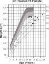
Growth curves for patients with TS. Heights of individuals with TS at the start of GH treatment (open symbols) and at final height (solid symbols). Heights from a reference TS population are shown with solid lines and overlaid on normative growth curves from the National Center for Health Statistics (shaded area), with normative 50th to 90th percentiles in dark shading and normative 10th to 50th percentiles in light shading. Reprinted from J Pediatr, 132(2), Rosenfeld RG, et al. 319-24, ©1998 Mosby, Inc. with permission from Elsevier. GH, growth hormone; TS, Turner syndrome.
Figure 4
Figure 4
Genetic testing flowchart for TS. A diagnosis of TS can be suspected based on fetal tests, including amniocentesis and chorionic villous sampling. However, all diagnoses require postnatal confirmation via 30-cell karyotyping to identify defects such as a missing X chromosome, the presence of a Y chromosome, or other X chromosome problems (eg, deletions, ring chromosomes, isochromosomes, translocations). In cases where TS is strongly suspected based on clinical features but the first karyotype comes back normal, additional karyotyping should be performed using additional tissues and/or more metaphase spreads. FISH can also be used to detect X-chromosome mosaicism. Cases that present as 45,X/46,XY and 45,X should undergo additional FISH for the DYZ3 locus, which will detect the presence of Y-chromosome material. If Y-chromosome material is present, the risk for gonadoblastoma is elevated and patients should be referred to a specialist for consultation for gonadectomy. Adapted from Ackermann A, Bamba V. J Clin Transl Endocrinol 2014;1:61-5. FISH, fluorescence in situ hybridization; TS, Turner syndrome.
Figure 5
Figure 5
Standardized curves for height and weight of non-GH-treated patients with PWS. (A, B) Standardized curves for height of non-GH-treated males (A) and females (B) with PWS. Heights of individuals with PWS are shown with solid lines and overlaid on the normative percentile ranges (shaded area) with normative 97th to 50th percentiles in dark shading and 50th to 3rd percentiles in light shading. Adapted from Butler MG et al. Pediatrics 2015;135(1):e126-35. (C, D) Standardized curves for weight of non-GH-treated males (C) and females (D) with PWS. Weights of individuals with PWS are shown with solid lines and overlaid on the normative percentile ranges (shaded area) with normative 97th to 50th percentiles in dark shading and 50th to 3rd percentiles in light shading. Adapted from Butler MG et al. Pediatrics 2015;135(1):e126-35. GH, growth hormone; PWS, Prader-Willi syndrome.
Figure 6
Figure 6
Genetic testing flowchart for PWS. Testing should begin with a DNA methylation analysis, to confirm that only maternally imprinted genes from the 15q11-q13 region are present. High-resolution SNP arrays, FISH, MS-MLPA, and/or ddPCR can be used to detect chromosome 15 deletion subtypes. SNP arrays can also be used to detect maternal UPD 15 subtypes or imprinting center defects. A streamlined approach combining DNA methylation, copy number analysis, and exome sequencing has also been reported for molecular diagnosis of PWS (208). Adapted from Butler MG, Duis J. Front Pediatr 2020;8:154. Chr, chromosome; ddPCR, droplet digital polymerase chain reaction; FISH, fluorescence in situ hybridization; MS-MLPA, methylation-specific multiplex ligation-dependent probe amplification; PWS, Prader-Willi syndrome; SNP, single nucleotide polymorphism; UPD 15, uniparental disomy 15; WES, whole-exome sequencing; WGS, whole-genome sequencing.
Figure 7
Figure 7
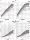
Standardized curves for height and weight of GH-treated patients with PWS. Curves for height (top) and weight (bottom) of GH-treated males (A) and females (B) with PWS. Heights and weights of GH-treated individuals with PWS are shown with solid lines and overlaid on the normative percentile ranges (shaded area) with normative 97th to 50th percentiles in dark shading and 50th to 3rd percentiles in light shading. Butler MG et al. Clin Pediatr (Phila) 2016;55(10):957-974. ©2016, SAGE Publications. Reprinted by Permission of SAGE Publications. GH, growth hormone; PWS, Prader-Willi syndrome.

References

    1. Romano AA, Allanson JE, Dahlgren J, Gelb BD, Hall B, Pierpont ME, et al. Noonan syndrome: clinical features, diagnosis, and management guidelines. Pediatrics (2010) 126(4):746–59. doi: 10.1542/peds.2009-3207 - DOI - PubMed
    1. Roberts AE, Allanson JE, Tartaglia M, Gelb BD. Noonan syndrome. Lancet (2013) 381(9863):333–42. doi: 10.1016/S0140-6736(12)61023-X - DOI - PMC - PubMed
    1. Bhambhani V, Muenke M. Noonan syndrome. Am Fam Phys (2014) 89(1):37–43. - PMC - PubMed
    1. Allen MJ, Sharma S. Noonan syndrome. In: StatPearls. FL: Treasure Island; (2021).
    1. Rauen KA. The RASopathies. Annu Rev Genomics Hum Genet (2013) 14:355–69. doi: 10.1146/annurev-genom-091212-153523 - DOI - PMC - PubMed

Publication types