

Optical coherence tomography of the retina combined with color Doppler ultrasound of the tibial nerve in the diagnosis of diabetic peripheral neuropathy
- PMID: 36339439
- PMCID: PMC9634106
- DOI: 10.3389/fendo.2022.938659
Optical coherence tomography of the retina combined with color Doppler ultrasound of the tibial nerve in the diagnosis of diabetic peripheral neuropathy
Abstract
Objective: To investigate the value of the retinal nerve fiber layer (RNFL) thickness in the optic disc and the cross-sectional area (CSA) of lower limb nerves in the diagnosis of diabetic peripheral neuropathy (DPN) separately and in combination.
Methods: A total of 140 patients with type 2 diabetes were enrolled, including 51 patients with DPN (DPN group) and 89 patients without DPN (NDPN group). Clinical data and biochemical parameters were collected. Electromyography/evoked potential instrument was performed for nerve conduction study. Optical coherence tomography was performed to measure the RNFL thickness of the optic disc. Color Doppler ultrasound was performed to measure CSA of lower limb nerves.
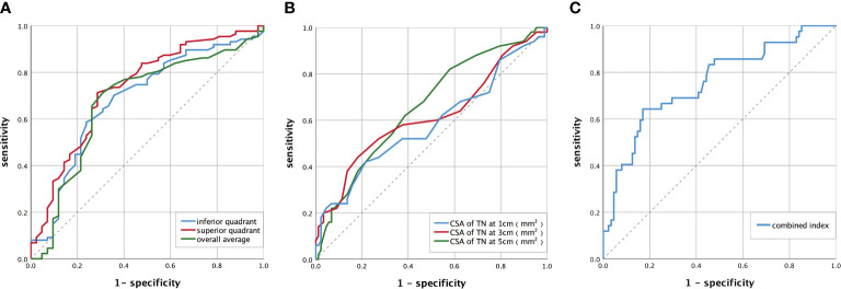
Results: The RNFL thickness was lower and the CSA of the tibial nerve (TN) in the DPN group was larger than that in the NDPN group. The album/urine creatinine ratio, diabetic retinopathy, and CSA of TN at 3 cm were positively correlated with DPN. The RNFL thickness in the superior quadrant of the optic disc was negatively correlated with DPN. For RNFL thickness to diagnose DPN, the area under the curve (AUC) of the superior quadrant was the largest, which was 0.723 (95% confidence interval [CI]: 0.645-0.805), and the best cutoff value was 127.5 μm (70.5% sensitivity, 72.1% specificity). For CSA of TN to diagnose DPN, the AUC of the distance of 5 cm was the largest, which was 0.660 (95% CI: 0.575-0.739), and the best cutoff value was 13.50 mm2 (82.0% sensitivity, 41.6% specificity). For the combined index, the AUC was greater than that of the above two indicators, which was 0.755 (95% CI: 0.664-0.846), and the best cutoff value was 0.376 (64.3% sensitivity, 83.0% specificity).
Conclusions: Patients with DPN have a reduction of the RNFL thickness and an increase in the CSA of TN, and these two changes are related to DPN. The RNFL thickness of the optic disc and the CSA of TN can be used as diagnostic indicators of DPN, and the combination of the two indicators has a higher diagnostic value.
Keywords: color doppler ultrasound; diabetes mellitus; diagnosis; optical correlation tomography; peripheral neuropathy.
Copyright © 2022 Chen, Wu, Li, Zhang, Huang, Zhuang, Bai, Chen and Lin.
Conflict of interest statement
The authors declare that the research was conducted in the absence of any commercial or financial relationships that could be construed as a potential conflict of interest.
Figures


References
-
- Saeedi P, Petersohn I, Salpea P, Malanda B, Karuranga S, Unwin N, et al. . Global and regional diabetes prevalence estimates for 2019 and projections for 2030 and 2045: Results from the international diabetes federation diabetes atlas. Diabetes Res Clin Pract (2019) 157:107843. doi: 10.1016/j.diabres.2019.107843 - DOI - PubMed
-
- Zhang Y, Zhang M, Lu D-b. Analysis of the causes and characteristic of amputation in diabetic patients with peripheral neuropathy. Med J West China (2011) 7:1241–3. doi: 10.3969/.issn.1672-3511.2011.07.014 - DOI
-
- Gylfadottir SS, Christensen DH, Nicolaisen SK, Andersen H, Callaghan BC, Itani M, et al. . Diabetic polyneuropathy and pain, prevalence, and patient characteristics: A cross-sectional questionnaire study of 5,514 patients with recently diagnosed type 2 diabetes. Pain (2020) 161(3):574. doi: 10.1097/j.pain.0000000000001744 - DOI - PMC - PubMed
Publication types
MeSH terms
LinkOut - more resources
Full Text Sources
Medical