

The unusual first sign of presentation of renal cell carcinoma: a rare case report
- PMID: 36339908
- PMCID: PMC9634463
- DOI: 10.21037/acr-22-16
The unusual first sign of presentation of renal cell carcinoma: a rare case report
Abstract
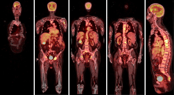
Background: Renal cell carcinoma (RCC) usually is characterized by a slow pattern of growth, although with an unpredictable evolution and metastatic potential, favored by its extensive vascularity and related high angioinvasive profile. The most common sites of metastases from kidney cancer are lung, lymph nodes, bone and liver; whereas orbital metastases are very uncommon. In more than 25% of cases, orbital metastases are the first manifestation of a primary tumor of unknown origin. The clinical features of orbital metastases from kidney cancer are non-specific and could divert attention from the real problem.
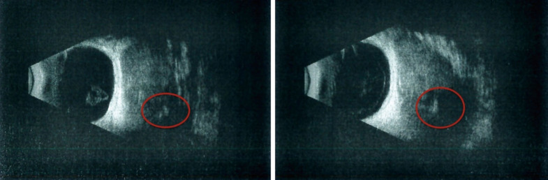
Case description: In this article, we describe the case of a 72-year-old male patient reporting a painful mass on the right orbit, with exophthalmos and ptosis, as the first and unique signs of a previously undetected advanced RCC. Due to the clinical conditions, the patient underwent palliative radiation therapy delivered to the orbital lesion with the scope to relieve pain; subsequently started systemic therapy with pazopanib at the dose of 800 mg daily. Unfortunately, he did not achieve any benefit from systemic therapy, his conditions progressively worsened, and he finally passed away after four months of treatment due to rapid disease progression.
Conclusions: Despite its rarity, differential diagnosis of an orbital lesion should always consider the possibility of metastasis from RCC, performing an appropriate radiological evaluation.
Keywords: Case report; orbital metastasis; renal carcinoma; undiagnosed cancer.
2022 AME Case Reports. All rights reserved.
Conflict of interest statement
Conflicts of Interest: All authors have completed the ICMJE uniform disclosure form (available at https://acr.amegroups.com/article/view/10.21037/acr-22-16/coif). SDP reports consulting fees for Consulting or advisory Role: GSK, MSD, Seagen, Daiichi Sankyo, Lilly, Clovis, Celgene, Astrazeneca, Novartis, Pfizer, Roche; and Speaker’s Bureau: Celgene, Astrazeneca, Novartis, Pfizer, Roche. MG reports consulting fees for Consulting or advisory Role: Astrazeneca, MSD, Seagen, Daiichi Sankyo, Lilly, Celgene, Novartis, Pfizer; Speaker’s Bureau: Lilly, Celgene, Novartis, Pfizer, Istituto Gentili, Eisai Europe Ltd., Roche; Travel, accommodation, expenses: Novartis, Pfizer, Roche. The other authors have no conflicts of interest to declare.
Figures


References
Publication types
LinkOut - more resources
Full Text Sources