. 2022 Oct 19:10:977725.
doi: 10.3389/fcell.2022.977725. eCollection 2022.

PDGF-BB signaling via PDGFR-β regulates the maturation of blood vessels generated upon vasculogenic differentiation of dental pulp stem cells

Affiliations

PDGF-BB signaling via PDGFR-β regulates the maturation of blood vessels generated upon vasculogenic differentiation of dental pulp stem cells

Zhaocheng Zhang et al. Front Cell Dev Biol. .

Abstract

A functional vascular network requires that blood vessels are invested by mural cells. We have shown that dental pulp stem cells (DPSC) can undergo vasculogenic differentiation, and that the resulting vessels anastomize with the host vasculature and become functional (blood carrying) vessels. However, the mechanisms underlying the maturation of DPSC-derived blood vessels remains unclear. Here, we performed a series of studies to understand the process of mural cell investment of blood vessels generated upon vasculogenic differentiation of dental pulp stem cells. Primary human DPSC were co-cultured with primary human umbilical artery smooth muscle cells (HUASMC) in 3D gels in presence of vasculogenic differentiation medium. We observed DPSC capillary sprout formation and SMC recruitment, alignment and remodeling that resulted in complex vascular networks. While HUASMC enhanced the number of capillary sprouts and stabilized the capillary network when co-cultured with DPSC, HUASMC by themselves were unable to form capillary sprouts. In vivo, GFP transduced human DPSC seeded in biodegradable scaffolds and transplanted into immunodeficient mice generated functional human blood vessels invested with murine smooth muscle actin (SMA)-positive, GFP-negative cells. Inhibition of PDGFR-β signaling prevented the SMC investment of DPSC-derived capillary sprouts in vitro and of DPSC-derived blood vessels in vivo. In contrast, inhibition of Tie-2 signaling did not have a significant effect on the SMC recruitment in DPSC-derived vascular structures. Collectively, these results demonstrate that PDGF-BB signaling via PDGFR-β regulates the process of maturation (mural investment) of blood vessels generated upon vasculogenic differentiation of human dental pulp stem cells.

Keywords: angiogenesis; pericytes; self-renewal; smooth muscle cells; tissue regeneration; vasculogenesis.


Conflict of interest statement

The authors declare that the research was conducted in the absence of any commercial or financial relationships that could be construed as a potential conflict of interest.

Figures
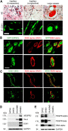
FIGURE 1
FIGURE 1
Vasculogenic differentiation of dental pulp stem cells (DPSC) in vivo. 1000000 GFP-transduced DPSC cells were seeded in scaffolds and transplanted into the subcutaneous space of the dorsum of SCID mice for 4 weeks. Scaffolds were retrieved and fixed with 10% buffered formaldehyde for HE and immunofluorescence staining. (A) HE staining showed capillaries and large blood vessels. DPSC-derived endothelial cells were positively stained with CD31. (B) Endothelial cells lining blood vessels were positive for GFP (i.e., generated upon vasculogenic differentiation of DPSC-GFP cells) while mural cells (pericytes/SMC) were positive for SMA-α, but not GFP (i.e., mural cells were recruited form the mouse host). (C) Immunofluorescence showing that endothelial cells were stained with CD31 while mural cells with SMA-α. Scale bar: 50 µm. (D,E) Western blots from cells (DPSC, SHED, HUASMC, and HDMEC) cultured in α-MEM supplemented with 5%FBS for the endothelial cell markers VEGFR2, Tie-2 and CD31 (D), and for mural cell markers PDGFR-β and SMA-α (E). HDMEC cells were used as positive control for endothelial cells, and HUASMC as controls for mural cells. GAPDH was used as loading control.
FIGURE 2
FIGURE 2
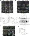
PDGF-BB and bFGF induce capillary sprouting of DPSCs. (A–D) 10000 DPSC or HUASMC were seeded in 12 well plates coated with growth factor reduced Matrigel and cultured with either endothelial differentiation medium-2 for microvascular cells (EGM2-MV) + 50 ng/ml VEGF as a positive controls (A), or α-MEM supplemented with 5% FBS +20 ng/ml bFGF +20 ng/ml PDGF (B) for indicated time points. Sprouting formation was photographed and the number of sprout-like structures was counted. (C,D) Graphs depicting the numbers of sprouting from (A) and (B), respectively. Scale bar: 100 µm. (E) DPSC cells were cultured with α-MEM supplemented with 5% FBS in presence of 20 ng/ml bFGF, PDGF-BB or bFGF + PDGF-BB for 5 days. Western blots were performed for VEGFR2, Tie-2, CD31, Bmi-1, and GAPDH (loading control). (F) DPSC were plated in Matrigel-coated wells and cultured with α-MEM supplemented with 5% FBS in presence of 20 ng/ml bFGF, PDGF-BB or bFGF + PDGF-BB for indicated time points. Sprout-like structures were photographed and counted. Scale bar: 100 µm. (G) Graph depicting the numbers of sprout-like structures from (F).
FIGURE 3
FIGURE 3
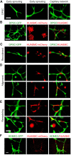
Maturation of DPSC-derived vascular networks in vitro. (A–E) 10000 DPSC (green) and HUASMC (red) were seeded in 12 well plates coated with growth factor reduced Matrigel and cultured with α-MEM supplemented with 5% FBS in presence of 20 ng/ml bFGF + PDGF-BB for 8–12 days, the sprouts were photographed with fluorescence microscope. (A) DPSC showed early branching, then sprouting and finally a capillary network with increasing time. (B) Photomicrographs of DPSC cells forming a capillary network (left); HUASMC cells that were unable to form capillary networks (middle); and HUASMC aligning with DPSC when cultured together (right). (C) Early stage-recruitment of HUASMC during initial spouting. (D) Photomicrographs showing HUASMC aligned and connecting two separate DPSC-derived branches to form initial capillary networks. (E) HUASMC stabilized the capillary-like structure and filled-up gaps present in the capillary networks. Scale bar: 100 µm. (F) 50000–100000 GFP-transduced HDMEC and mCherry-transduced HUASMC were seeded in 12 well plates coated with growth factor reduced Matrigel and cultured with EGM2-MV + 50 ng/ml VEGF for 24 h. Capillary-like networks showing HUASMC aligned with HDMEC-derived capillary-like sprouts were photographed. Scale bar: 100 µm.
FIGURE 4
FIGURE 4
Tie-2 does not contribute to mural investment of DPSC-derived capillaries in vitro. (A) HDMEC were starved overnight and cultured with α-MEM supplemented with 5% FBS and 0–400 ng/ml Ang-1 in presence of 0–5 µM SB203580 (Tie-2 inhibitor) for 30 min. Western blots were performed for p-Tie-2, Tie-2. (B,C) 10000–40000 DPSC (B) or HUASMC (C) were cultured with α-MEM supplemented with 5%FBS (B) or SMC growth medium (C) in the presence of 0–5 µM SB203580. After 5 days, the number of cells were counted under microscopic evaluation. Graphs depicting the number of DPSC (B) and HUASMC (C) in response to the Tie-2 inhibitor. (D) 10000 DPSC or HUASMC were seeded in 12 well plates coated growth factor reduced Matrigel and cultured with EGM2-MV + 50 ng/ml VEGF in presence of 0–10 µM SB203580 for indicated time points. Microphotographs of networks composed of DPSC-derived capillary-like structures lined with HUASMC cells.
FIGURE 5
FIGURE 5
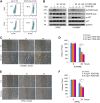
PDGFR-β signaling induces motility of DPSC cells and smooth muscle cells in vitro. (A) Flow plots depicting the expression of PDGFR-β in DPSC and SHED. IgG-AF488 was used as isotype control. (B) DPSC and HUASMC were starved overnight and pre-incubated with 0–100 nM Ki11502 (PDGFR inhibitor) for 2 h, and then treated with α-MEM supplemented with 5% FBS in presence of 20 ng/ml PDGF-BB for 30 min. Western blots were performed for p-PDGFR-β, PDGFR-β, p-AKT, and AKT. (C–F) HUASMC (C) and DPSC (E) were cultured in 6-well plates, starved overnight, scratched with a sterile 1,000 µl loading tip, then treated with α-MEM supplemented with 5% FBS in presence of 0–50 ng/ml PDGF-BB for indicated time points. Scale bar: 100 µm. (D,F) Graphs depicting the scratch width over time in response to PDGF-BB in HUASMC (C) and DPSC (E). Three independent experiments using triplicate wells/experimental condition were performed to verify reproducibility of the data. Asterisk indicates p < 0.05.
FIGURE 6
FIGURE 6
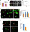
Blockade of PDGFR-β signaling inhibits maturation of DPSC-derived blood vessels in vitro and in vivo. (A) 10000–50000 DPSC or HUASMC were cultured with α-MEM supplemented with 5% FBS in presence of 0–25 nM Ki11502. After 4 days, the number of cells were counted under microscopic evaluation. (B,C) 10000 DPSC were plated in growth factor-reduced Matrigel-coated wells and cultured with EGM2-MV + 50 ng/ml VEGF in presence of 0 or 25 nM Ki11502 (PDGFR inhibitor) for indicated time points. Photomicrographs of capillary-like sprouts. Scale bar: 100 µm. (C) Graph depicting the number of capillary-like sprouts from (B). (D,E) DPSC (green) and HUASMC (red) were seeded in Matrigel-coated plates and cultured with α-MEM supplemented with 5% FBS in presence of 20 ng/ml bFGF and PDGF-BB in the presence of 25 nM Ki11502 for indicated time points. Vascular networks were photographed. Scale bar: 100 µm. (E) Graph depicting the percentage of HUASMC cells in direct contact with DPSC cells as a fraction of the total number of HUASMC cells in the microscopic field. Asterisks depict p < 0.001. (F,G) 1000000 GFP-transduced DPSC cells were seeded in scaffolds and transplanted into the subcutaneous space of immunodeficient mice. After 3 weeks, mice (n = 5 per group) were treated with vehicle (control) or with 5 mg/kg Ki11502 by oral gavage, once a day for 7 days. After 4 weeks, mice were euthanized, scaffolds were removed, fixed, and prepared for HE and immunofluorescence staining. (F) Immunofluorescence for GFP (DPSC, green) and SMA-α (mouse mural cells, red) double staining. (G) Graph depicting the percentage of blood vessels covered by SMA-α cells as compared to total blood vessels from (F). Asterisk depicts p < 0.001.

Similar articles

Cited by

References

    1. Armulik A., Genove G., Betsholtz C. (2011). Pericytes: Developmental, physiological, and pathological perspectives, problems, and promises. Dev. Cell 21, 193–215. 10.1016/j.devcel.2011.07.001 - DOI - PubMed
    1. Armulik A., Genove G., Ma¨e M., Nisancioglu M. H., Wallgard E., Niaudet C., et al. (2010). Pericytes regulate the blood-brain barrier. Nature 468, 557–561. 10.1038/nature09522 - DOI - PubMed
    1. Augustin H. G., Koh G. Y., Thurston G., Alitalo K. (2009). Control of vascular Morphogenesis and homeostasis through the angiopoietin-Tie system. Nat. Rev. Mol. Cell Biol. 10, 165–177. 10.1038/nrm2639 - DOI - PubMed
    1. Benjamin L. E., Hemo I., Keshet E. (1998). A plasticity window for blood vessel remodelling is defined by pericyte coverage of the preformed endothelial network and is regulated by PDGF-B and VEGF. Development 125, 1591–1598. 10.1242/dev.125.9.1591 - DOI - PubMed
    1. Bento L. W., Zhang Z., Imai A., Nör F., Dong Z., Shi S., et al. (2013). Endothelial differentiation of SHED requires MEK1/ERK signaling. J. Dent. Res. 92, 51–57. 10.1177/0022034512466263 - DOI - PMC - PubMed