. 2022 Oct 21;7(43):38436-38447.
doi: 10.1021/acsomega.2c03649. eCollection 2022 Nov 1.

Annona squamosa L. Extract-Loaded Niosome and Its Anti-Ehrlich Ascites' Carcinoma Activity

Affiliations

Annona squamosa L. Extract-Loaded Niosome and Its Anti-Ehrlich Ascites' Carcinoma Activity

Amr A Abd-Elghany et al. ACS Omega. .

Abstract

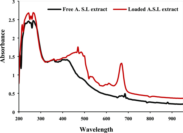
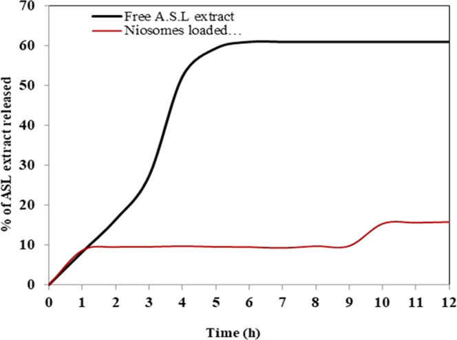
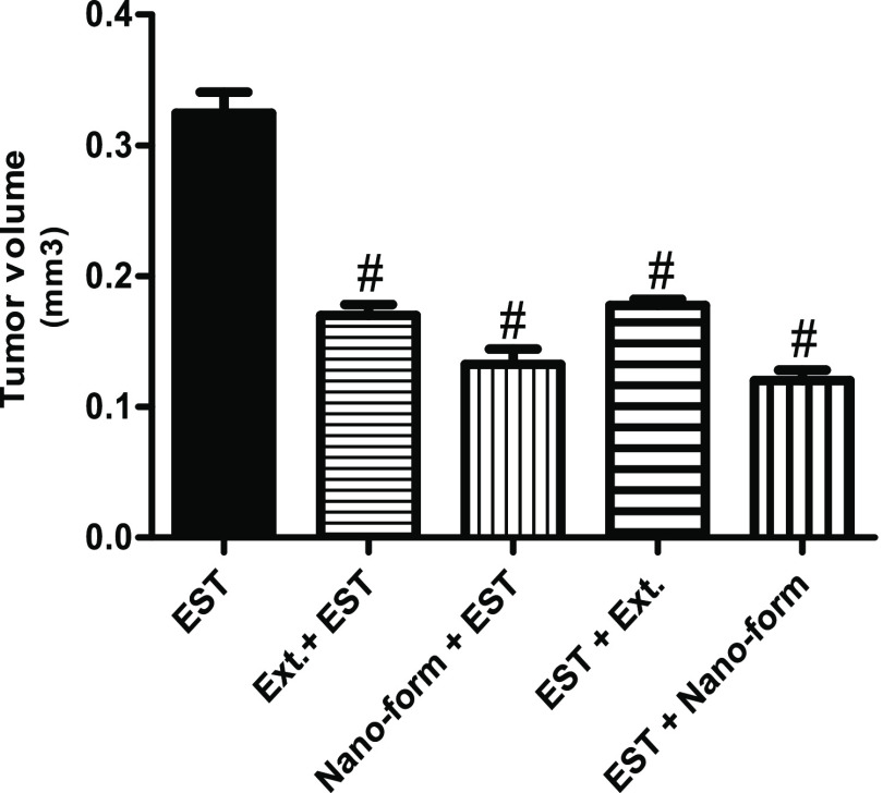
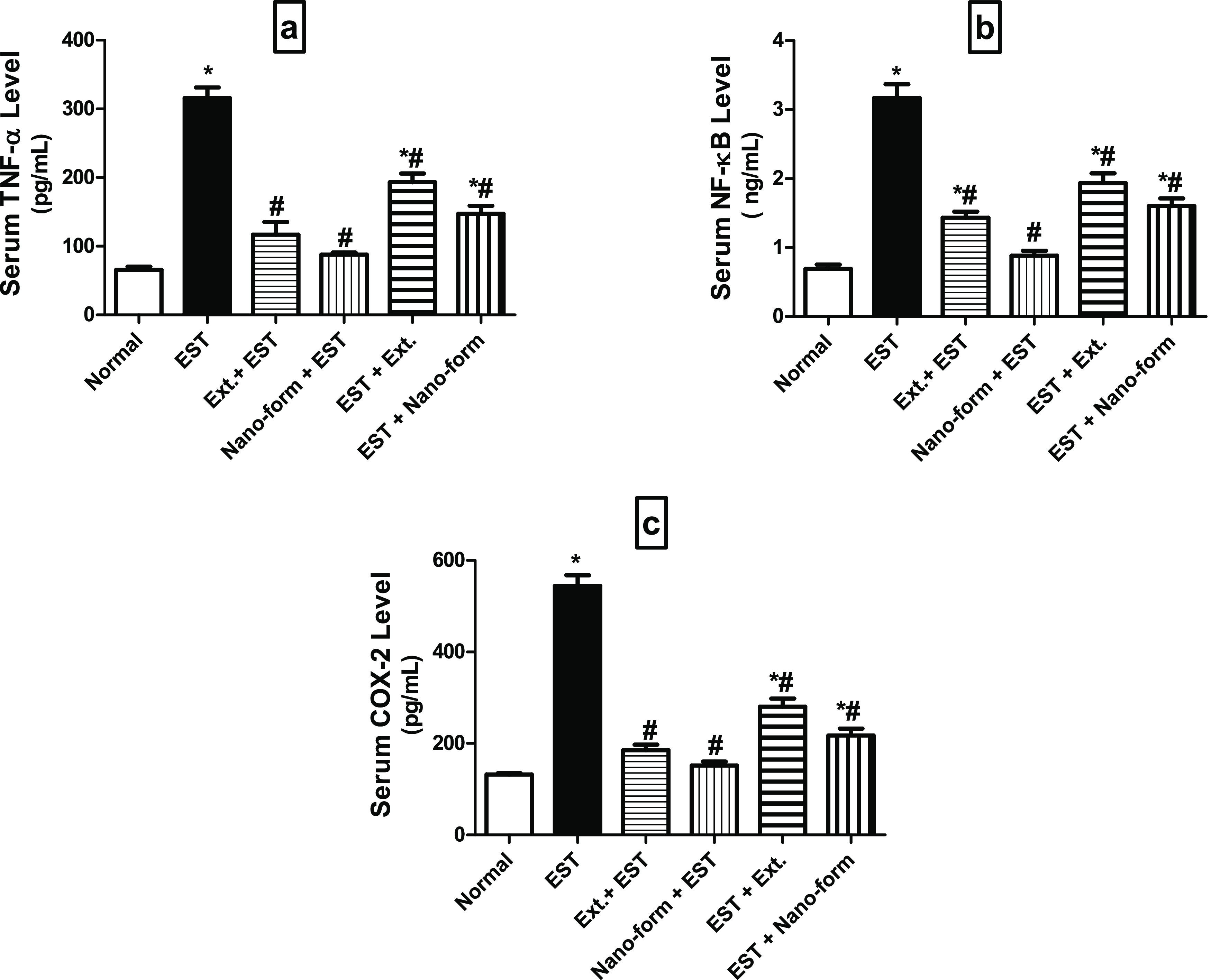
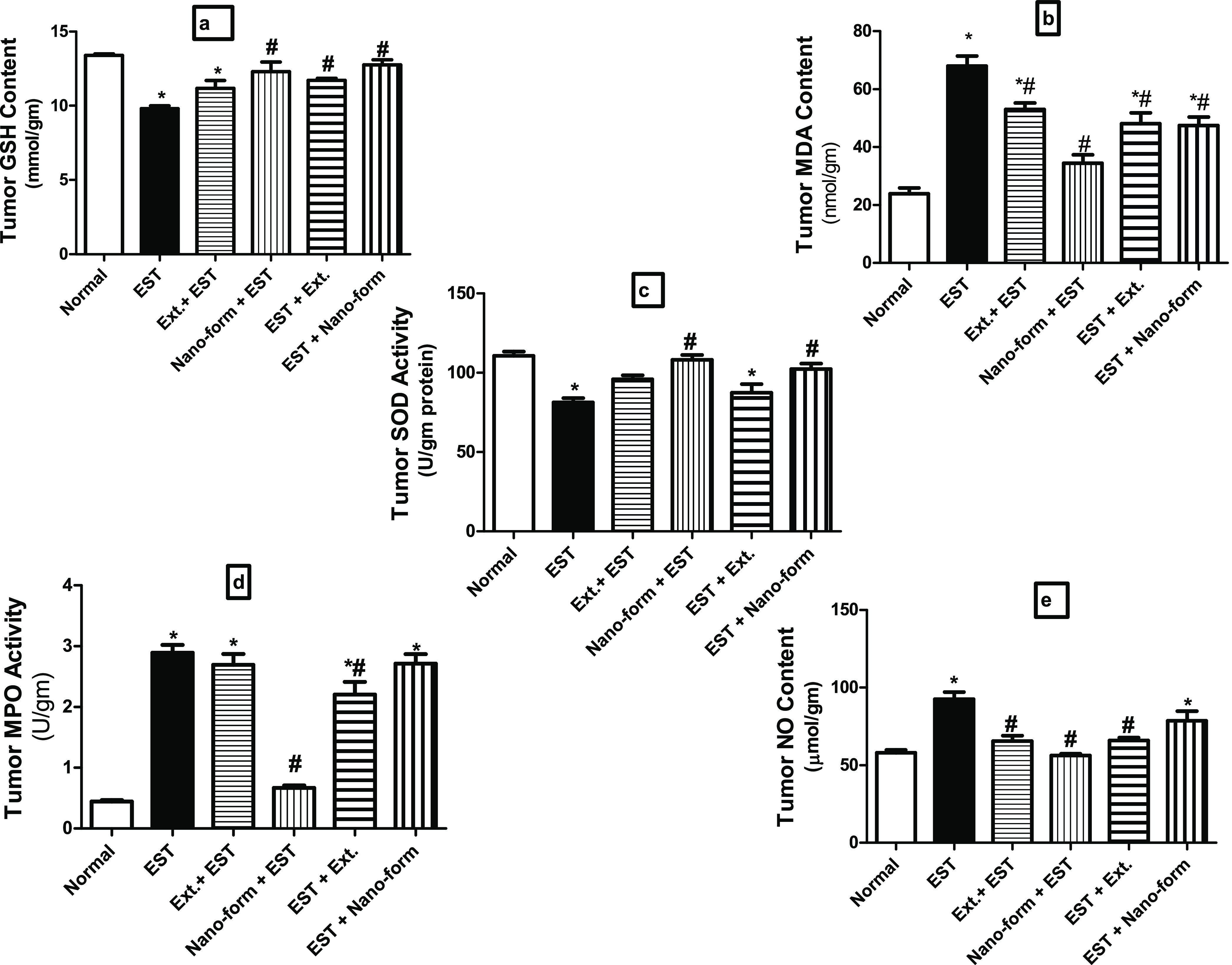
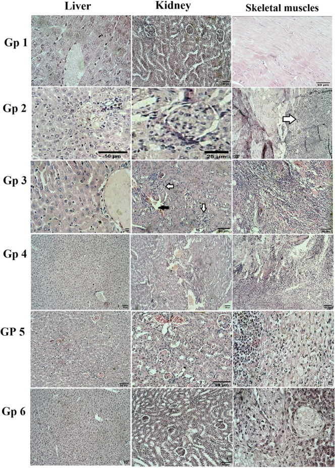
Current research is focused on cancer treatments other than chemotherapy medications, particularly those derived from natural sources. The goal of this work was to look at the anticancer and biomarker properties of a methanolic extract of Annona squamosa leaves and their extract-loaded noisome. A. squamosa leaves extract and their leaves extract-loaded noisome were prepared. Transmission electron microscopy was used to screen the size of the niosomes loaded with the A. squamosa L. leaves extract. The tumor size, blood picture (hemoglobin, red blood cells, white blood cells), liver functions, kidney function, oxidative stress, and inflammatory markers were evaluated to assess the potential anticancer activity of the A. squamosa leaves extract and A. squamosa leaves extract-loaded noisome in Ehrlich ascites carcinoma. A. squamosa L. leaves extract was found to be an effective anticancer treatment. The protective effect of the loaded extract showed more significant results. All treated groups showed a lower tumor volume compared to the positive control. Liver and kidney functions were improved, and inflammatory markers were decreased. Oxidative stress was improved in tumor, liver, and kidney tissues. A. squamosa leaves contain major anticancer compounds that in general help most enzymes of the liver and kidney and other injured organs to return to their normal levels.


Conflict of interest statement

The authors declare no competing financial interest.

Figures

Figure 1
Figure 1
UV absorption spectrum of ASLE and ASLELN.
Figure 2
Figure 2
TEM images of the niosome loaded with the ASL extract.
Figure 3
Figure 3
In vitro release curves of the free ASL extract and loaded ASL extract from the cellulose acetate dialysis bag.
Figure 4
Figure 4
Effect of ASLE and ASLELN on the development of Ehrlich’s volume. Each value indicates the mean ± SEM. #indicates significantly dissimilar from the EST control group at p < 0.05. Statistical analysis was performed by one-way ANOVA followed by Tukey multiple comparison test.
Figure 5
Figure 5
Levels of inflammatory markers (a) TNF-α, (b) NF-kB, and (c) COX-2 in the serum of different studied groups. Each value indicates the mean ± SEM. *Significantly different from the normal group at p < 0.05. #Significantly different from the control EST group at p < 0.05. Statistical analysis was performed by one-way ANOVA followed by Tukey multiple comparison test.
Figure 6
Figure 6
Levels of oxidant and antioxidant markers (a) GSH content, (b) MDA, (c) SOD activity, (d) MPO activity, and (e) NO content in tumor homogenate of different studied groups. Each value indicates the mean ± SEM. *Different significantly from the normal group at p < 0.05. #Different Significantly from the control EST group at p < 0.05. One way of exhibiting statistical analysis was performed by ANOVA followed by Tukey Multiple Comparison Test.
Figure 7
Figure 7
Histopathology photomicrographs for mic (liver, kidney, and skeletal muscles) tissues. Group 1 showed: liver tissue with the normal architecture of hepatocytes around the central vein (H&E, 10 μm), kidney tissue with normal renal tissue (H&E, 50 μm), skeletal muscles tissue with normal muscle fibers striation (H&E, 50 μm). Group 2 showed: liver tissue congested central vein with infiltration of activated Kupffer′s cells (H&E, 50 μm), a glomerular tuft of the kidney with slightly hyperplastic glomerular cells (H&E, 25 μm), skeletal muscles tissue with the invasion of the neoplastic cells to the surrounding skeletal muscles with marked necrosis (H&E, 50 μm). Group 3 showed: hepatic tissue of the central vein with infiltration of activated Kupffer′s cells (H&E, 10 μm), the obliteration of Bowman’s space in the kidney tissue with the hyperplastic glomerular cells (white arrows) with dilatation and congestion of renal blood vessel (black arrow) (H&E, 50 μm), muscle bundles with the invading neoplastic cells diffusing all around its adjacent tissue with marked necrosis (H&E, 50 μm). Group 4 showed: hepatic tissue with infiltration of activated Kupffer′s cells between hepatocytes (H&E, 50 μm), kidney have the obliteration of Bowman’s space with the hyperplastic glomerular cells with dilatation and congestion of renal blood vessel (H&E, 50 μm), the neoplastic cells diffusing all around its adjacent tissue with necrosis of skeletal muscles (H&E, 50 μm). Group 5 showed: that the liver has congested central veins with an activated Kupffer′s cells infiltrating in between (H&E, 50 μm), kidney with the obliteration of Bowman’s space with the hyperplastic glomerular cells with dilatation and congestion of renal blood vessel (H&E, 50 μm), muscle bundles with the invading neoplastic cells diffusing all around its adjacent tissue with marked necrosis (H&E, 10 μm). Group 6 showed: Image of the liver with the normal architecture of the hepatic lobules (H&E, 50 μm), Normal renal tissue architecture (H&E, 50 μm), and muscle bundles showing the invading neoplastic cells diffusing all around its adjacent tissue with marked necrosis (H&E, 10 μm).

References

    1. Siegel R. L.; Miller K. D.; Fuchs H. E.; Jemal A. Cancer Statistics, 2022. Ca-Cancer J. Clin. 2022, 72, 7–33. 10.3322/caac.21708. - DOI - PubMed
    1. Wu J.; Zhou T.; Wang Y.; Jiang Y.; Wang Y. Mechanisms and Advances in Anti-Ovarian Cancer with Natural Plants Component. Molecules 2021, 26, 594910.3390/molecules26195949. - DOI - PMC - PubMed
    1. Abd-Elghany A. A.; Mohamad E. A. Ex-vivo transdermal delivery of Annona squamosa entrapped in niosomes by electroporation. J. Radiat. Res. Appl. Sci. 2020, 13, 164–173. 10.1080/16878507.2020.1719329. - DOI
    1. Pardhasaradhi B.; Reddy M.; Ali A. M.; Kumari A. L.; Khar A. Antitumour activity of Annona squamosa seed extracts is through the generation of free radicals and induction of apoptosis. Indian J. Biochem. Biophys. 2004, 41, 167–172. - PubMed
    1. Wang Z.; Li Z.; Ye Y.; Xie L.; Li W. Oxidative stress and liver cancer: etiology and therapeutic targets. Oxid. Med. Cell. Longevity 2016, 2016, 789157410.1155/2016/7891574. - DOI - PMC - PubMed