. 2022 Oct 29;3(4):101798.
doi: 10.1016/j.xpro.2022.101798. eCollection 2022 Dec 16.

Biosensor Cell-Fit-HD4D for correlation of single-cell fate and microscale energy deposition in complex ion beams

Affiliations

Biosensor Cell-Fit-HD4D for correlation of single-cell fate and microscale energy deposition in complex ion beams

Julian Schlegel et al. STAR Protoc. .

Abstract

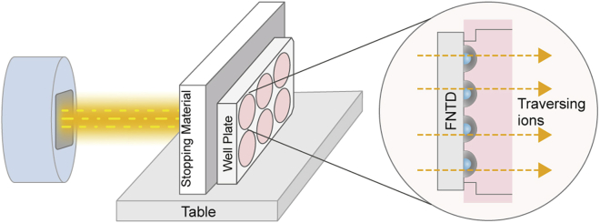
We present a protocol for the biosensor Cell-Fit-HD4D. It enables long-term monitoring and correlation of single-cell fate with subcellular-deposited energy of ionizing radiation. Cell fate tracking using widefield time-lapse microscopy is uncoupled in time from confocal ion track imaging. Registration of both image acquisition steps allows precise ion track assignment to cells and correlation with cellular readouts. For complete details on the use and execution of this protocol, please refer to Niklas et al. (2022).

Keywords: Biophysics; Biotechnology and bioengineering; Cancer; Microscopy; Molecular biology; Molecular/Chemical probes; Single cell.


Conflict of interest statement

The authors declare no competing interests.

Figures

None
Graphical abstract
Figure 1
Figure 1
Cell coating of Cell-Fit-HD4D (A and B) Exemplary comparison of a healthy cell layer (A) and cells showing abnormal morphology as a consequence of suboptimal construction of Cell-Fit-HD4D (B). Images were taken three days after seeding. Residual glue in contact with the medium leads to impaired proliferation as well as enlargement and elongation, indicating physiologic stress. Here, higher cell numbers were seeded for demonstration.
Figure 2
Figure 2
Readout protocol for the biosensor Cell-Fit-HD4D In the initial readout the so-called initial stack, the FNTD and the tumor cells are scanned in a single step. Thus, the earliest cellular response irradiation and the spinels (indicated by black discs), acting as landmarks for later image registration are recorded. The initial stack typically comprises an image stack of ∼ 30 imaging planes (2.). In a second step live-cell imaging comprising an image stack of ∼3 imaging planes of the biological compartment (cell layer) is performed by widefield microscopy (3.) After imaging, the Cell-Fit-HD4D is transferred to confocal microscopy to read out the FNTD. Using image registration routines, each ion track (red arrows) can be reconstructed into the cell layer recorded in the initial readout. The tilt between cell layer plane and confocal planes indicates the separate acquisition on two different microscopes, followed by post-acquisition stitching and image registration. Figure and figure caption were taken from (Niklas et al., 2022).
Figure 3
Figure 3
Exemplary irradiation setup for Cell-Fit-HD4D The biosensor is aligned perpendicular to the incoming ion beam. Additional stopping material in front of the well plate comprising the FNTD wafer coated with a single cell layer enables to locate the cell in the desired position of a Bragg peak. Figure and figure caption were taken from (Niklas et al., 2022). The irradiation setup was similar to (Dokic et al., 2016).
Figure 4
Figure 4
Fluorescent readout signal of the cell layer (A) Representative fluorescent images of the cell cycle inhibitor p21, 53BP1 and DAPI. Cell-Fit-HD4D allows to identify p21 induction in a subpopulation of cells in response to irradiation. (B) Histogram of p21 mean intensities in single cell nuclei (IR: C-12 irradiation 1 Gy, spread-out Bragg peak, Ctrl: control) including thresholding (66 a.u.) for p21-positive cells. Figure and figure caption were taken from (Niklas et al., 2022).
Figure 5
Figure 5
Exemplary intensity-to-LET conversion (A) The LET-distribution by Monte Carlo simulation considering all ion types in the spread-out-Bragg peak (SOBP) of a carbon ion beam of 1 Gy is shown. The peak at 50 keV/μm correspond mainly to primary ions (carbon ions), the other peaks correspond to fragments. (B) The individual distributions (carbon ions: C, fragments: B, Be, H, He, Li) constituting the peak at 50 keV/μm are depicted. The carbon ions dominate. (C) Intensity distribution of ion traversals detected by the FNTD is shown. Intensity-based thresholding was applied to distinguish between primary ions (carbon ions, C) and fragments. (D) Only the carbon ions (Experiment) were considered in the in the LET calculation. The intensity distribution of the carbon ions were mathematically transferred by LETj=101ab to match the peak at 50 keV/μm in the LET distribution in (A) with I: track intensity of the ion, LETj: LET of the ion j, a and b: parameters adjusted to match intensity- and LET distribution. According to this transfer the intensity values were converted into LET values. For detailed description we refer to (Klimpki et al., 2016; Rahmanian et al., 2017; Greilich et al., 2018; Niklas et al., 2022).
Figure 6
Figure 6
Spatio-temporal correlation of physical energy deposition in individual cell nuclei with cellular response (A) Ion tracks (blue crosses) are reconstructed into the cell layer in the very first time point (T0) after irradiation. The corresponding processed data sets were recorded in the initial readout. Cell nuclei were identified by cell tracking software. (B) Cell nucleus tracking and radiation induced DNA damage foci (RIF) segmentation of the data set gained by live-cell imaging allows to assign each mother cell (identification: MotherID) and their corresponding daughter cells (identification: DaugherID) a set of biological parameters. In our case radiation induced foci (RIF) of the mother cell within 24 h after irradiation, number of RIFs and p21 level in the daughter cells at 96 h after irradiation. (C) The ion traversals are reconstructed into a binary mask of the maximum intensity z projections gained by the initial readout. By assessment of the track intensity and hence LET (keV/μm) of individual ion traversal identified by TrackID, a set of physical beam parameters (∑LET: sum of single ions LET in a cell nucleus) can be extracted for individual cell nucleus. (D) Combining the biological and physical data sets generates unique spatiotemporal correlation of the energy deposition in a cell nucleus and its response. Exemplarily correlations, i.e., correlation of RIF numbers 5.6 h after irradiation with number of carbon ion hits in the corresponding nucleus as well as cell division tree are depicted. SD: standard deviation, RIF: radiation induced DNA damage foci.

References

    1. Akselrod M., Kouwenberg J. Fluorescent nuclear track detectors - review of past, present and future of the technology. Radiat. Meas. 2018;117:35–51. doi: 10.1016/j.radmeas.2018.07.005. - DOI
    1. Akselrod M.S., Sykora G.J. Fluorescent nuclear track detector technology – a new way to do passive solid state dosimetry. Radiat. Meas. 2011;46:1671–1679. doi: 10.1016/j.radmeas.2011.06.018. - DOI
    1. Arganda-Carreras I., Kaynig V., Rueden C., Eliceiri K.W., Schindelin J., Cardona A., Sebastian Seung H. Trainable Weka segmentation: a machine learning tool for microscopy pixel classification. Bioinformatics. 2017;33:2424–2426. doi: 10.1093/bioinformatics/btx180. - DOI - PubMed
    1. Dimitrova N., Chen Y.C.M., Spector D.L., de Lange T. 53BP1 promotes non-homologous end joining of telomeres by increasing chromatin mobility. Nature. 2008;456:524–528. doi: 10.1038/nature07433. - DOI - PMC - PubMed
    1. Dokic I., Mairani A., Niklas M., Zimmermann F., Chaudhri N., Krunic D., Tessonnier T., Ferrari A., Parodi K., Jäkel O., et al. Next generation multi-scale biophysical characterization of high precision cancer particle radiotherapy using clinical proton, helium-carbon- and oxygen ion beams. Oncotarget. 2016;7:56676–56689. doi: 10.18632/oncotarget.10996. - DOI - PMC - PubMed

Publication types