

Measurement of D-dimer in cerebrospinal fluid using a luminescent oxygen channeling immunoassay
- PMID: 36341102
- PMCID: PMC9632730
- DOI: 10.3389/fneur.2022.951802
Measurement of D-dimer in cerebrospinal fluid using a luminescent oxygen channeling immunoassay
Abstract
Background: Measurement of D-dimer in cerebrospinal fluid (CSF) allows insight into coagulation system activation in the central nervous system and can be utilized to monitor intracranial hemorrhage as well as acute phase processes beyond hemostasis in inflammatory and neoplastic diseases. So far, the measurability of D-dimer in low and very low concentrations in CSF was limited in conventional immunoassays. Novel high-sensitivity chemiluminescent immunoassays such as the luminescent oxygen channeling immunoassay (LOCI®) are getting increasingly available but have not been validated in CSF. The aim of this study was to investigate the accuracy and linearity of the LOCI® in assessing D-dimer in CSF.
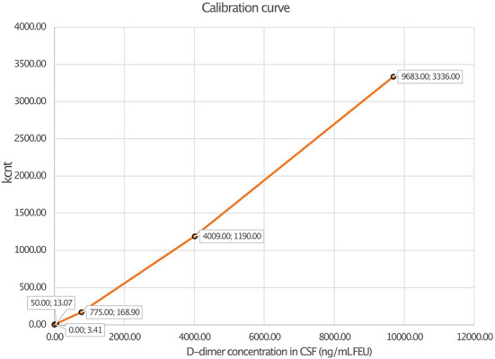
Methods: INNOVANCE LOCI hs D-dimer reagent cartridge was used for the measurement of D-dimer in CSF of patients with different neurological diseases. For the evaluation of linearity, dilution series were performed in a pooled CSF sample with the determination of intra-assay precision (CV, coefficient of variation) in 3 individual samples with 20 replicates. Furthermore, D-dimer concentrations measured by LOCI® were compared with the respective results of a routinely available clinical latex-enhanced immunoassay (HemosiIL D-Dimer HS 500).
Results: Linear regression analysis of the LOCI® method revealed a r 2 of 1.00 (p < 0.001) with a regression coefficient B of 1.012 ± 0.003 (CI: 1.005-1.019, p < 0.001) and an intercept of -1.475 ± 1.309 (CI: -4.493 to 1.543); the median intra-assay CV was 0.69% (range: 0.68-0.75). In total, 185 CSF samples were measured by LOCI® technology, showing a mean concentration of 204.84 ± 2,214.93 ng/ml. D-dimer concentration between LOCI and latex-enhanced immunoassay differed by a factor of 10.6 ± 13.6 on average with a maximum deviation by a factor of 61.3; the maximum deviation was found at low concentrations.
Conclusion: D-dimer in CSF of patients with neurological disease can be reliably measured by the LOCI® method with high linearity and accuracy at low concentrations.
Keywords: biomarker; coagulation; diagnostics; immunoassay; laboratory.
Copyright © 2022 Kohlhase, Schaefer, Miesbach, Hintereder, Kirchmayr, Zwinge, Yalachkov, Foerch and Schaller-Paule.
Conflict of interest statement
The authors declare that the research was conducted in the absence of any commercial or financial relationships that could be construed as a potential conflict of interest.
Figures


References
-
- Schaefer JH, Yalachkov Y, Friedauer L, Kirchmayr K, Miesbach W, Wenger KJ, et al. Measurement of prothrombin fragment 1+2 in cerebrospinal fluid to identify thrombin generation in inflammatory central nervous system diseases. Mult Scler Relat Disord. (2022) 60:103720. 10.1016/j.msard.2022.103720 - DOI - PubMed
LinkOut - more resources
Full Text Sources