Review
. 2022 Oct 19:13:997786.
doi: 10.3389/fimmu.2022.997786. eCollection 2022.

Microglia morphophysiological diversity and its implications for the CNS

Affiliations
Review

Microglia morphophysiological diversity and its implications for the CNS

Andrés Vidal-Itriago et al. Front Immunol. .

Abstract

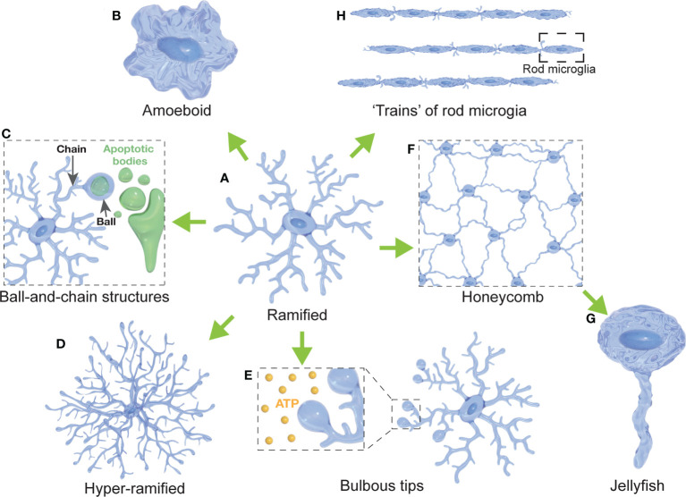
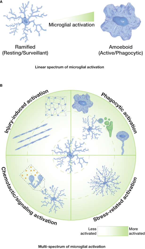
Microglia are mononuclear phagocytes of mesodermal origin that migrate to the central nervous system (CNS) during the early stages of embryonic development. After colonizing the CNS, they proliferate and remain able to self-renew throughout life, maintaining the number of microglia around 5-12% of the cells in the CNS parenchyma. They are considered to play key roles in development, homeostasis and innate immunity of the CNS. Microglia are exceptionally diverse in their morphological characteristics, actively modifying the shape of their processes and soma in response to different stimuli. This broad morphological spectrum of microglia responses is considered to be closely correlated to their diverse range of functions in health and disease. However, the morphophysiological attributes of microglia, and the structural and functional features of microglia-neuron interactions, remain largely unknown. Here, we assess the current knowledge of the diverse microglial morphologies, with a focus on the correlation between microglial shape and function. We also outline some of the current challenges, opportunities, and future directions that will help us to tackle unanswered questions about microglia, and to continue unravelling the mysteries of microglia, in all its shapes.

Keywords: microglia; microglia activation; microglia diversity; microglia-neuron interactions; microglial morphology.


Conflict of interest statement

The authors declare that the research was conducted in the absence of any commercial or financial relationships that could be construed as a potential conflict of interest.

Figures

Figure 1
Figure 1
Diversity of microglial morphologies. (A) Ramified microglia are highly branched with multiple primary and secondary processes (often considered surveillant). (B) Amoeboid microglia present with a highly rounded morphology compared to their ramified states (often with a high phagocytic and migratory capacity). (C) Microglia can form ball-and-chain structures at the tip of their processes to phagocytose small amounts of material (such as synapses or apoptotic bodies). (D) Hyper-ramified microglia present with increased branching of their processes (often observed in acute and chronic stress models). (E) Microglia display bulbous budding at the end of some of their processes (considered to be important for ATP sensing). (F) Several microglial cells form a network resembling a honeycomb (reported in response to BBB leakage). (G) Jellyfish morphologies have been reported as a morphological transition of honeycomb microglia after extensive astrocytic death in the glia limitans (in response to TBI). (H) Rod microglia are characterized by an elongated, narrowed soma without planar processes that can form trains of rod microglial cells (in response to injury).
Figure 2
Figure 2
Spectrums of microglial activation. (A) Classical or ‘linear spectrum’ of microglial activation where ramified microglia are considered non-activated/resting, and amoeboid microglia are considered fully activated. Other microglial morphologies would reflect an intermediate state. (B) Proposed ‘multi-spectrum’ that reflects changes in microglial morphologies and function in a radial pattern, with context-dependent microglial activation.

Similar articles

Cited by

References

    1. Sierra A, de Castro F, del Río-Hortega J, Rafael Iglesias-Rozas J, Garrosa M, Kettenmann H. The “Big-bang” for modern glial biology: Translation and comments on pío del río-hortega 1919 series of papers on microglia. Glia (2016) 64:1801–40. doi: 10.1002/glia.23046 - DOI - PubMed
    1. Li Q, Barres BA. Microglia and macrophages in brain homeostasis and disease. Nat Rev Immunol (2018) 18:225–42. doi: 10.1038/nri.2017.125 - DOI - PubMed
    1. Nimmerjahn A, Kirchhoff F, Helmchen F. Resting microglial cells are highly dynamic surveillants of brain parenchyma in vivo . Science (2005) 308:1314–8. doi: 10.1126/science.1110647 - DOI - PubMed
    1. Tremblay ME, Stevens B, Sierra A, Wake H, Bessis A, Nimmerjahn A. The role of microglia in the healthy brain. J Neurosci (2011) 31:16064–9. doi: 10.1523/JNEUROSCI.4158-11.2011 - DOI - PMC - PubMed
    1. Hammond TR, Dufort C, Dissing-Olesen L, Giera S, Young A, Wysoker A, et al. . Single-cell RNA sequencing of microglia throughout the mouse lifespan and in the injured brain reveals complex cell-state changes. Immunity (2019) 50:253–71. doi: 10.1016/j.immuni.2018.11.004 - DOI - PMC - PubMed

Publication types

LinkOut - more resources