The Incidence, Characteristics and Outcomes of Vertebral Artery Injury Associated with Cervical Spine Trauma: A Systematic Review
- PMID: 36341773
- PMCID: PMC10189332
- DOI: 10.1177/21925682221137823
The Incidence, Characteristics and Outcomes of Vertebral Artery Injury Associated with Cervical Spine Trauma: A Systematic Review
Abstract
Study design: Systematic Review.
Objectives: Vertebral Artery Injury (VAI) is a potentially serious complication of cervical spine fractures. As many patients can be asymptomatic at the time of injury, the identification and diagnosis of VAI can often prove difficult. Due to the high rates of morbidity and mortality associated with VAI, high clinical suspicion is paramount. The purpose of this review is to elucidate incidence, diagnosis, treatment and outcomes of VAI associated with cervical spine injuries.
Methods: A systematic search of electronic databases was performed using 'PUBMED', 'EMBASE','Medline (OVID)', and 'Web of Science, for articles pertaining to traumatic cervical fractures with associated VAI.
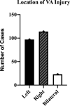
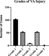
Results: 24 studies were included in this systematic review. Data was included from 48 744 patients. In regards to the demographics of the focus groups that highlighted information on VAI, the mean average age was 46.6 (32.1-62.6). 75.1% (169/225) were male and 24.9% (56/225) were female. Overall incidence of VAI was 596/11 479 (5.19%). 190/420 (45.2%) of patients with VAI had fractures involving the transverse foramina. The right vertebral artery was the most commonly injured 114/234 (48.7%). V3 was the most common section injured (16/36 (44.4%)). Grade I was the most common (103/218 (47.2%)) injury noted. Collective acute hospital mortality rate was 32/226 (14.2%), ranging from 0-26.2% across studies.
Conclusion: VAI secondary to cervical spine trauma has a notable incidence and high associated mortality rates. The current available literature is limited by a low quality of evidence. In order to optimise diagnostic protocols and treatment strategies, in addition to reducing mortality rates associated with VAI, robust quantitative and qualitative studies are needed.
Keywords: blunt trauma; cervical spinal trauma; spinal orthopaedics; vertebral artery injury.
Conflict of interest statement
The author(s) declared no potential conflicts of interest with respect to the research, authorship, and/or publication of this article.
Figures


References
-
- Standring S. Gray’s Anatomy E-Book: The Anatomical Basis of Clinical Practice. Elsevier Health Sciences; 2015.
-
- Cloud GC, Markus HS. Diagnosis and management of vertebral artery stenosis. QJM. 2003;96(1):27-54. - PubMed
-
- Chung D, Sung JK, Cho DC, Kang DH. Vertebral artery injury in destabilized midcervical spine trauma; predisposing factors and proposed mechanism. Acta Neurochir. 2012;154(11):2091-2098. - PubMed
LinkOut - more resources
Full Text Sources
Research Materials

