. 2022 Dec 15:457:116295.
doi: 10.1016/j.taap.2022.116295. Epub 2022 Oct 29.

Social isolation exacerbates acute ozone inhalation induced pulmonary and systemic health outcomes

Affiliations

Social isolation exacerbates acute ozone inhalation induced pulmonary and systemic health outcomes

Andres R Henriquez et al. Toxicol Appl Pharmacol. .

Abstract

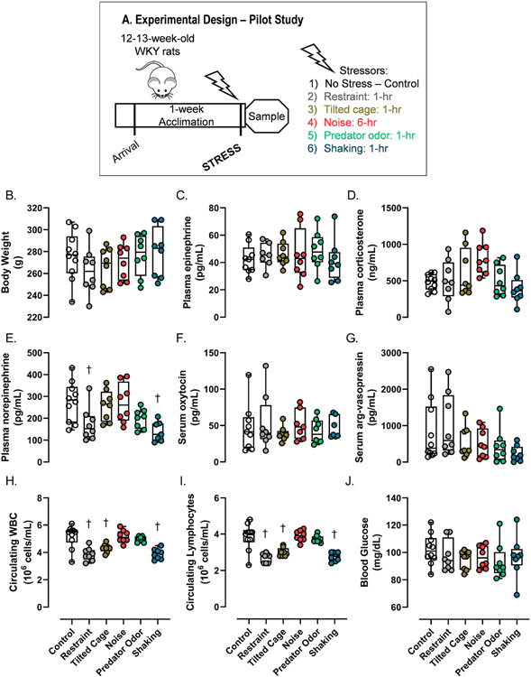
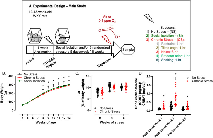
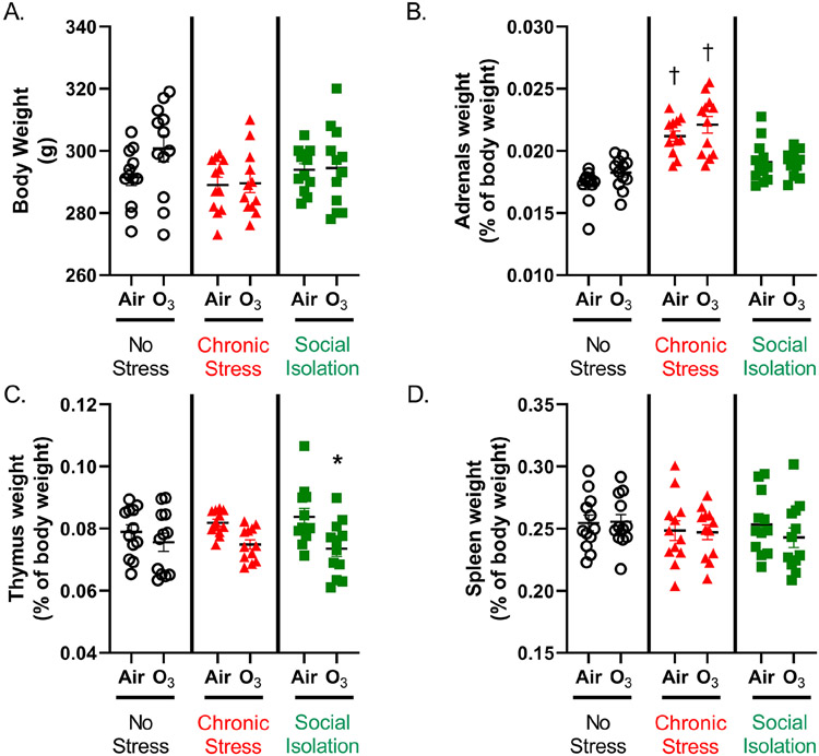
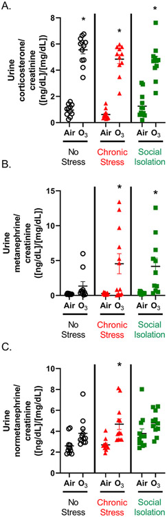
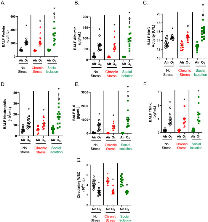
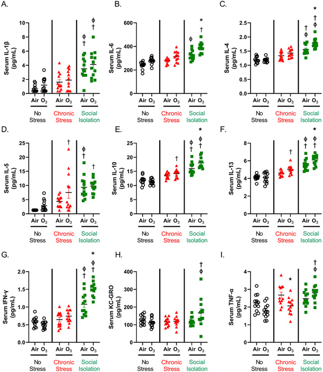
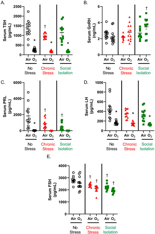
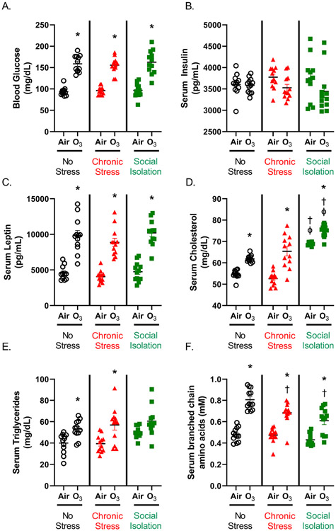
Psychosocially-stressed individuals might have exacerbated responses to air pollution exposure. Acute ozone exposure activates the neuroendocrine stress response leading to systemic metabolic and lung inflammatory changes. We hypothesized chronic mild stress (CS) and/or social isolation (SI) would cause neuroendocrine, inflammatory, and metabolic phenotypes that would be exacerbated by an acute ozone exposure. Male 5-week-old Wistar-Kyoto rats were randomly assigned into 3 groups: no stress (NS) (pair-housed, regular-handling); SI (single-housed, minimal-handling); CS (single-housed, subjected to mild unpredicted-randomized stressors [restraint-1 h, tilted cage-1 h, shaking-1 h, intermittent noise-6 h, and predator odor-1 h], 1-stressor/day*5-days/week*8-weeks. All animals then 13-week-old were subsequently exposed to filtered-air or ozone (0.8-ppm) for 4 h and immediately necropsied. CS, but not SI animals had increased adrenal weights. However, relative to NS, both CS and SI had lower circulating luteinizing hormone, prolactin, and follicle-stimulating hormone regardless of exposure (SI > CS), and only CS demonstrated lower thyroid-stimulating hormone levels. SI caused more severe systemic inflammation than CS, as evidenced by higher circulating cytokines and cholesterol. Ozone exposure increased urine corticosterone and catecholamine metabolites with no significant stressor effect. Ozone-induced lung injury, and increases in lavage-fluid neutrophils and IL-6, were exacerbated by SI. Ozone severely lowered circulating thyroid-stimulating hormone, prolactin, and luteinizing hormone in all groups and exacerbated systemic inflammation in SI. Ozone-induced increases in serum glucose, leptin, and triglycerides were consistent across stressors; however, increases in cholesterol were exacerbated by SI. Collectively, psychosocial stressors, especially SI, affected the neuroendocrine system and induced adverse metabolic and inflammatory effects that were exacerbated by ozone exposure.

Keywords: Catecholamines; Glucocorticoids; Mild chronic stress model; Neuroendocrine; Ozone; Pituitary hormones; Social isolation; Stress response; Systemic inflammation.


Conflict of interest statement

Declaration of Competing Interest The authors declare that they have no known competing financial interests or personal relationships that could have appeared to influence the work reported in this paper.

Figures

Figure 1.
Figure 1.
Changes in stress associated biomarkers immediately after a single application of each individual stressor for the pilot study in 12-13 week old male WKY rats. A) experimental protocol; B) body weight; C) plasma epinephrine; D) plasma corticosterone; E) plasma norepinephrine; F) serum oxytocin; G) serum arginine-vasopressin; H) circulating white blood cells; I) circulating lymphocytes; J) blood glucose. Each group of rats were subjected to one of the stressors, and immediately following stressor application, rats were euthanized for biomarkers assessment. The groups included were: 1) No stress- control, 2) restraint: rats are placed in nose-only inhalation tube for 1-hr; 3) tilted cage: each rat is placed in a 45° tilted in wire-mesh bottom cage for 1-hr (C); 4) noise: rats are exposed to white noise, of 85 dB through speakers above each cage for 5-25 min, with 5-45 min between noise bursts for 6-hrs; 5) predator odor: rats are housed in an isolated room where fox urine odorous ingredient, 2,5-dihydro-2,4,5-trimethylthiazoline is allowed to dissipate through an open petri dish, 1-hr; and 6) shaking: each rat is placed in cage with 4 dividers, placed on a modified orbital plate shaker set at 100 rpm for 1-hr. Each graph shows median, 25th-75th percentiles and min-max for n=10 animals/group for no-stress controls and n=8 for each stressor. A “†” indicates significant stressor effect relative to no stress control (p ≤ 0.05). One urine sample was not collected for CS group. One animal each from control, restraint and noise group for epinephrine and one from control group for oxytocin were removed by outlier test.
Figure 2.
Figure 2.
Experimental protocol for stressors application and temporal assessment of body weight, body composition as well as urinary corticosterone until prior to air or ozone exposure. A. Experimental design. Variable unpredicted stresses were applied over 8 weeks (from 5 to 13 weeks of age) in male WKY rats. Responsiveness to a single ozone inhalation exposure was evaluated upon the termination of stress protocols. Necropsy and tissue collection were carried out immediately after air or ozone exposure (within 2 hr). No stress (NS) animals were double housed with crinkled paper provided in cages for environmental enrichment and were handled regularly for body weights, body composition and urine sample collection, but no stressors were applied. For chronic stress (CS), animals were single-housed without environmental enrichment, and each stressor was applied in a random manner Monday-Friday, 5 days/week for 8 weeks. These rats were handled daily. Social isolation (SI) animals were single-housed without environmental enrichment with limited handling only for weighing and cage changes. Temporal changes in body weight (B), body fat % assessed through body composition analysis (C) and urine corticosterone levels (D) during 8-week of stress maneuver in NS, CS and SI groups. Body weights were measured each week for all animals (B). For NS and CS body composition was assessed at week 4 and prior to week 8 (C). Note that body composition was not assessed for SI. Urine corticosterone was measured in NS and CS after the 4th week of stress protocol and after the last stress application on the 7th week of the protocol (Friday of week 7), and finally after the weekend (Monday at the start of week 8) on Monday to determine the reversibility of stressor effect observed on Friday. Data were normalized to creatinine levels (D). Curves values indicate means ± SEM for n=24 animals/group. An “*” indicates significant ozone effect relative to matched air group. A (“†” indicates significant stressor effect relative to matched NS group (p ≤ 0.05).
Figure 3.
Figure 3.
Body and organ weights at necropsy in no-stress (NS), chronic stress (CS) and social isolation (SI) groups after exposure to air or ozone. A) body weights at necropsy; B) adrenal weights; C) thymus weights; D) spleen weights. Data are shown as mean ± SEM for n=12 animals per group. An “*” indicates significant ozone effect relative to matched air group. A “†” indicates significant stressor effect relative to matched exposure NS group (p ≤ 0.05).
Figure 4.
Figure 4.
Acute exposure to ozone increased urine levels of stress hormones in no-stress (NS), chronic stress (CS) and social isolation (SI) groups. A) urine corticosterone; B) urine metanephrine; C) urine normetanephrine. Urine samples were collected directly from urinary bladder at the time of necropsy. Data were normalized to urine creatinine levels. Data are shown as mean ± SEM for n=12 animals per group. An “*” indicates significant ozone effect relative to matched air group (p ≤ 0.05). Corticosterone data for one animal in SI air group; and metanephrine data for one animal in NS air group and one in NS ozone group were removed by outlier test.
Figure 5.
Figure 5.
BALF markers of lung injury, inflammation and circulating white blood cells after a single acute ozone exposure in no-stress (NS), chronic stress (CS) and social isolation (SI) groups of animals. Lung injury and inflammation were determined in the bronchoalveolar lavage fluid (BALF) collected at the time of necropsy. A) BALF protein; B) BALF albumin; C) BALF NAG (N-acetyl-β-D-glucosaminidase) activity; D) BALF neutrophils; E) BALF IL-6; F) BALF TNF-α; G) circulating white blood cells. Data are shown as mean ± SEM for n=12 animals per group. An “*” indicates significant ozone effect relative to matched air group. A “†” indicates significant stress effect relative to matched exposure NS group. A “φ” indicates significant SI effect relative to matched exposure CS group (p ≤ 0.05). BALF protein and albumin data for two animals in NS air group; one in CS air group; one in SI air group; one for BALF protein in NS ozone group, one for BALF NAG in NS ozone group; one BALF IL-6 in CS ozone group and one for TNF-α in SI ozone group were removed by outlier test.
Figure 6.
Figure 6.
Circulating cytokine changes indicating systemic inflammation following ozone exposure in no-stress (NS), mild chronic stress (CS) and social isolation (SI) groups. Cytokines were determined in the serum samples collected during necropsy. A) IL-1β; B) IL-6; C) IL-4; D) IL-5; E) IL-10; F) IL-13; G) IFN-γ; H) KC-GRO; I) TNF-α. Data are shown as mean ± SEM for n=12 animals per group. An “*” indicates significant ozone effect relative to matched air group. A “†” indicates significant stress effect relative to matched exposure NS group. A “φ” indicates significant SI effect relative to matched exposure CS group (p ≤ 0.05). Serum IL-1β data for one animal in NS air group; and IL-5 data for two animals in NS air group were removed by outlier test.
Figure 7.
Figure 7.
Changes in circulating pituitary hormones following ozone exposure in no-stress (NS), mild chronic stress (CS) and social isolation (SI) groups. Pituitary hormones were analyzed in serum samples collected during necropsy. A) Thyroid stimulating hormone (TSH); B), gonadotropin releasing hormone (GnRH); C) prolactin (PRL); D) luteinizing hormone (LH); E) follicle stimulating hormone (FSH). Data are shown as mean ± SEM for n=12 animals per group. An “*” indicates significant ozone effect relative to matched air group. A “†” indicates significant stress effect relative to matched exposure NS group (p ≤ 0.05). Serum PRL data for two animals in each, NS ozone, CS ozone and SI ozone groups; and FSH data for one animal in NS air group and one animal in CS air group were removed by outlier test.
Figure 8.
Figure 8.
Circulating metabolites and metabolic hormones following ozone exposure in no-stress (NS), mild chronic stress (CS) and social isolation (SI) groups. Serum levels of hormones and metabolites were determined in the samples collected during necropsy. A) blood glucose; B) serum insulin; C) serum leptin; D) serum cholesterol; E) serum triglycerides; F) serum branched chain amino acids (BCAA). Data are shown as mean ± SEM for n=12 animals per group. An “*” indicates significant ozone effect relative to matched air group. A “†” indicates significant stress effect relative to matched exposure NS group. A “φ” indicates significant SI effect relative to matched exposure CS group (p ≤ 0.05). Serum insulin and leptin data for one animal in NS air group; and cholesterol data for one animal in SI air group were removed by outlier test.

Similar articles

Cited by

References

    1. Ailshire JA, Crimmins EM. Fine particulate matter air pollution and cognitive function among older US adults. Am J Epidemiol. 2014. Aug 15;180(4):359–66. doi: 10.1093/aje/kwu155. Epub 2014 Jun 24. - DOI - PMC - PubMed
    1. Ailshire J, Karraker A, Clarke P. Neighborhood social stressors, fine particulate matter air pollution, and cognitive function among older U.S. adults. Soc Sci Med. 2017. Jan; 172:56–63. doi: 10.1016/j.socscimed.2016.11.019. Epub 2016 Nov 14. - DOI - PMC - PubMed
    1. Arcego DM, Krolow R, Lampert C, Noschang C, Ferreira AG, Scherer E, Wyse AT, Dalmaz C. Isolation during the prepubertal period associated with chronic access to palatable diets: effects on plasma lipid profile and liver oxidative stress. Physiol Behav. 2014. Jan 30;124:23–32. doi: 10.1016/j.physbeh.2013.10.029. Epub 2013 Oct 30. - DOI - PubMed
    1. Atrooz F, Alkadhi KA, Salim S. Understanding stress: Insights from rodent models. Curr. Res Neurobiol 2: 100013, 2021. - PMC - PubMed
    1. Attademo L, Bernardini F, Spatuzzi R. Suicidality in Individuals with Schizoid Personality Disorder or Traits: A Clinical Mini-Review of a Probably Underestimated Issue. Psychiatr Danub. 2021. Fall;33(3):261–265. doi: 10.24869/psyd.2021.261. - DOI - PubMed