. 2022 Nov 22;88(22):e0154622.
doi: 10.1128/aem.01546-22. Epub 2022 Nov 7.

Hungatella hathewayi, an Efficient Glycosaminoglycan-Degrading Firmicutes from Human Gut and Its Chondroitin ABC Exolyase with High Activity and Broad Substrate Specificity

Affiliations

Hungatella hathewayi, an Efficient Glycosaminoglycan-Degrading Firmicutes from Human Gut and Its Chondroitin ABC Exolyase with High Activity and Broad Substrate Specificity

Parkash Singh Rawat et al. Appl Environ Microbiol. .

Abstract

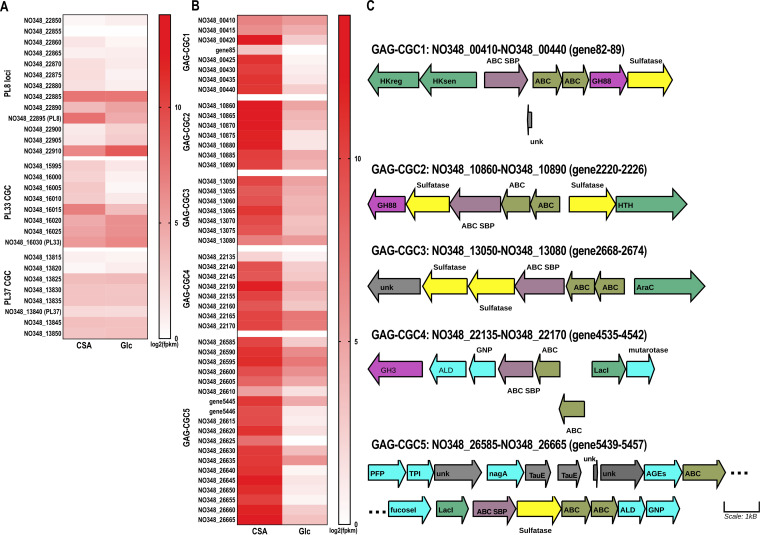
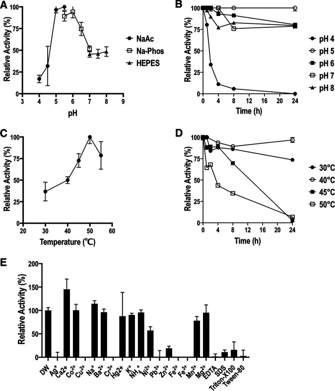
The degradation of glycosaminoglycans (GAGs) by intestinal bacteria is critical for their colonization in the human gut and the health of the host. Both colonic Bacteroides and Firmicutes have been reported to degrade GAGs; however, the enzymatic details of the latter remain largely unknown. Our bioinformatic analyses of fecal Firmicutes revealed that their genomes, especially Hungatella hathewayi strains, are an abundant source of putative GAG-specific catabolic enzymes. Subsequently, we isolated a Firmicutes strain, H. hathewayi N2-326, that can catabolize various GAGs. While H. hathewayi N2-326 was as efficient in utilizing chondroitin sulfate A (CSA) and dermatan sulfate as Bacteroides thetaiotaomicron, a well-characterized GAG degrader, it outperformed B. thetaiotaomicron in assimilating hyaluronic acid. Unlike B. thetaiotaomicron, H. hathewayi N2-326 could not utilize heparin. The chondroitin lyase activity of H. hathewayi N2-326 was found to be present predominantly in the culture supernatant. Genome sequence analysis revealed three putative GAG lyases, but only the HH-chondroitin ABC lyase was upregulated in the presence of CSA. In addition, five CAZyme gene clusters containing GAG metabolism genes were significantly upregulated when grown on CSA. Further characterization of the recombinant HH-chondroitin ABC lyase revealed that it cleaves GAGs predominantly in an exo-mode to produce unsaturated disaccharides as the primary hydrolytic product while exhibiting a higher specific activity than reported chondroitin ABC lyases. HH-chondroitin ABC lyase represents the first characterized chondroitin lyase from intestinal Firmicutes and offers a viable commercial option for the production of chondroitin, dermatan, and hyaluronan oligosaccharides and also for potential medical applications. IMPORTANCE An increased understanding of GAG metabolism by intestinal bacteria is critical in identifying the driving factors for the composition, modulation, and homeostasis of the human gut microbiota. In addition, GAG-depolymerizing polysaccharide lyases are highly desired enzymes for the production of GAG oligosaccharides and as therapeutics. At present, the dissection of the enzymatic machinery for GAG degradation is highly skewed toward Bacteroides. In this study, we have isolated an efficient GAG-degrading Firmicutes bacterium from human feces and characterized the first chondroitin ABC lyase from a Firmicutes, which complements the fundamental knowledge of GAG utilization in the human colon. The genomic and transcriptomic analysis of the bacterium shows that Firmicutes might use a distinct approach to catabolize GAGs from that used by Bacteroides. The high specific activity of the characterized chondroitin ABC lyase aids future attempts to develop a commercial chondroitinase for industrial and medicinal applications.

Keywords: Bacteroides; Firmicutes; Hungatella hathewayi; chondroitin ABC lyase; glycosaminoglycans.


Conflict of interest statement

The authors declare no conflict of interest.

Figures

FIG 1
FIG 1
Number of GAG-specific CAZymes in the genomic assemblies of 114 intestinal Firmicutes strains obtained from the BioProject PRJNA482748. The number of GAG-specific CAZymes from the prominent GAG-degrading bacterium B. thetaiotaomicron VPI-5482 (BT) is also plotted for comparison. GH, glycoside hydrolase; CBM, carbohydrate-binding module containing protein; PL, polysaccharide lyase. The position of B. thetaiotaomicron VPI-5482 (BT) in the plot is highlighted by an arrow, while all H. hathewayi strains are boxed in a rectangle.
FIG 2
FIG 2
Comparative growth of B. thetaiotaomicron VPI-5482 (A), E. coli (B), and H. hathewayi N2-326 (C) on various GAGs. The bacteria were grown in YCFA supplemented with 1.5% GAG substrates, including CSA, DS, HA, and Hep, or in unsupplemented YCFA (No-GAG medium Control, NG) for 120 h. Error bars indicate standard errors (SE) of three replicates for E. coli and B. thetaiotaomicron and six replicates for H. hathewayi N2-326.
FIG 3
FIG 3
Quantitative measurement of GAG consumption by H. hathewayi N2-326 (HH), B. thetaiotaomicron (BT), and E. coli. DMMB assay was used to determine CSA (A), DS (B), and heparin (D) consumption, while CTAB turbidimetric assay was used to quantitate HA (C) utilization. Error bars indicate standard errors (SE) of three replicates.
FIG 4
FIG 4
Putative polysaccharide lyase containing genomic loci in the genome of H. hathewayi N2-326. The neighboring genes of PL8 (HH-chondroitin ABC lyase) gene (A) are not related to carbon metabolism, whereas both PL33 (B) and PL37 (C) genes are surrounded by other CAZymes, ABC transport genes, and transcription regulators. sigF, RNA polymerase sporulation sigma factor SigF; unk, protein of unknown function; SpoVxx, sporulation-related protein; Penicillin BP, penicillin-binding protein; YigZ, YigZ translation regulator; DedA, DedA family membrane protein; GT1, glycosyl transferase family 1 protein; GH, glycoside hydrolase family protein; ABC SBP, ABC family substrate-binding protein; Hksen, histidine kinase sensor protein; HKreg, histidine kinase response regulator; AraC, AraC/YesN two-component DNA-binding response regulator.
FIG 5
FIG 5
Transcriptomics analysis of the H. hathewayi N2-326 strain grown in YCFA-CSA (CSA group) or YCFA-Glc (Glc group). (A) Heat map showing the differential expression [log2(FPKM) of CSA group versus Glc group] of the genomic regions containing three putative GAG-degrading polysaccharide lyases of the bacterium. PL8-loci, genes in the neighborhood of the PL8 gene (hh-cs-abc); PL33 CGC, putative CGC of the PL33-heparinase gene; PL37-CGC, putative CGC of the PL37 gene. (B) Heat map showing the differential expression of selected gene clusters. GAG-CGCs 1–5, the five significantly upregulated GAG-CGCs in the CSA group versus Glc group. (C) Illustrative representation of the significantly upregulated, putative GAG-CGCs 1–5. Hkreg, histidine kinase response regulator; HKsen, histidine kinase sensor protein; ABC SBP, ABC transport family substrate-binding protein; ABC, ABC permease; unk, unknown; GH, glycosyl hydrolase family protein; HTH, helix-turn-helix DNA binding protein; AraC, AraC/YesN two-component DNA-binding response regulator; ALD, aldolase; GNP, glucosamine-6-phosphate isomerase/deaminase; LacI, LacI family transcription regulator; PFP, pyrophosphate-fructose 6-phosphate 1-phosphotransferase; TPI, triose-phosphate isomerase; nagA, N-acetylglucosamine-6-phosphate deacetylase; TauE, TauE/SafE family sulfite exporter; AGEs, N-acylglucosamine 2-epimerase; fucoseI, fucose isomerase.
FIG 6
FIG 6
Domain organization, purification, and phylogenetic analysis of HH-chondroitin ABC lyase. (A) Domain architecture of HH-chondroitin ABC lyase. The protein sequence was analyzed by NCBI’s CD search tool and illustrated to scale using Domain graph software. The gray region of the protein does not have any conserved domains. The red region at the N terminus corresponds to the secretory signal peptide. PspC, PspC_subgroup_1 superfamily, bacterial surface protein domain. (B) SDS-PAGE analysis of the expression and purification of HH-chondroitin ABC lyase in E. coli BL21(DE3) Star. Lane M, protein molecular mass standards; lane 1, cell extract; lane 2, size exclusion chromatography purified HH-chondroitin ABC lyase. (C) Phylogenetic tree of HH-chondroitin ABC lyase with all characterized polysaccharide lyase 8 and 29 family proteins in the CAZy database. The evolutionary history was inferred by using the Maximum Likelihood method and JTT matrix-based model.
FIG 7
FIG 7
Biochemical characterization of HH-chondroitin ABC lyase. (A) Optimum pH of HH-chondroitin ABC lyase was determined using 50 mM sodium acetate (NaAc/HAc, pH 4.0 to 5.0), Na2HPO4/NaH2PO4 (Na-phos, pH 5.5 to 7.0), and HEPES (pH 7.0 to 8.0) buffer systems at 37°C. (B) The pH stability of HH-chondroitin ABC lyase was determined by preincubating the enzyme in 50 mM buffers in the pH range of 4.0 to 8.0 for various times (0 to 24 h), and then the residual activities were measured. (C) Optimum temperature determinations were performed in 50 mM NaAc/HAc buffer, pH 5.5, at temperatures ranging from 30 to 55°C. (D) The thermal stability of HH-chondroitin ABC lyase was determined by preincubating the enzyme in the temperature range of 30°C to 50°C for various times (0 to 24 h), and then the residual activities were determined under optimum conditions. (E) The effect of metal ions (5 mM), EDTA (5 mM), and 1% detergents (SDS, Triton X-100, and Tween 80) was examined under optimum conditions. All reactions (B to E) were conducted under optimum conditions, i.e., 50 mM NaAc/HAc with 5 mM CaCl2, pH 5.5, 50°C for 10 min. Error bars indicate standard errors (SE) of three replicates.
FIG 8
FIG 8
HPLC analysis of the degradation products by incubating HH-chondroitin ABC lyase with 5.0 mg/mL of CSA (A) and HA (B). The reactions were performed with 17 ng HH-chondroitin ABC lyase in 50 mM sodium acetate buffer, pH 5.5, with 5 mM CaCl2 at 40°C. Samples were taken at 0, 10 min, 30 min, 2 h, and 24 h. The degradation products were analyzed with HPLC and detected at 232 nm. ΔDi, Δtetra, Δhexa, and Δocta represent unsaturated disaccharide, unsaturated tetrasaccharide, unsaturated hexasaccharide, and unsaturated octasaccharide GAG degradation products, respectively.
FIG 9
FIG 9
A presumptive model of glycosaminoglycans (CS, DS, and HA) utilization in Hungatella hathewayi N2-326. Dietary and endogenous GAGs are captured by HH-chondroitin ABC lyase in the gut lumen. The captured polysaccharides are broken down to oligo- and disaccharides, which are bound by the extracellular substrate-binding proteins of the ABC transport family. The bound substrate is internalized to the cell interior through the ABC family permeases, where substrate-specific sulfatases and GH88 proteins degrade them further to monosaccharides, which are funneled to the downstream metabolic pathways to provide energy and building blocks for the cell. Divided diamonds represent unsaturated hexuronic acid, while squares represent HexNAc sugar residues.

Similar articles

Cited by

References

    1. Iozzo RV. 1987. Turnover of heparan sulfate proteoglycan in human colon carcinoma cells. A quantitative biochemical and autoradiographic study. J Biol Chem 262:1888–1900. 10.1016/S0021-9258(19)75723-8. - DOI - PubMed
    1. Hooper LV, Midtvedt T, Gordon JI. 2002. How host-microbial interactions shape the nutrient environment of the mammalian intestine. Annu Rev Nutr 22:283–307. 10.1146/annurev.nutr.22.011602.092259. - DOI - PubMed
    1. Simon-Assmann P, Bouziges F, Vigny M, Kedinger M. 1989. Origin and deposition of basement membrane heparan sulfate proteoglycan in the developing intestine. J Cell Biol 109:1837–1848. 10.1083/jcb.109.4.1837. - DOI - PMC - PubMed
    1. Oshiro M, Ono K, Suzuki Y, Ota H, Katsuyama T, Mori N. 2001. Immunohistochemical localization of heparan sulfate proteoglycan in human gastrointestinal tract. Histochem Cell Biol 115:373–380. 10.1007/s004180100271. - DOI - PubMed
    1. Bode L, Salvestrini C, Park PW, Li J-P, Esko JD, Yamaguchi Y, Murch S, Freeze HH. 2008. Heparan sulfate and syndecan-1 are essential in maintaining murine and human intestinal epithelial barrier function. J Clin Invest 118:229–238. 10.1172/JCI32335. - DOI - PMC - PubMed

Publication types

MeSH terms

Supplementary concepts