. 2022 Dec 21;10(6):e0273222.
doi: 10.1128/spectrum.02732-22. Epub 2022 Nov 7.

Use of a Bacterial Artificial Chromosome to Generate Recombinant SARS-CoV-2 Expressing Robust Levels of Reporter Genes

Affiliations

Use of a Bacterial Artificial Chromosome to Generate Recombinant SARS-CoV-2 Expressing Robust Levels of Reporter Genes

Chengjin Ye et al. Microbiol Spectr. .

Abstract

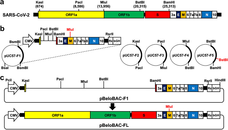
Reporter-expressing recombinant virus represents an excellent option and a powerful tool to investigate, among others, viral infection, pathogenicity, and transmission, as well as to identify therapeutic compounds that inhibit viral infection and prophylactic vaccines. To combat the ongoing coronavirus disease 2019 (COVID-19) pandemic, we have established a robust bacterial artificial chromosome (BAC)-based reverse genetics (RG) system to rapidly generate recombinant severe acute respiratory syndrome coronavirus 2 (rSARS-CoV-2) to study the contribution of viral proteins in viral pathogenesis. In addition, we have engineered reporter-expressing recombinant viruses in which we placed the reporter genes upstream of the viral nucleocapsid (N) gene to promote high levels of reporter gene expression, which facilitates the study of SARS-CoV-2 in vitro and in vivo. To date, we have shared our BAC-based RG system with more than 100 laboratories around the world, which has helped to expedite investigations with SARS-CoV-2. However, genetic manipulation of the BAC containing the entire SARS-CoV-2 genome (~30,000 nt) is challenging. Herein, we provide the technical details to engineer rSARS-CoV-2 using the BAC-based RG approach. We describe (i) assembly of the full-length (FL) SARS-CoV-2 genome sequences into the empty pBeloBAC, (ii) verification of pBeloBAC-FL, (iii) cloning of a Venus reporter gene into pBeloBAC-FL, and (iv) recovery of the Venus-expressing rSARS-CoV-2. By following this protocol, researchers with knowledge of basic molecular biology and gene engineering techniques will be able to generate wild-type (WT) and reporter-expressing rSARS-CoV-2. IMPORTANCE We have established a bacterial artificial chromosome (BAC)-based RG system to generate recombinant severe acute respiratory syndrome coronavirus 2 (rSARS-CoV-2) and to engineer reporter-expressing recombinant viruses to assess viral infection in vitro and in vivo. To date, we have shared our BAC-based RG system with more than 100 laboratories around the world, which has helped to expedite investigations with SARS-CoV-2. However, genetic manipulation of the BAC containing the full-length SARS-CoV-2 genome of ~30,000 nucleotides is challenging. Here, we provide all the detailed experimental steps required for the successful generation of wild-type (WT) recombinant SARS-CoV-2 (rSARS-CoV-2). Likewise, we provide a comprehensive protocol on how to generate and rescue rSARS-CoV-2 expressing high levels of a Venus fluorescent reporter gene from the locus of the viral nucleocapsid (N) protein. By following these protocols, researchers with basic knowledge in molecular biology will be able to generate WT and Venus-expressing rSARS-CoV-2 within 40 days.

Keywords: BAC; COVID-19; SARS-CoV-2; bacterial artificial chromosome; coronavirus; pBeloBAC; recombinant virus; reporter genes; reverse genetics.


Conflict of interest statement

The authors declare a conflict of interest. C.Y. and L.M.-S. are co-inventors on a patent application directed to reverse genetics approaches to generate recombinant SARS-CoV-2.

Figures

FIG 1
FIG 1
Overview of a BAC-based RG system for generation of rSARS-CoV-2. (a) Schematic representation of the SARS-CoV-2 genome. Unique restriction sites used for viral genome assembly are indicated. (b) Commercially synthesized fragments (F1 to F5) in pUC57 plasmids. Restriction sites used to release each of the viral fragments are indicated. The MluI and BstBI sites in red are the restriction sites that have been removed by silent mutation and used as genetics tags. (c) Assembly of the entire SARS-CoV-2 genome into the empty pBeloBAC to generate the full-length rescue plasmid (pBeloBAC-FL).
FIG 2
FIG 2
Assembly of the entire SARS-CoV-2 genome into pBeloBAC. (a) Fragments (F1 to F5) released from pUC57 plasmids. (b) PCR screening for colonies positive for pBeloBAC-F1. (c) Restriction digestion analysis of pBeloBAC-F13 using PacI and MluI. (d) Restriction digestion analysis of pBeloBAC-F134 using PacI and MluI (F3) and MluI and BstBI (F4). (e) Restriction digestion analysis of pBeloBAC-F1342 using KasI and PacI (F2), PacI and MluI (F3), and MluI and BstBI (F4). (f) Restrict digestion confirmation of pBeloBAC-FL using KasI and PacI (F2), PacI and MluI (F3), MluI and BstBI (F4), and BstBI and BamHI (F5).
FIG 3
FIG 3
Recovery of rSARS-CoV-2 from pBeloBAC-FL. (a) Protocol used for recovery of rSARS-CoV-2 from pBeloBAC-FL using Lipofectamine 2000 transfection in Vero E6 cells. (b) CPE caused by rSARS-CoV-2/WT in Vero E6 cells. Bars, 100 μm. (c) Confirmation of the rescued rSARS-CoV-2/WT by IFA using a mouse antibody against viral N protein and a TRITC-labeled donkey anti-mouse IgG secondary antibody. The nucleus was stained with DAPI. (d) Sanger sequencing of the M gene of the natural isolate SARS-CoV-2 (top) and the recombinant SARS-CoV-2 (bottom) showed that the MluI site was removed by silent mutation.
FIG 4
FIG 4
Overview of the experimental approach to generate rSARS-CoV-2 expressing Venus from the N protein locus. (a) Schematic representation of the experimental flow to assemble Venus into pUC57-FA and subclone it into pBeloBAC-FL. (b) Schematic depiction of pBeloBAC-FL/Venus-2A.
FIG 5
FIG 5
Assembly of F1 containing Venus-2A into pBeloBAC for generation of rSARS-CoV-2/Venus-2A. (a) PCR amplification of Venus-2A and pUC57F1. (b) Purification of the F1/Venus-2A released from pUC57-F1/Venus-2A and the pBeloBAC-FL backbone after BamHI and RsrII digestion to remove the parental F1 segment for subcloning of F1/Venus-2A. (c) PCR screening of pBeloBAC-FL/Venus-2A colonies.
FIG 6
FIG 6
Characterization of rSARS-CoV-2/Venus-2A. (a) Fluorescent (Venus) and IFA (N) confirmation of rSARS-CoV-2/Venus-2A rescue. rSARS-CoV-2/WT was included as an internal control. (b) Detection of fluorescent plaques formed by rSARS-CoV-2/Venus-2A with a ChemiDoc imaging system. Immunostaining with the N protein 1C7C7 MAb of the plaques formed by rSARS-CoV-2/Venus-2A and rSARS-CoV-2/WT.

Similar articles

Cited by

References

    1. de Wit E, van Doremalen N, Falzarano D, Munster VJ. 2016. SARS and MERS: recent insights into emerging coronaviruses. Nat Rev Microbiol 14:523–534. doi:10.1038/nrmicro.2016.81. - DOI - PMC - PubMed
    1. van der Hoek L, Pyrc K, Jebbink MF, Vermeulen-Oost W, Berkhout RJM, Wolthers KC, Wertheim-van Dillen PME, Kaandorp J, Spaargaren J, Berkhout B. 2004. Identification of a new human coronavirus. Nat Med 10:368–373. doi:10.1038/nm1024. - DOI - PMC - PubMed
    1. Liu DX, Liang JQ, Fung TS. 2021. Human coronavirus-229E, -OC43, -NL63, and -HKU1 (Coronaviridae), p 428–440. In Bamford DH, Zuckerman M (ed), Encyclopedia of virology. Elsevier, Amsterdam, The Netherlands.
    1. Sohrabi C, Alsafi Z, O'Neill N, Khan M, Kerwan A, Al-Jabir A, Iosifidis C, Agha R. 2020. World Health Organization declares global emergency: a review of the 2019 novel coronavirus (COVID-19). Int J Surg 76:71–76. doi:10.1016/j.ijsu.2020.02.034. - DOI - PMC - PubMed
    1. Arshad Ali S, Baloch M, Ahmed N, Arshad Ali A, Iqbal A. 2020. The outbreak of coronavirus disease 2019 (COVID-19)—an emerging global health threat. J Infect Public Health 13:644–646. doi:10.1016/j.jiph.2020.02.033. - DOI - PMC - PubMed

Publication types