. 2022 Nov 1;11(11):6.
doi: 10.1167/tvst.11.11.6.

Iris Color Matters-A Contractility Analysis With Dynamic Volume-Rendered Optical Coherence Tomography Pupillometry

Affiliations

Iris Color Matters-A Contractility Analysis With Dynamic Volume-Rendered Optical Coherence Tomography Pupillometry

Philippe Valmaggia et al. Transl Vis Sci Technol. .

Abstract

Purpose: To analyze natural variability in pupillary contractility with dynamic volume-rendered optical coherence tomography (OCT) pupillometry regarding iris color, age, and sex in healthy Caucasian participants.

Methods: The intrapupillary spaces (IPSs) derived from anterior segment swept-source OCT of 71 healthy eyes were retrospectively analyzed. Baseline scotopic and photopic volumes and the functional parameters of pupillary ejection fraction (PEF), three-dimensional (3D) contractility, and relative light response (RLR) were measured on the swept-source OCT volumes. The effect on these parameters of iris color (brown, green, and blue), age, and sex was assessed.

Results: More pigmented irises were more contractile than less pigmented irises. Iris color significantly affected scotopic baseline IPSs (brown, 10.39 ± 4.86 mm3; green, 9.68 ± 3.31 mm3; blue, 6.75 ± 4.27 mm3; P = 0.018), PEF (brown, 90.8% ± 2.7%; green, 89.1% ± 2.5%; blue, 85.0% ± 9.3%; P = 0.010), 3D contractility (brown, 9.52 ± 4.59 mm3; green, 8.66 ± 3.07 mm3; blue, 6.44 ± 4.87 mm3; P = 0.016), and RLR (brown, 11.90 ± 4.03; green, 9.75 ± 2.73; blue, 8.52 ± 3.88; P = 0.026). Absolute scotopic volume (P = 0.022) and 3D contractility (P = 0.024) decreased with age. Sex showed no correlations.

Conclusions: The natural variability of pupillary contractility can be analyzed with dynamic OCT pupillometry. Iris color and age can impact pupillary response with this method.

Translational relevance: Iris contractility parameters can be measured using a commercially available OCT system, allowing for quantification of the aqueous humor volume inside the pupil.


Conflict of interest statement

Disclosure: P. Valmaggia, Swiss National Science Foundation (Grant 323530_199395) (F), AlumniMedizin Basel (F); N. Inglin, None; P. Kaiser, Supercomputing Systems AG, Zurich, Switzerland (E); H.P.N. Scholl, Swiss National Science Foundation (Project funding: “Developing novel outcomes for clinical trials in Stargardt disease using structure/function relationship and deep learning” #310030_201165, and National Center of Competence in Research Molecular Systems Engineering: “NCCR MSE: Molecular Systems Engineering (phase II)” #51NF40-182895) (F), the Wellcome Trust (PINNACLE study) (F), and the Foundation Fighting Blindness Clinical Research Institute (ProgStar study) (F), Astellas Pharma Global Development, Inc./Astellas Institute for Regenerative Medicine (S), Boehringer Ingelheim Pharma GmbH & Co (S), Gyroscope Therapeutics Ltd. (S), Janssen Research & Development, LLC (Johnson & Johnson) (S), Novartis Pharma AG (CORE) (S), Okuvision GmbH (S), and Third Rock Ventures, LLC (S), Gerson Lehrman Group (C), Guidepoint Global, LLC (C), and Tenpoint Therapeutics Limited (C), Data Monitoring and Safety Board/Committee of Belite Bio (CT2019-CTN-04690-1), ReNeuron Group Plc/Ora Inc. (NCT02464436), F. Hoffmann-La Roche Ltd (VELODROME trial, NCT04657289; DIAGRID trial, NCT05126966) and member of the Steering Committee of Novo Nordisk (FOCUS trial; NCT03811561) (N); P.M. Maloca, Roche (C), MIMO AG (O), VisionAI (O)

Figures

Figure 1.
Figure 1.
Study flowchart including the umbrella study population. The present study investigated the subgroup of healthy participants, highlighted in blue. PEXG, pseudoexfoliation glaucoma; NAION, non-arteritic anterior ischemic optic neuropathy; n, number of participants; e, number of eyes.
Figure 2.
Figure 2.
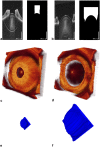
Image processing for dynamic volume-rendered OCT pupillometry. (a, b) B-scans in miosis (a) and mydriasis (b) with the corresponding IPS segmentations. (c, d) Volume-rendered OCT acquisitions in photopic (c) and scotopic (d) conditions, anterior visualization. (e, f) Corresponding volume-rendered IPS segmentations, posterior visualization. The volume rendering visualizes the structural differences between the photopic (e) and scotopic (f) states and corresponds to the differences in aqueous humor inside the IPS.
Figure 3.
Figure 3.
Summary statistics of pupillary contractility parameters with regard to iris color, sex, and age. The three analyzed eye colors and sex are presented as boxplots with medians and interquartile ranges, and the influence of age is shown as scatterplots. (a) Scotopic volume corresponds to the volume measured in a darkened room. (b) Photopic volume corresponds to the volume under lighting conditions. (c–e) Functional contractility parameters are described for PEF (c), 3D contractility (d), and RLR (e).
Figure 4.
Figure 4.
Correlation matrix chart for the variables Vol pho, Vol sco, PEF, Con, RLR, and age. Plots on the matrix diagonal show histograms with density estimators in red. Numbers in the upper half of the matrix are Pearson correlation coefficients, with asterisks indicating results of tests of no correlation based on Pearson's product moment correlation coefficient. Plots in the lower half of the matrix are bivariate scatterplots with fitted lines in red. Vol pho, photopic volume; Vol sco, scotopic volume; Con, 3D contractility.

References

    1. Huang D, Swanson E, Lin C, et al. .. Optical coherence tomography. Science. 1991; 254(5035): 1178–1181, 10.1126/science.1957169. - DOI - PMC - PubMed
    1. Fujimo, Fujimoto J, Swanson E. The development, commercialization, and impact of optical coherence tomography. Invest Ophthalmol Vis Sci. 2016; 57(9): OCT1–OCT13, 10.1167/iovs.16-19963. - DOI - PMC - PubMed
    1. Izatt JA, Hee MR, Swanson EA, et al. .. Micrometer-scale resolution imaging of the anterior eye in vivo with optical coherence tomography. Arch Ophthalmol. 1994; 112(12): 1584–1589, 10.1001/archopht.1994.01090240090031. - DOI - PubMed
    1. Maloca PM, de Carvalho ER, Hasler PW, et al. .. Dynamic volume-rendered optical coherence tomography pupillometry. Acta Ophthalmol. 2022; 100(6): 654–664, 10.1111/aos.15063. - DOI - PubMed
    1. Laughlin SB. Retinal information capacity and the function of the pupil. Ophthalmic Physiol Opt. 1992; 12(2): 161–164, 10.1111/J.1475-1313.1992.TB00281.X. - DOI - PubMed

Publication types