Review
. 2022 Dec;69(12):3224-3231.
doi: 10.1109/TUFFC.2022.3220568. Epub 2022 Nov 24.

Ultrasound Retinal Stimulation: A Mini-Review of Recent Developments

Review

Ultrasound Retinal Stimulation: A Mini-Review of Recent Developments

Gengxi Lu et al. IEEE Trans Ultrason Ferroelectr Freq Control. 2022 Dec.

Abstract

Ultrasound neuromodulation is an emerging technology. A significant amount of effort has been devoted to investigating the feasibility of noninvasive ultrasound retinal stimulation. Recent studies have shown that ultrasound can activate neurons in healthy and degenerated retinas. Specifically, high-frequency ultrasound can evoke localized neuron responses and generate patterns in visual circuits. In this review, we recapitulate pilot studies on ultrasound retinal stimulation, compare it with other neuromodulation technologies, and discuss its advantages and limitations. An overview of the opportunities and challenges to develop a noninvasive retinal prosthesis using high-frequency ultrasound is also provided.


Figures

Fig. 1.
Fig. 1.
Schematic diagram of the Argus II retinal prosthesis system. Image from [6].
Fig. 2.
Fig. 2.
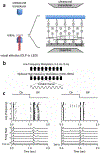
43 MHz ultrasound evokes localized retinal neuron activities. (a) Schematic diagram of experiment setup. (b) The 43 MHz sinusoidal wave was modulated at frequencies ranging from 10 Hz to 1 MHz with a duty cycle of 50%. This wave was then modulated again at 0.5 Hz with a duty cycle of 50%. (c) Raster plots and peri-stimulus time histogram (PSTH) from two neurons. Y-axis shows the order of modulation frequency (0 means DC, 6 means 1 MHz modulation frequency). Images from [15].
Fig. 3.
Fig. 3.
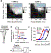
ARF is the physical mechanism for ultrasound retinal stimulation. (a) The left plot shows the normalized neuron firing rate with 43 MHz ultrasound stimulation (n = 22). X axis represents the pulse duration, whereas the y axis represents the pulse power. The right plot shows the same results of using 15 MHz ultrasound (n = 9). Brighter color indicates stronger neuron responses. Cross-hatching areas indicate the parameters that were not tested. (b) Radiation force model of retinal response. Average radiation pressures of three frequencies are shown versus radial distance. (c) Normalized neuron response for three different frequencies is compared to the radiation force model output (n > 20 cells in each case). Images from [16].
Fig. 4.
Fig. 4.
Ultrasound retinal stimulation can evoke neuron activities in visual circuits of rats with photoreceptor degeneration. Patterned ultrasound stimulation is demonstrated to achieve shape perception. (a and c) Light response and (b and d) US response from normal-sighted rats and RCS blind rats. The first row shows the schematic diagram of a healthy retina and a photoreceptor-degenerated retina. The second row shows the received multi-unit neuron activities. The third row shows the average spike counts of all channels (n = 12). (e) Schematic diagram: the 4.4-MHz helical transducer fires a beam pattern of “C” to the retina in vivo. (f) The spatial positions of each electrode in the 56-channel MEA placed on the surface of SC. (g) The acoustic field of the helical transducer measured by hydrophone shows a pattern “C” at the focal panel. (h) The ultrasound evoked neuron activities at SC were mapped by the 56-ch MEA, showing a corresponding pattern of “C”. Images from [18].

References

    1. Wong WL, Su X, Li X, Cheung CMG, Klein R, Cheng C-Y, and Wong TY, “Global prevalence of age-related macular degeneration and disease burden projection for 2020 and 2040: a systematic review and meta-analysis,” The Lancet Global Health, vol. 2, no. 2, pp. e106–e116, 2014. - PubMed
    1. Ferrari S, Di Iorio E, Barbaro V, Ponzin D, S Sorrentino F, and Parmeggiani F, “Retinitis pigmentosa: genes and disease mechanisms,” Current genomics, vol. 12, no. 4, pp. 238–249, 2011. - PMC - PubMed
    1. Humayun MS, Dorn JD, Da Cruz L, Dagnelie G, Sahel J-A, Stanga PE, Cideciyan AV, Duncan JL, Eliott D, and Filley E, “Interim results from the international trial of Second Sight’s visual prosthesis,” Ophthalmology, vol. 119, no. 4, pp. 779–788, 2012. - PMC - PubMed
    1. Gaillet V, Cutrone A, Artoni F, Vagni P, Pratiwi AM, Romero SA, Di Paola DL, Micera S, and Ghezzi D, “Spatially selective activation of the visual cortex via intraneural stimulation of the optic nerve,” Nature biomedical engineering, vol. 4, no. 2, pp. 181–194, 2020. - PubMed
    1. Chen X, Wang F, Fernandez E, and Roelfsema PR, “Shape perception via a high-channel-count neuroprosthesis in monkey visual cortex,” Science, vol. 370, no. 6521, pp. 1191–1196, 2020. - PubMed

Publication types