. 2022 Nov 22;19(6):066014.
doi: 10.1088/1741-2552/aca0c2.

High-density spinal cord stimulation selectively activates lower urinary tract nerves

Affiliations

High-density spinal cord stimulation selectively activates lower urinary tract nerves

Maria K Jantz et al. J Neural Eng. .

Abstract

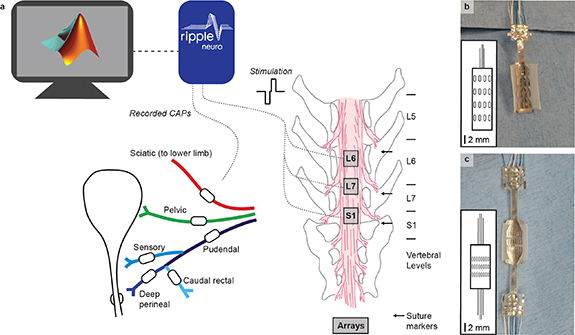
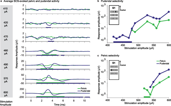
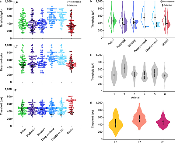
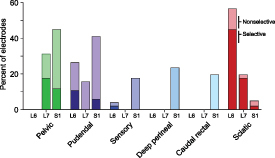
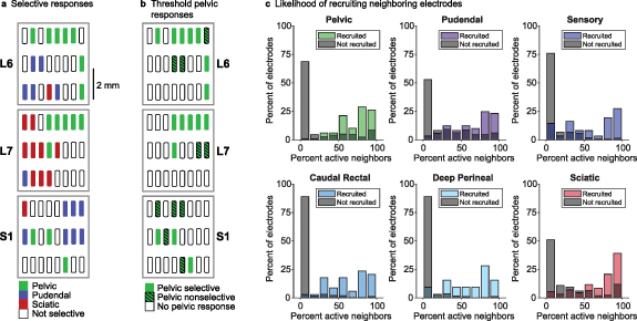
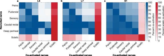
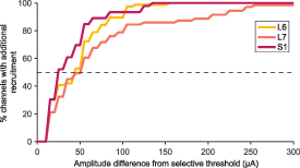
Objective.Epidural spinal cord stimulation (SCS) is a potential intervention to improve limb and autonomic functions, with lumbar stimulation improving locomotion and thoracic stimulation regulating blood pressure. Here, we asked whether sacral SCS could be used to target the lower urinary tract (LUT) and used a high-density epidural electrode array to test whether individual electrodes could selectively recruit LUT nerves.Approach. We placed a high-density epidural SCS array on the dorsal surface of the sacral spinal cord and cauda equina of anesthetized cats and recorded the stimulation-evoked activity from nerve cuffs on the pelvic, pudendal and sciatic nerves.Main results. Here we show that sacral SCS evokes responses in nerves innervating the bladder and urethra and that these nerves can be activated selectively. Sacral SCS always recruited the pelvic and pudendal nerves and selectively recruited both of these nerves in all but one animal. Individual branches of the pudendal nerve were always recruited as well. Electrodes that selectively recruited specific peripheral nerves were spatially clustered on the arrays, suggesting anatomically organized sensory pathways.Significance.This selective recruitment demonstrates a mechanism to directly modulate bladder and urethral function through known reflex pathways, which could be used to restore bladder and urethral function after injury or disease.

Keywords: autonomic nervous system; bladder; neuromodulation; peripheral nervous system; spinal cord stimulation.


Figures

Figure 1.
Figure 1.
Experimental setup. (a) Nerve cuffs, shown as white ovals on each nerve, were placed on multiple peripheral nerves and a high-density electrode array was placed at three locations over the sacral cord and cauda equina. Suture markers were placed at the center of the dorsal spinal processes prior to the laminectomy and are labeled with arrows. Nerve cuffs on the pelvic nerve (green), pudendal nerve (blue), and pudendal branches (shades of blue) had an inner diameter between 500 μm and 1000 μm. The sciatic nerve (red) cuff had an inner diameter of 3 mm. Recording and stimulation were completed through a MATLAB interface with a Ripple Grapevine system using a closed-loop response detection algorithm. (b) In animals 1 and 2, a 16 channel epidural array with four electrode columns spaced laterally across the cord and four electrode rows spaced rostrocaudally was used. The inset shows the array layout to scale, with the wire bundle represented in the same orientation as the photo. The electrodes on the 16 channel array were each 0.45 mm × 1.35 mm and were spaced 0.69 mm apart laterally and 1.64 mm apart rostrocaudally. (c) In animals 3–6, a 24 channel epidural array with eight columns and three rows was used. The electrodes on the 24 channel array were each 0.29 mm × 1.0 mm and were spaced 0.23 mm apart laterally and 0.78 apart rostrocaudally.
Figure 2.
Figure 2.
Examples of selective pelvic and pudendal nerve recruitment in animal 3 during stimulation on two different electrodes. (a) Stimulation-triggered averages of the pelvic (green) and pudendal (purple) nerve compound nerve action potentials at selected stimulation amplitudes. The traces include 1 ms preceding the stimulus pulse. Windows in which responses were detected are indicated under each trace as colored bars. At 410 μA, no response was detected in either nerve. At 420 μA, a selective response was detected in the pudendal nerve. At 480 μA the pelvic nerve was also recruited. 600 μA was the maximum stimulation amplitude for this trial and evoked large compound action potentials in both nerves. Note the different y-axis scales for each stimulation amplitude. (b) Peak-to-peak compound action potential amplitude of the pelvic and pudendal nerves for the electrode illustrated in (a) that was selective for the pudendal nerve from 420 μA up to 480 μA (selective range highlighted with a bar along the x-axis). Only the pudendal and pelvic nerve traces are shown here, but this electrode did not recruit any other instrumented nerves at threshold. The y-axis is shown on a log scale. The specific stimulation electrode is highlighted in the inset. (c) Peak-to-peak compound action potential amplitude of the pelvic and pudendal nerves for a nearby electrode (see inset) that selectively recruited the pelvic nerve from 520 μA up to 540 μA (selective range highlighted with a bar along the x-axis). The y-axis is shown on a log scale.
Figure 3.
Figure 3.
Recruitment thresholds for all animals, nerves and locations. (a) Recruitment thresholds for each nerve at each location across all animals. Trials that recruited nerves non-selectively at the threshold amplitude for that nerve are marked with unfilled circles and trials that recruited nerves selectively at the threshold amplitude are marked with filled circles. (b) Recruitment thresholds for each nerve across all locations and animals. Trials that recruited nerves non-selectively are marked with unfilled violins, left, and trials that recruited nerves selectively are marked with filled violins, right. (c) Recruitment thresholds for each animal across all nerves and locations. (d) Recruitment thresholds for each location across all nerves and animals. In the violin plots, the circle indicates the median value and the thick vertical bar indicates the interquartile range.
Figure 4.
Figure 4.
Recruitment of all nerves at threshold at each spinal level. Selective and nonselective nerve recruitment at each location at the threshold amplitude. The darkest color (bottom) in each stacked bar is the median percentage of electrodes that recruited each nerve selectively at the threshold amplitude. The lighter shade (top) represents the percentage of electrodes that recruited each nerve non-selectively at the threshold amplitude. Thus, the cumulative total of the bars represents the total recruitment of each nerve at the threshold amplitude.
Figure 5.
Figure 5.
Spatial arrangement of evoked responses. (a) Selective recruitment for the pelvic nerve, pudendal nerve, and sciatic nerve in a representative animal (animal 3). (b) Pelvic nerve recruitment for the same animal as panel a, demonstrating that the pelvic nerve was frequently recruited non-selectively on electrodes adjacent to selective electrodes. (c) When an electrode activated a particular nerve at the threshold amplitude, neighboring electrodes were likely to activate that nerve as well (colored bars). The y-axis is normalized to the total number of electrodes that activated a specific nerve. When that nerve had not been activated, surrounding electrodes were much less likely to activate neighboring electrodes (gray bars). The y-axis for the gray bars is normalized to the total number of electrodes that did not activate a specific nerve.
Figure 6.
Figure 6.
Coactivation of all nerves. Results are shown for each electrode placement at the (a) L6 vertebral segment, (b) L7 vertebral segment and (c) S1 vertebral segment. When a nerve first became active (vertical axis), other nerves were often co-activated or were recruited at lower amplitudes (horizontal axis). Sciatic comparisons are colored differently for clarity. As an example, in (a) when the pelvic nerve was first recruited, the sciatic nerve was frequently also active. However, when the sciatic nerve was first recruited, the pelvic nerve was less likely to be active.
Figure 7.
Figure 7.
Distribution of dynamic ranges for each array location. The dynamic range of each selective electrode is the amount of additional stimulation current necessary to evoke activity in an additional nerve, over and above the initial selective response. Many nerves recruited selectively had a dynamic range of less than 50 μA. Median dynamic range is marked with a dotted line.

Similar articles

Cited by

References

    1. Hunskaar S, Arnold E P, Burgio K, Diokno A C, Herzog A R, Mallett V T. Epidemiology and natural history of urinary incontinence. Int. Urogynecol. J. 2000;11:301–19. doi: 10.1007/s001920070021. - DOI - PubMed
    1. Hu T-W, Wagner T H, Bentkover J D, Leblanc K, Zhou S Z, Hunt T. Costs of urinary incontinence and overactive bladder in the United States: a comparative study. Urology. 2004;63:461–5. doi: 10.1016/j.urology.2003.10.037. - DOI - PubMed
    1. Brown J, McGhan W, Chokroverty S. Comorbidities associated with overactive bladder. Am. J. Manage. Care. 2000;6:S574–9. - PubMed
    1. Simpson L A, Eng J J, Hsieh J T C, Wolfe D L. The health and life priorities of individuals with spinal cord injury: a systematic review. J. Neurotrauma. 2012;29:1548–55. doi: 10.1089/neu.2011.2226. - DOI - PMC - PubMed
    1. Benevento B T, Sipski M L. Neurogenic bladder, neurogenic bowel, and sexual dysfunction in people with spinal cord injury. Phys. Ther. 2002;82:601–12. doi: 10.1093/ptj/82.6.601. - DOI - PubMed

Publication types