. 2023 May;19(5):1512-1532.
doi: 10.1080/15548627.2022.2139921. Epub 2022 Nov 7.

Tegument protein UL21 of alpha-herpesvirus inhibits the innate immunity by triggering CGAS degradation through TOLLIP-mediated selective autophagy

Affiliations

Tegument protein UL21 of alpha-herpesvirus inhibits the innate immunity by triggering CGAS degradation through TOLLIP-mediated selective autophagy

Zicheng Ma et al. Autophagy. 2023 May.

Abstract

Alpha-herpesvirus causes lifelong infections and serious diseases in a wide range of hosts and has developed multiple strategies to counteract the host defense. Here, we demonstrate that the tegument protein UL21 (unique long region 21) in pseudorabies virus (PRV) dampens type I interferon signaling by triggering the degradation of CGAS (cyclic GMP-AMP synthase) through the macroautophagy/autophagy-lysosome pathway. Mechanistically, the UL21 protein scaffolds the E3 ligase UBE3C (ubiquitin protein ligase E3C) to catalyze the K27-linked ubiquitination of CGAS at Lys384, which is recognized by the cargo receptor TOLLIP (toll interacting protein) and degraded in the lysosome. Additionally, we show that the N terminus of UL21 in PRV is dominant in destabilizing CGAS-mediated innate immunity. Moreover, viral tegument protein UL21 in herpes simplex virus type 1 (HSV-1) also displays the conserved inhibitory mechanisms. Furthermore, by using PRV, we demonstrate the roles of UL21 in degrading CGAS to promote viral infection in vivo. Altogether, these findings describe a distinct pathway where alpha-herpesvirus exploits TOLLIP-mediated selective autophagy to evade host antiviral immunity, highlighting a new interface of interplay between the host and DNA virus.Abbreviations: 3-MA: 3-methyladenine; ACTB: actin beta; AHV-1: anatid herpesvirus 1; ATG7: autophagy related 7; ATG13: autophagy related 13; ATG101: autophagy related 101; BHV-1: bovine alphaherpesvirus 1; BNIP3L/Nix: BCL2 interacting protein 3 like; CALCOCO2/NDP52: calcium binding and coiled-coil domain 2; CCDC50: coiled-coil domain containing 50; CCT2: chaperonin containing TCP1 subunit 2; CGAS: cyclic GMP-AMP synthase; CHV-2: cercopithecine herpesvirus 2; co-IP: co-immunoprecipitation; CQ: chloroquine; CRISPR: clustered regulatory interspaced short palindromic repeat; Cas9: CRISPR-associated system 9; CTD: C-terminal domain; Ctrl: control; DAPI: 4',6-diamidino-2-phenylindole; DBD: N-terminal DNA binding domain; DMSO: dimethyl sulfoxide; DYNLRB1: dynein light chain roadblock-type 1; EHV-1: equine herpesvirus 1; gB: glycoprotein B; GFP: green fluorescent protein; H&E: hematoxylin and eosin; HSV-1: herpes simplex virus 1; HSV-2: herpes simplex virus 2; IB: immunoblotting; IRF3: interferon regulatory factor 3; lenti: lentivirus; MAP1LC3/LC3: microtubule associated protein 1 light chain 3; MARCHF9: membrane associated ring-CH-type finger 9; MG132: cbz-leu-leu-leucinal; NBR1: NBR1 autophagy cargo receptor; NC: negative control; NEDD4L: NEDD4 like E3 ubiquitin protein ligase; NH4Cl: ammonium chloride; OPTN: optineurin; p-: phosphorylated; PFU: plaque-forming unit; Poly(dA:dT): Poly(deoxyadenylic-deoxythymidylic) acid; PPP1: protein phosphatase 1; PRV: pseudorabies virus; RB1CC1/FIP200: RB1 inducible coiled-coil 1; RNF126: ring finger protein 126; RT-PCR: real-time polymerase chain reaction; sgRNA: single guide RNA; siRNA: small interfering RNA; SQSTM1/p62: sequestosome 1; STING1: stimulator of interferon response cGAMP interactor 1; TBK1: TANK binding kinase 1; TOLLIP: toll interacting protein; TRIM33: tripartite motif containing 33; UL16: unique long region 16; UL21: unique long region 21; UL54: unique long region 54; Ub: ubiquitin; UBE3C: ubiquitin protein ligase E3C; ULK1: unc-51 like autophagy activating kinase 1; Vec: vector; VSV: vesicular stomatitis virus; VZV: varicella-zoster virus; WCL: whole-cell lysate; WT: wild-type; Z-VAD: carbobenzoxy-valyl-alanyl-aspartyl-[O-methyl]-fluoromethylketone.

Keywords: Alpha-herpesvirus; CGAS; UL21 protein; selective autophagy; type I interferon signaling.


Conflict of interest statement

The authors declare that there are no conflicts of interest.

Figures

Figure 1.
Figure 1.
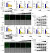
PRV UL21 promotes viral replication and pathogenicity. (A) Plaque morphology of PRV-WT, ΔUL21, and ΔUL21R in Vero cells. Plaque sizes of PRV-WT, ΔUL21, and ΔUL21R were plotted as a percentage of the average PRV-WT plaque sizes. (B) Viral replication in PK-15, HEK-293 T, MEF, and N2a cells. Cells were infected with PRV-WT, ΔUL21, or ΔUL21R at 0.01, 0.05 or 3 plaque forming unit (PFU)/cell. At 48 (0.01, 0.05 PFU/cell) or 24 (3 PFU/cell) hpi, cells were harvested, and the total virus yields were determined in Vero cells by using a plaque assay. (C) Mouse survival was recorded and shown as a percentage over time. Mice housed in 4 groups (10 mice/group) were mock-infected or infected with PRV-WT, ΔUL21, or ΔUL21R (1 × 104 pfu/mice) through intraperitoneal injection. (D) Four groups of mice (4 mice/group) were treated as in (C) and sacrificed at 3 dpi. The viral DNA loads in the brain, lung, and brainstem were quantified by RT-PCR. (E) hematoxylin and eosin (H&E) stained brain and lung sections from the indicated mice 3 dpi (1 × 104 pfu/mice) (scale bar: 100 μm). The data are representative of at least three independent experiments with similar results (mean ± SD of n = 3 biological replicates in B, n = 4 biological replicates in D). * p < 0.05, ** p < 0.01 (two-tailed unpaired t test).
Figure 2.
Figure 2.
PRV UL21 inhibits poly(dA:dT) and CGAS-STING1-mediated type I interferon pathway. (A and E) PRV UL21 inhibits poly(dA:dT) or CGAS-STING1 stimulated IFNB promoter activity. (A) HEK-293 T cells seeded in 24-well plates were co-transfected with pIFNB-Luc (50 ng) and pRL-TK (10 ng) along with increasing doses of Flag-UL21 (100, 300, or 500 ng). At 24 hpt, cells were stimulated with poly(dA:dT) at a concentration of 200 ng/mL. After another 24 h, cells were harvested for a luciferase assay. (E) HEK-293 T cells seeded in 24-well plates were co-transfected with pIFNB-Luc (50 ng) and pRL-TK (10 ng), HA-CGAS (500 ng), or MYC-STING1 (50 ng) alone with the along with increasing doses of Flag-UL21 (100, 300, or 500 ng). At 48 hpt, the cells were harvested for a luciferase assay. (B and F) PRV UL21 inhibits poly(dA:dT) or HA-CGAS + MYC-STING1 stimulated IFNB, ISG15, IFIT2, and IFIT1 transcripts. HEK-293 T cells were treated similar to (A) and (E), but without pIFNB-Luc and pRL-TK transfection. The cells were harvested for RNA extraction and RT-PCR detection of IFNB, ISG15, IFIT2, and IFIT1, and RNA18S rRNA mRNA levels. (C and G) HEK-293 T cells were treated as in (B) and (F). The supernatants were harvested and incubated with fresh confluent HEK-293 T cells. After 24 h, the cells were infected with VSV-GFP at an MOI of 0.01. At 24 hpi, GFP signaling was detected by microscopy (scale bar: 100 μm). (D and H) HEK-293 T cells were treated the same as in (B) and (F). The cell lysates were harvested for western blot analysis with antibodies against Flag-tag, IRF3, p-IRF3 (phosphorylated interferon regulatory factor 3), p-TBK1 (TBK1, phosphorylated TANK binding kinase 1), and ACTB/β-actin. Data are representative of at least three independent experiments with similar results (mean ± SD of n = 3 biological replicates in A, B, E, and F). * p < 0.05, ** p < 0.01 (two-tailed unpaired t test).
Figure 3.
Figure 3.
The autophagy-lysosome pathway is associated with CGAS degradation induced by PRV UL21. (A) Ectopic expression of PRV UL21 decreases the CGAS protein level. HEK-293 T cells seeded in 6-well plates were transfected with HA-CGAS (2 μg) along with indicated plasmids (empty vector, Flag-GFP, or Flag-UL21) (2 μg). At 24 hpt, the cells were harvested for western blot analysis with antibodies against HA-tag, Flag-tag, and ACTB. (B) PRV UL21 degrades CGAS in a dose-dependent manner. HEK-293 T cells seeded in 6-well plates were transfected with HA-CGAS (2 μg) together with empty vector or increasing doses of Flag-UL21 (0.5, 1, or 2 μg). At 24 hpt, the cells were harvested for western blot analysis with antibodies against HA-tag, Flag-tag, and ACTB. (C) Effects of Z-VAD, MG132, and 3-MA on CGAS degradation induced by PRV UL21. HEK-293 T cells seeded in 6-well plates were transfected with HA-CGAS (2 μg) together with empty vector or Flag-UL21 (2 μg). At 12 hpt, cells were treated with DMSO or Z-VAD (20 μM), MG132 (10 μM), or 3-MA (5 mM). After another 12 h, cells were harvested for western blot analysis with antibodies against HA-tag, Flag-tag, and ACTB. (D) Effects of NH4Cl, 3-MA, and CQ on CGAS degradation induced by PRV UL21. HEK-293 T cells seeded in 6-well plates were transfected with HA-CGAS (2 μg) together with empty vector or Flag-UL21 (2 μg). At 12 hpt, cells were treated with DMSO or NH4Cl (20 mM), 3-MA (5 mM), or CQ (50 μM). After another 12 h, cells were harvested for western blot analysis with antibodies against HA-tag, Flag-tag, and ACTB. Data are representative of at least three independent experiments with similar results.
Figure 4.
Figure 4.
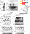
PRV UL21 degrades CGAS through the autophagy pathway. (A) Ectopic expression of PRV UL21 induces autophagy and decreases the endogenous level CGAS protein. HEK-293 T cells seeded in 6-well plates were transfected with Flag-UL21 (2 μg). At 24 hpt, the cells were harvested for western blot analysis with antibodies against LC3, CGAS, Flag-tag, and ACTB. (B) PRV UL21 induces GFP-LC3 puncta formation. HeLa cells seeded in 6-well plates were transfected with GFP-LC3 (2 μg) along with empty vector or Flag-UL21 (2 μg). At 24 hpt, cells were stained with anti-Flag (red) and subjected to analysis by confocal microscopy. Nuclei were stained with DAPI (blue) (scale bar: 10 μm). (C and D) PRV UL21 is important in virus-induced autophagy and CGAS degradation. HEK-293 T (C) or PK-15 (D) cells were mock-infected or infected with PRV-WT, ΔUL21, or ΔUL21R at 3 PFU/cell. At 12 hpi, the cells were harvested for western blot analysis with antibodies against LC3, CGAS, STING1, UL21, UL54, and ACTB. Densitometric quantification of LC3-II:I. The protein bands shown in panel C and D were quantified using NIH ImageJ software. The data are presented as the relative amount of LC3-II: I normalized to the total level of ACTB in each sample. (E) ATG7 deficiency inhibits PRV UL21-mediated CGAS degradation. HEK-293 T cells were transfected with siRNA targeting to ATG7 (siATG7) (10 mM) or control siRNA (siNC) (10 mM). After 12 h, cells were transfected with the HA-CGAS (2 μg) together with empty vector (2 μg) or Flag-UL21 (2 μg). After another 24 h, the cells were harvested for western blot analysis with antibodies against HA-tag, ATG7, Flag-tag, and ACTB. (F and G) ATG7 deficiency inhibits PRV UL21 induced CGAS degradation in viral infection. HEK-293 T (F) or PK-15 (G) cells were transfected with siATG7 (10 mM) or siNC (10 mM). After 12 h, cells were mock-infected or infected with PRV, ΔUL21, or ΔUL21R at 3 PFU/cell. At 12 hpi, the cells were harvested for western blot analysis with antibodies against CGAS, ATG7, UL21, UL54, and ACTB. Densitometric quantification of CGAS. The protein bands shown in panel F and G were quantified using NIH ImageJ software. The data are presented as the relative amount of CGAS normalized to the total level of ACTB in each sample. (H and I) The effects of CGAS on viral replication. Wild-type (WT), CGAS−/−, or CGAS−/–rescued HEK-293 T cells (H), and WT, CGAS−/−, or CGAS−/–rescued PK-15 cells (I) were mock-infected or infected with PRV-WT, ΔUL21, or ΔUL21R at 0.01 PFU/cell. At 48 hpi, cells and supernatants were harvested, and the total virus yields were determined in Vero cells by a plaque assay. Data are representative of at least three independent experiments with similar results (mean ± SD of n = 3 biological replicates in H and I). * p < 0.05, ** p < 0.01 (two-tailed unpaired t test).
Figure 5.
Figure 5.
TOLLIP is the selective autophagic cargo mediating UL21 induced CGAS degradation. (A) PRV UL21 interacts with CGAS. HEK-293 T cells were transfected with Flag-UL21 (3 μg) together with indicated plasmids (empty vector [3 μg], HA-CGAS [3 μg], or HA-GFP [3 μg]). At 24 hpt, cells were processed for immunoprecipitation (IP) with anti-Flag magnetic beads. Whole-cell lysates (WCLs) and precipitated proteins were probed with antibodies against Flag-tag, HA-tag, and ACTB. (B) PRV UL21 colocalizes with CGAS in the cytoplasm. HeLa cells were transfected with HA-CGAS (2 μg) along with empty vector or Flag-UL21 (2 μg). At 24 hpt, cells were stained with anti-HA (red) and anti-Flag (green) and subjected to analysis by confocal microscopy. Nuclei were stained with DAPI (blue) (scale bar: 10 μm). (C) PRV UL21 interacts with SQSTM1 and TOLLIP. HEK-293 T cells were transfected with MYC-UL21 (3 μg) together with indicated plasmids (empty vector [3 μg], Flag-NBR1 [3 μg], Flag-OPTN [3 μg], Flag-SQSTM1 [3 μg], Flag-CALCOCO2 [3 μg], Flag-BNIP3L [3 μg], or Flag-TOLLIP [3 μg]). At 24 hpt, cells were processed for IP with anti-MYC magnetic beads. WCLs and precipitated proteins were probed with antibodies against Flag-tag, MYC-tag, and ACTB. (D) CGAS interacts with SQSTM1 and TOLLIP. HEK-293 T cells were transfected with HA-CGAS (3 μg) together with indicated plasmids (empty vector [3 μg], Flag-NBR1 [3 μg], Flag-OPTN [3 μg], Flag-SQSTM1 [3 μg], Flag-CALCOCO2 [3 μg], Flag-BNIP3L [3 μg], or Flag-TOLLIP [3 μg]). At 24 hpt, the cells were processed for IP with anti-HA magnetic beads. WCLs and precipitated proteins were probed with antibodies against Flag-tag, HA-tag, and ACTB. (E) UL21 strengthen the interaction between TOLLIP and CGAS. HEK-293 T cells were transfected with HA-CGAS (3 μg) together with indicated plasmids (Flag-SQSTM1 [3 μg] with MYC-UL21 [3 μg], or empty vector [3 μg]) or (Flag-TOLLIP [3 μg] with MYC-UL21 [3 μg] or empty vector [3 μg]). At 24 hpt, cells were processed for IP with anti-HA magnetic beads. WCLs and precipitated proteins were probed with antibodies against Flag-tag, MYC-tag, HA-tag, and ACTB. (F) SQSTM1 is not associated with UL21 induced CGAS degradation. Wild type (WT) or SQSTM1−/− HEK-293 T cells seeded in 6-well plates were transfected with HA-CGAS (2 μg) together with or without MYC-UL21 (2 μg) and with or without Flag-SQSTM1 (2 μg). At 24 hpt, cells were harvested for western blot analysis with antibodies against HA-tag, MYC-tag, SQSTM1, and ACTB. (G and H) TOLLIP deficiency inhibits UL21 induced CGAS degradation. Wild-type (WT) or TOLLIP−/− HEK-293 T (G) and PK-15 (H) cells seeded in 6-well plates were transfected with HA-CGAS (2 μg) together with or without MYC-UL21 (2 μg) and with or without Flag-TOLLIP (2 μg). At 24 hpt, cells were harvested for western blot analysis with antibodies against HA-tag, MYC-tag, TOLLIP, and ACTB. (I) TOLLIP deficiency inhibits UL21-mediated CGAS degradation in PRV infection. WT and TOLLIP−/− HEK-293 T cells were mock-infected or infected with PRV-WT, ΔUL21, or the ΔUL21R at 3 PFU/cell. At 12 hpi, cells were harvested for western blot analysis with antibodies against CGAS, TOLLIP, UL21, UL54 and ACTB. Data are representative of at least three independent experiments with similar results.
Figure 6.
Figure 6.
UL21 induces K27-linked ubiquitination of CGAS at K384 site. (A) UL21 induces the ubiquitination of CGAS. HEK-293 T cells were transfected with Flag-UL21 (3 μg) and HA-Ub (3 μg) together with empty vector (3 μg) or MYC-UL21 (3 μg). At 24 hpt, cells were processed for immunoprecipitation (IP) with anti-Flag magnetic beads. Whole-cell lysates (WCLs) and precipitated proteins were probed with antibodies against HA-tag, MYC-tag, Flag-tag, and ACTB. (B) UL21 catalyzes K27-linked ubiquitination of CGAS. HEK-293 T cells were transfected with Flag-CGAS (3 μg), MYC-UL21 (3 μg), empty vector (3 μg), ubiquitination-HA (3 μg) or its mutants (HA-K6 [3 μg], HA-K11 [3 μg], HA-K27 [3 μg], HA-K29 [3 μg], HA-K33 [3 μg], HA-K48 [3 μg], or HA-K63 [3 μg]). At 24 hpt, cells were processed for IP with anti-Flag magnetic beads. WCLs and precipitated proteins were probed with antibodies against HA-tag, MYC-tag, Flag-tag, and ACTB. (C) Schematic representation of wild-type CGAS and its deletion mutants. HEK-293 T cells were transfected with the empty vector (2 μg), Flag-CGAS (2 μg) or its truncated mutants (Flag-CGAS [1–160 aa; 2 μg], Flag-CGAS [160–522 aa; 2 μg], Flag-CGAS [1–330 aa; 2 μg], Flag-CGAS [160–330 aa; 2 μg], Flag-CGAS [330–522 aa; 2 μg], and Flag-CGAS [290–400 aa; 2 μg]). At 24 hpt, cells were harvested for western blot analysis with antibodies against Flag-tag and ACTB. (D) UL21 interacts with CGAS at the internal domain 290–400 aa. HEK-293 T cells were transfected with the MYC-UL21 (3 μg) together with the indicated plasmids (empty vector [3 μg], Flag-CGAS [3 μg], Flag-CGAS [1–160 aa; 3 μg], Flag-CGAS [160–522 aa; 3 μg], Flag-CGAS [1–330 aa; 3 μg], Flag-CGAS [160–330 aa; 3 μg], Flag-CGAS [330–522 aa; 3 μg], or Flag-CGAS [290–400 aa; 3 μg]). At 24 hpt, cells were processed for IP with anti-Flag magnetic beads. WCLs and precipitated proteins were probed with antibodies against MYC-tag, Flag-tag, and ACTB. (E) UL21 catalyzes K27-linked ubiquitination of CGAS at K384 site. HEK-293 T cells were transfected with MYC-UL21 (3 μg) and HA-Ub-K27 (3 μg) together with the indicated plasmids (empty vector [3 μg], Flag-CGAS-WT [3 μg], Flag-CGAS-K301R [3 μg], Flag-CGAS-K327 R [3 μg], Flag-CGAS-K347R [3 μg], Flag-CGAS-K362R [3 μg], Flag-CGAS-K365R [3 μg], Flag-CGAS-K384R [3 μg], or Flag-CGAS-K394R [3 μg]). At 24 hpt, cells were processed for IP with anti-Flag magnetic beads. WCLs and precipitated proteins were probed with antibodies against HA-tag, Flag-tag, MYC-tag, and ACTB. (F and G) Effects of UL21 on human and porcine CGAS mutant expression. HEK-293 T (F) or PK-15 (G) cells seeded in 6-well plates were transfected with MYC-UL21 (2 μg) together with Flag-CGAS/Flag-CGAS [P] or its mutants (2 μg). At 24 hpt, cells were harvested for western blot analysis with antibodies against MYC-tag, Flag-tag, and ACTB. Data are representative of at least three independent experiments with similar results.
Figure 7.
Figure 7.
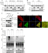
UBE3C is the E3 ligase that mediates CGAS ubiquitination induced by UL21. (A and B) UBE3C mediates CGAS degradation induced by UL21. Wild-type (WT) or UBE3C−/− HEK-293 T (A) and PK-15 (B) cells seeded in 6-well plates were transfected with HA-CGAS (2 μg) together with or without MYC-UL21 (2 μg), and with or without Flag-UBE3C (2 μg). At 24 hpt, cells were harvested for western blot analysis with antibodies against HA-tag, MYC-tag, UBE3C, and ACTB. (C) UL21 interacts with UBE3C. HEK-293 T cells were transfected with MYC-UL21 (3 μg) together with indicated plasmids (empty vector [3 μg], Flag-NBR1 [as the negative control; 3 μg], or Flag-UBE3C [3 μg]). At 24 hpt, cells were processed for immunoprecipitation (IP) with anti-MYC magnetic beads. Whole-cell lysates (WCLs) and precipitated proteins were probed with antibodies against Flag-tag, MYC-tag, and ACTB. (D) UL21 colocalizes with UBE3C. HeLa cells were transfected with Flag-UBE3C (2 μg) along with empty vector (2 μg) or MYC-UL21 (2 μg). At 24 hpt, cells were stained with anti-Flag (red) and anti-MYC (green) subjected to analysis by confocal microscopy. Nuclei were stained with DAPI (blue) (scale bar: 10 μm). (E) UBE3C is necessary for the K27-linked ubiquitination of CGAS induced by UL21. Wild-type (WT) or UBE3C−/− HEK-293 T cells were transfected with MYC-UL21 (2 μg), Flag-CGAS (2 μg), HA-Ub (K27 only) (2 μg), or HA-Ub (K27R) (K27 lysine residues mutated to arginine) (2 μg). At 24 hpt, cells were processed for IP with anti-Flag magnetic beads. WCLs and precipitated proteins were probed with antibodies against Flag-tag, MYC-tag, HA-tag, and ACTB. Data are representative of at least three independent experiments with similar results.
Figure 8.
Figure 8.
The N terminus of UL21 triggers CGAS degradation. (A) Conservation of UL21 across alpha-herpesviruses; (detailed information is shown in Materials and methods). Construction of UL21 truncated mutants. HEK-293 T cells were transfected with a Flag-vector (2 μg), Flag-UL21 (2 μg), or its truncated mutants (Flag-UL21 [1–300 aa; 2 μg], Flag-UL21 [200–532 aa; 2 μg], Flag-UL21 [1–200 aa; 2 μg], Flag-UL21 [300–532 aa; 2 μg]) for 24 h, and then the cell lysates were collected for western blot with antibodies against Flag-tag and ACTB. (B) CGAS binds to the 1–200 aa of UL21. HEK-293 T cells were transfected with HA-CGAS (3 μg) together with indicated plasmids (empty vector [3 μg], Flag-UL21 [3 μg], Flag-UL21 [1–300 aa; 3 μg], Flag-UL21 [200–532 aa; 3 μg], Flag-UL21 [1–200 aa; 3 μg], or Flag-UL21 [300–532 aa; 3 μg]). At 24 hpt, cells were processed for IP with anti-HA magnetic beads. WCLs and precipitated proteins were probed with antibodies against Flag-tag, HA-tag, and ACTB. (C) UL21 (1–200 aa) colocalizes with CGAS in the cytoplasm. HeLa cells were transfected with HA-CGAS (2 μg) along with empty vector (2 μg), Flag-UL21 (2 μg), Flag-UL21 (1–200 aa) (2 μg), or Flag-UL21 (200–532 aa) (2 μg). At 24 hpt, cells were stained with anti-HA (red) and anti-Flag (green) subjected to analysis by confocal microscopy. Nuclei were stained with DAPI (blue) (scale bar: 10 μm). (D) UL21 (1–200 aa) inhibits CGAS-STING1 stimulated IFNB promoter activity. HEK 293 T cells seeded in 24-well plates were co-transfected with pIFNB-Luc (50 ng) and pRL-TK (10 ng), HA-CGAS (500 ng), MYC-STING1 (50 ng) along with empty vector, Flag-UL21 (500 ng), Flag-UL21 (1–200 aa) (500 ng), or Flag-UL21 (200–532 aa) (500 ng). At 48 hpt, cells were harvested for a luciferase assay and western blot analysis with antibodies against HA-tag, MYC-tag, Flag-tag, and ACTB. (E) The N terminus of UL21 induces CGAS degradation mediated by autophagy. HEK-293 T cells seeded in 6-well plates were transfected with indicated plasmids (empty vector, FL, N, or C; 2 μg). At 24 hpt, cells were harvested for western blot analysis with antibodies against LC3, CGAS, Flag-tag, and ACTB. (F) HEK-293 T cell seeded in 6-well plates were transfected with HA-CGAS (2 μg) together with indicated plasmids (empty vector, FL, N, or C; 2 μg). At 12 hpt, cells were treated with DMSO or 3-MA (5 mM). After another 12 h, cells were harvested for western blot analysis with antibodies against HA-tag, Flag-tag, and ACTB. (G and H) TOLLIP and UBE3C mediate CGAS degradation induced by the N terminus of UL21. WT or TOLLIP−/− (G) and UBE3C−/− (H) HEK-293 T cells seeded in 6-well plates were transfected with HA-CGAS (2 μg) together with or without MYC-TOLLIP (2 μg), UBE3C (2 μg), and with indicated plasmids (empty vector, FL, N, or C; 2 μg). At 24 hpt, cells were harvested for western blot analysis with antibodies against HA-tag, Flag-tag, TOLLIP, UBE3C, and ACTB. FL represents Flag-UL21; N represents Flag-UL21 (1–200 aa); C represents Flag-UL21 (200–532 aa). (I and J) TOLLIP and UBE3C mediate CGAS degradation induced by the N terminus of UL21 in PRV infection. WT, TOLLIP−/− (I), or UBE3C−/− (J) HEK-293 T cells were mock-infected or infected with PRV-WT, ΔUL21, ΔUL21 (1–200 aa), ΔUL21 (200–532 aa), or ΔUL21R at 3 PFU/cell. At 12 hpi, cells were harvested for western blot analysis with antibodies against CGAS, UL21, TOLLIP, UBE3C, UL54, and ACTB. Data are representative of at least three independent experiments with similar results (mean ± SD of n = 3 biological replicates in D). * p < 0.05, ** p < 0.01 (two-tailed unpaired t test).
Figure 9.
Figure 9.
CGAS is essential for controlling PRV infection. (A) RT-PCR analysis of Ifnb, Isg15, Ifit2, Ifit1 in wild-type (WT) and cgas−/− BMDMs after PRV infection. WT and cgas−/− BMDMs cells were mock-infected or infected with PRV-WT, ΔUL21, ΔUL21 (1–200 aa), ΔUL21 (200–532 aa), or ΔUL21R at 3 PFU/cell. At 12 hpi, the cells were harvested for RNA extraction and RT-PCR detection of Ifnb, Isg15, Ifit2, and Ifit1 mRNA, and Rna18s rRNA levels. (B) Immunoblot analysis of lysates of WT or cgas−/− BMDMs after PRV infection. WT and cgas−/− BMDMs cells were mock infected or infected with PRV-WT, ΔUL21, ΔUL21 (1–200 aa), ΔUL21 (200–532 aa), or ΔUL21R at 3 PFU/cell. At 12 hpi, cells were harvested for western blot analysis with antibodies against CGAS, STING1, p-TBK1, TBK1, p-IRF3, IRF3, UL21, UL54, and ACTB. (C) Mouse survival was recorded and shown as a percentage over time. WT and cgas−/− mice separately housed in 6 groups (n = 8 mice/group) were mock-infected or infected with PRV-WT, ΔUL21, ΔUL21 (1–200 aa), ΔUL21 (200–532 aa), or ΔUL21R (1 × 104 pfu/mice) through intraperitoneal injection. (D) Viral DNA loads in the brain, lung, or brainstem were quantified by RT-PCR. WT and cgas−/− mice separately housed in 6 groups (n = 4 mice/group) were mock-infected or infected with PRV-WT, ΔUL21, ΔUL21 (1–200 aa), ΔUL21 (200–532 aa), or ΔUL21R (1 × 104 pfu/mice) through intraperitoneal injection. Three days post-infection, the brain, lung or brainstem tissues were harvested for RT-PCR detection of viral loads. (E) The Isg expression in lung tissues from the mice in (D). Total RNA was extracted from the lung tissues and used for RT-PCR detection of Ifnb, Isg15, Ifit2, and Ifit1 mRNA, and Rna18s rRNA levels. (F) H&E-stained brain and lung sections from the mice in (D) (scale bar: 100 μm). The data are representative of at least three independent experiments with similar results (mean ± SD of n = 3 biological replicates in A, n = 4 biological replicates in D, E and F). * p < 0.05, ** p < 0.01, ns: not significant (two-tailed unpaired t test).
Figure 10.
Figure 10.
A working model of the role of the UL21 protein of PRV in the regulation of CGAS mediated signaling. Black arrows indicate the CGAS-STING1-mediated type I IFN signaling pathway. Blue arrows indicate expression of the UL21 protein. Red arrows indicate the process of UL21-mediated autophagic degradation of CGAS.

References

    1. Sehrawat S, Kumar D, Rouse BT.. Herpesviruses: harmonious pathogens but relevant cofactors in other diseases? Front Cell Infect Microbiol. 2018;8:177. - PMC - PubMed
    1. Sharma V, Mobeen F, Prakash T. Comparative genomics of herpesviridae family to look for potential signatures of human infecting strains. Int J Genomics. 2016;2016:9543274. - PMC - PubMed
    1. Pomeranz LE, Reynolds AE, Hengartner CJ. Molecular biology of pseudorabies virus: impact on neurovirology and veterinary medicine. Microbiol Mol Biol Rev. 2005. Sep;69(3):462–500. - PMC - PubMed
    1. Loret S, Guay G, Lippe R. Comprehensive characterization of extracellular herpes simplex virus type 1 virions. J Virol. 2008. Sep;82(17):8605–8618. - PMC - PubMed
    1. Karasneh GA, Shukla D. Herpes simplex virus infects most cell types in vitro: clues to its success. Virol J. 2011. Oct 26;8(1):481. - PMC - PubMed

Publication types