. 2022 Nov 7;13(1):6729.
doi: 10.1038/s41467-022-34459-3.

Hippocampal convergence during anticipatory midbrain activation promotes subsequent memory formation

Affiliations

Hippocampal convergence during anticipatory midbrain activation promotes subsequent memory formation

Jia-Hou Poh et al. Nat Commun. .

Abstract

The hippocampus has been a focus of memory research since H.M's surgery abolished his ability to form new memories, yet its mechanistic role in memory remains debated. Here, we identify a candidate memory mechanism: an anticipatory hippocampal "convergence state", observed while awaiting valuable information, and which predicts subsequent learning. During fMRI, participants viewed trivia questions eliciting high or low curiosity, followed seconds later by its answer. We reasoned that encoding success requires a confluence of conditions, so that hippocampal states more conducive to memory formation should converge in state space. To operationalize convergence of neural states, we quantified the typicality of multivoxel patterns in the medial temporal lobes during anticipation and encoding of trivia answers. We found that the typicality of anticipatory hippocampal patterns increased during high curiosity. Crucially, anticipatory hippocampal pattern typicality increased with dopaminergic midbrain activation and uniquely accounted for the association between midbrain activation and subsequent recall. We propose that hippocampal convergence states may complete a cascade from motivation and midbrain activation to memory enhancement, and may be a general predictor of memory formation.


Conflict of interest statement

The authors declare no competing interests.

Figures

Fig. 1
Fig. 1. Task schematics & Memory performance.
a Prior to fMRI scanning, participants were shown a series of trivia questions. For each, they were told to indicate the likelihood that they knew the answer, and how curious they were about it. Questions were excluded if participants indicated a likelihood of knowing the answer above 90%. The remaining questions were separated into tertiles to be used for fMRI scanning, with the 1st and 3rd tertile categorized as Low and High Curiosity questions respectively (72 questions each). During fMRI scanning, participants were shown each trivia question along with a colored rectangle that indicated the duration and action contingency of the trial. On action-contingent trials, an arrow was presented after a 9 s or 13 s delay. Participants indicated the direction of the arrow with a button press. This was followed by the presentation of the trivia answer. On non-action contingent trials, the trivia answer was presented immediately after the delay interval. Following the scan, participants were shown each trivia question and were required to recall its associated answer. Analyses of answer Anticipation used activation evoked by each question (the Question Interval). Analyses of answer encoding used activation evoked by each answer (Answer Interval, including the response on action-contingent trials). b Box plots for memory recall performance across each condition. Memory recall was better for high- (orange) than low-curiosity (blue) trials based on two-tailed paired t-test (t(22) = 9.32, p < 0.001, d = 1.94, mean difference = 0.23, 95% CI = [0.18 0.28]; N = 23 participants).The center of each box corresponds to the mean value, the upper and lower hinges correspond to the first and third quartiles, while the whiskers correspond to the largest and smallest values within 1.5 times of the interquartile range. Each dot corresponds to the recall performance of each participant. ***p < 0.001.
Fig. 2
Fig. 2. Curiosity increased univariate activation in the midbrain VTA and perirhinal cortex during anticipation of trivia answers.
Anticipatory BOLD activation (i.e., during the Question interval preceding each trivia answer) was greater after High-Curiosity (orange) versus Low-Curiosity (blue) trivia questions in the midbrain VTA (p = 0.026) and perirhinal cortex (PRC) (p = 0.017; N = 23 participants). Hippocampus (HPC) and parahippocampal cortex (PHC) activation did not differentiate curiosity states. Red overlays on the brain images demarcate the ROIs (Brain template obtained from FSL and reproduced with permission). The large dots in each panel represent the group mean; small dots represent mean activation for each participant. Error bars represent the SEM. *p < 0.05.
Fig. 3
Fig. 3. Midbrain VTA univariate activation during anticipation of trivia answers uniquely explains subsequent recall.
Anticipatory BOLD activation (i.e., during the Question interval preceding each trivia answer) in the midbrain VTA and medial temporal lobe ROIs was used to predict memory outcome for each trial in a mixed-effects logistic regression model (N = 23 participants). This method allows the identification of variance that is uniquely accounted for by each of the ROIs. Bar graphs in each panel represent the parameter estimate for each ROI in the full model. Among the ROIs, midbrain VTA activation was the only statistically significant predictor of subsequent recall of answers (p = 0.015). For visualization, the estimated predicted probability of recall relative to the individual’s mean probability (delta from within-subject mean, y-axis) is plotted against the univariate signal in each ROI (arbitrary units, a.u.; x-axis). Red overlays on the brain images demarcate the ROIs (Brain template obtained from FSL and reproduced with permission). Light gray lines depict the slope for each participant, while the solid black line depicts the mean slope across all participants. Error bars represent the SEM. *p < 0.05.
Fig. 4
Fig. 4. Anticipatory pattern typicality in the hippocampus is uniquely associated with curiosity and subsequent recall.
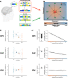
a For the convergence state analysis, patterns of activation for each trial were extracted from each of the ROIs (Brain icon created with BioRender.com). Convergence states were operationalized as pattern typicality, or the distance between the activation pattern of each trial and an independently defined centroid representing the prototypical state). A leave-one-run-out approach was used where the cluster centroid was identified using data from N-1 runs. This centroid was then used as the origin to quantify the distance for trials from the left-out run. Patterns showing higher typicality, (i.e., shorter distance from the centroid) are considered to exhibit greater convergence than patterns more eccentric from the centroid. b During the anticipation of High-Curiosity (orange) trivia answers, patterns of activation exhibited higher typicality in the hippocampus (HPC, p = 0.011), but not the surrounding medial temporal lobe cortices (PHC: Parahippocampal cortex; PRC: Perirhinal cortex) (N = 23 participants). The larger dots in each panel represents the group mean, while the smaller dots represent the mean distance for each participant. c We used a mixed-effects logistic regression model to predict memory outcome for each trial using the pattern typicality of the medial temporal lobe ROIs (N = 23 participants). Hippocampal typicality was the only significant predictor of subsequent recall (p = 0.008). Bar graph of each panel represents the parameter estimate of each ROI in the full model. For visualization, the estimated change in probability of recall (demeaned within subject) is plotted against the distance from centroid for each ROI. Light gray lines depict the slope for each participant, while the solid black line depicts the mean slope across all participants. Error bars represent the SEM. *p < 0.05, **p < 0.01.
Fig. 5
Fig. 5. Anticipatory midbrain VTA activation selectively modulates hippocampal pattern typicality.
a Greater activation in the midbrain VTA is associated with greater pattern typicality in the hippocampus during the anticipation of trivia answers (p < 0.001; N = 23 participants). The bar graph represents the parameter estimate for the association of VTA activation and hippocampal typicality. For visualization, the predicted distance from centroid for the hippocampus is plotted against univariate VTA BOLD activation. Light gray lines depict the slope for each participant, while the solid black line depicts the mean slope across all participants. Typicality is defined as a shorter distance from centroid. b Whole-brain analysis (controlling for univariate VTA activation) showed that anticipatory univariate activations in clusters that included the visual cortex and posterior hippocampus (overlaid on brain template obtained from FSL and reproduced with permission) were also positively associated with pattern typicality in the HPC (FWE p < 0.05). Error bars represent the SEM. ***p < 0.001.
Fig. 6
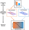
Fig. 6. Schematic of proposed relationships of hippocampal convergence with midbrain VTA neuromodulation.
During the anticipation of high curiosity answers, we propose that neuromodulation by the VTA promotes the consistent engagement of neural states/processes in the hippocampus that supports the formation of new memories. This can manifest as greater convergence in distributed patterns of activity. In the absence of neuromodulatory inputs from the VTA (during low curiosity), patterns of activity in the hippocampus can show greater variability. The increased convergence in hippocampal state during VTA neuromodulation may be supported by the stabilization of specific attractors and the suppression of noise-driven transitions between different possible attractor states. As hippocampal convergence and VTA activation were measured over simultaneous intervals, it is possible that hippocampal convergence supports univariate VTA activation; see text for detailed discussion.

Similar articles

Cited by

References

    1. Kentros CG, Agnihotri NT, Streater S, Hawkins RD, Kandel ER. Increased attention to spatial context increases both place field stability and spatial memory. Neuron. 2004;42:283–295. - PubMed
    1. Kennedy PJ, Shapiro ML. Motivational states activate distinct hippocampal representations to guide goal-directed behaviors. Proc. Natl Acad. Sci. 2009;106:10805–10810. - PMC - PubMed
    1. Bunzeck N, Doeller CF, Fuentemilla L, Dolan RJ, Düzel E. Reward motivation accelerates the onset of neural novelty signals in humans to 85 milliseconds. CURBIO. 2009;19:1294–1300. - PMC - PubMed
    1. Apitz T, Bunzeck N. Reward modulates the neural dynamics of early visual category processing. Neuroimage. 2012;63:1614–1622. - PubMed
    1. Forkmann K, et al. Pain-specific modulation of hippocampal activity and functional connectivity during visual encoding. J. Neurosci. 2013;33:2571–2581. - PMC - PubMed

Publication types