. 2022 Nov;4(11):1459-1473.
doi: 10.1038/s42255-022-00667-w. Epub 2022 Nov 7.

ADRA1A-Gαq signalling potentiates adipocyte thermogenesis through CKB and TNAP

Affiliations

ADRA1A-Gαq signalling potentiates adipocyte thermogenesis through CKB and TNAP

Janane F Rahbani et al. Nat Metab. 2022 Nov.

Abstract

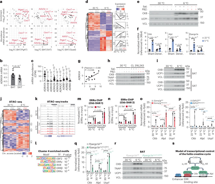
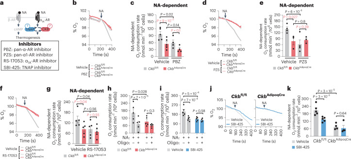
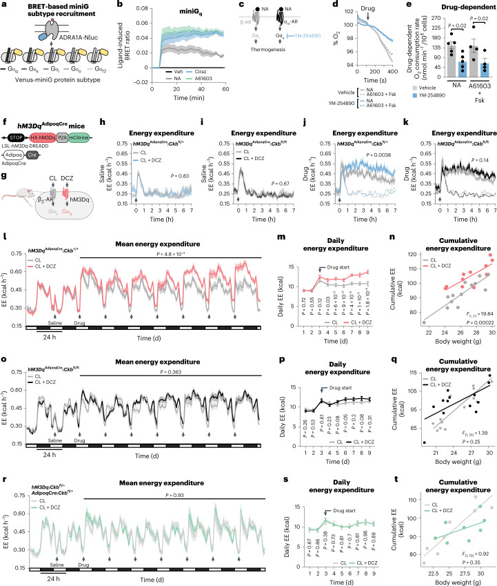
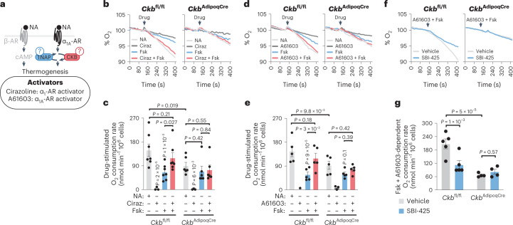
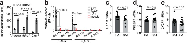
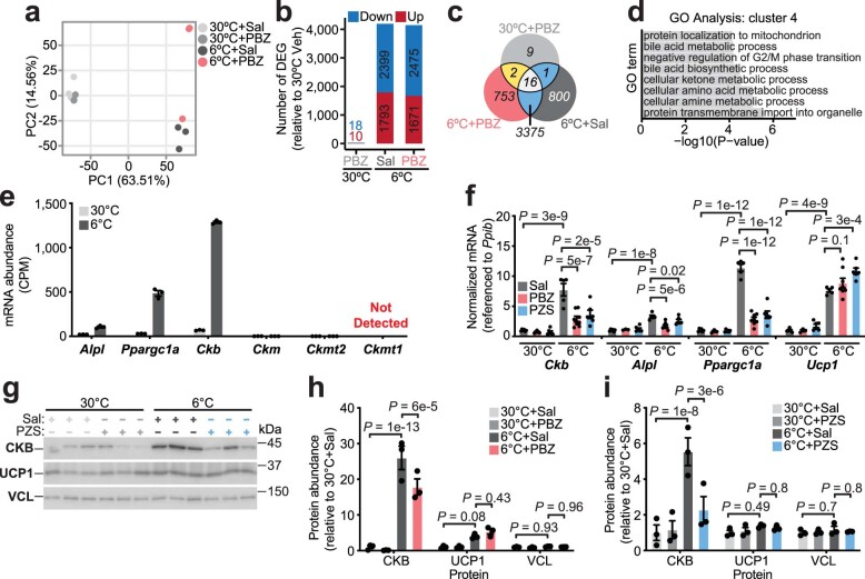
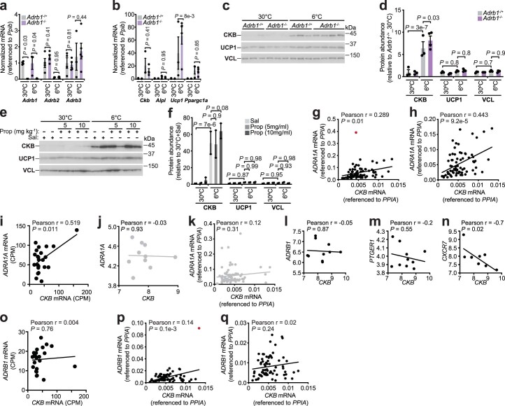
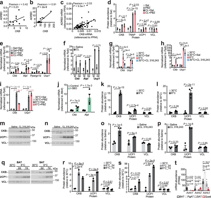
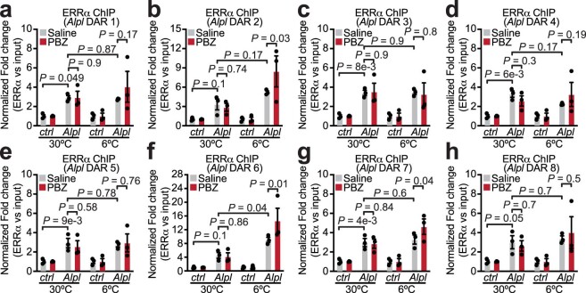
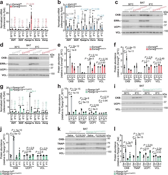
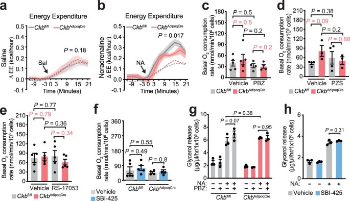
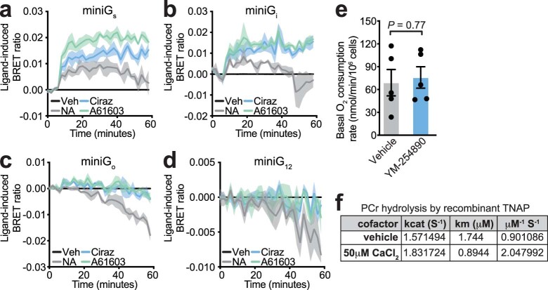
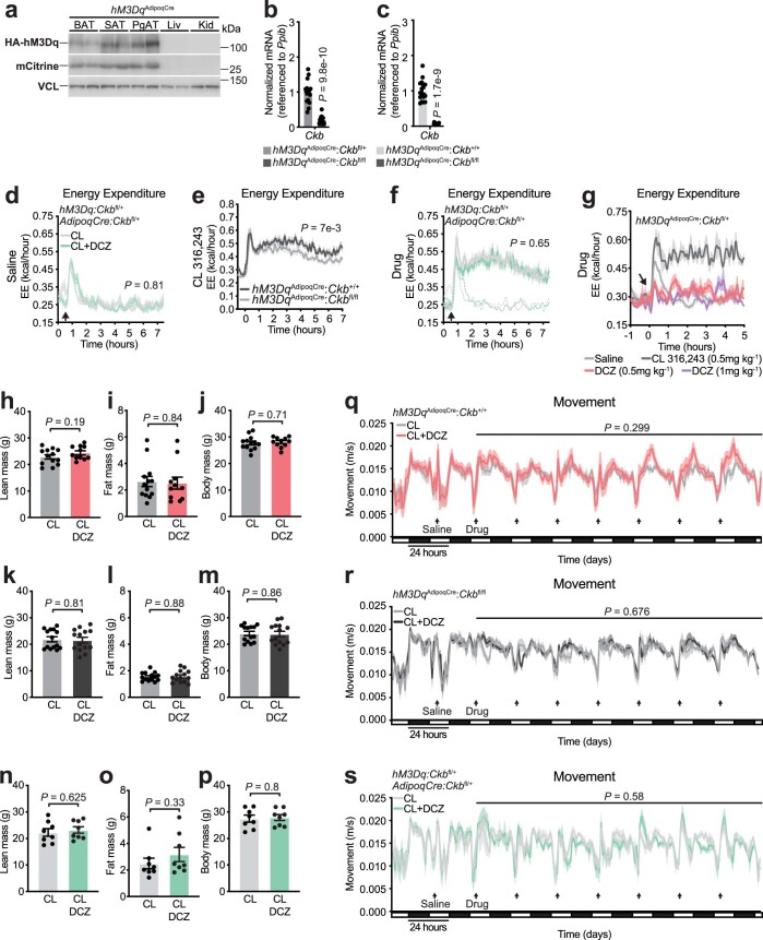
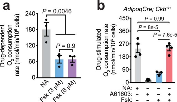
Noradrenaline (NA) regulates cold-stimulated adipocyte thermogenesis1. Aside from cAMP signalling downstream of β-adrenergic receptor activation, how NA promotes thermogenic output is still not fully understood. Here, we show that coordinated α1-adrenergic receptor (AR) and β3-AR signalling induces the expression of thermogenic genes of the futile creatine cycle2,3, and that early B cell factors, oestrogen-related receptors and PGC1α are required for this response in vivo. NA triggers physical and functional coupling between the α1-AR subtype (ADRA1A) and Gαq to promote adipocyte thermogenesis in a manner that is dependent on the effector proteins of the futile creatine cycle, creatine kinase B and tissue-non-specific alkaline phosphatase. Combined Gαq and Gαs signalling selectively in adipocytes promotes a continual rise in whole-body energy expenditure, and creatine kinase B is required for this effect. Thus, the ADRA1A-Gαq-futile creatine cycle axis is a key regulator of facultative and adaptive thermogenesis.


Conflict of interest statement

The authors declare no competing interests.

Figures

Fig. 1
Fig. 1. Regulation of CKB and TNAP expression.
a, BAT-enriched (top; false discovery rate (FDR) < 0.0005) and BAT-abundant (bottom; 10% most abundant) GPCRs. log2FC, log2 fold change; PgAT, perigonadal adipose tissue; Quad, quadriceps. b, ADRA1A expression in human BAT (n = 10) and SAT (n = 10), first cohort. c, mRNA expression in human BAT (n = 23), second cohort. CPM, counts per million. d, Heat map of DEGs in BAT following 24 h of 6 °C exposure. e, Western blot of BAT from mice treated as in d. f, qPCR with reverse transcription (RT–qPCR) of sham or denervated BAT 24 h following 5 °C (n = 4 per group). g, Pearson correlation in human BAT, first cohort (n = 10). h, Western blot from BAT from wild-type (C57BL6/N) male mice, after 48 h of CL 316,243 (1 mg kg−1 body weight) or saline (n = 3 biologically independent samples). i, Western blot from BAT and SAT from wild-type male mice 48 h after 6 °C exposure (n = 4 per group). j, Heat map showing ATAC–seq density of DARs proximal to DEGs from d (n = 3 per group). k, ATAC–seq tracks. Grey shading represents cold-stimulated DARs. l, Motifs of transcription factors enriched at DARs proximal to cluster 4 genes, and present at DARs proximal to both Ckb and Alpl. m,n, ChIP–qPCR of ERRα bound to Ckb DAR 1 (m) and DAR 2 (n) (n = 3 per group). o, RT–qPCR from BAT 24 h after 6 °C exposure (n = 5 per group, females). p, RT–qPCR from BAT following 7 d of 4 °C exposure (n = 3 for Ebf1/2AdipoqCre at 4 °C; n = 4 for all other groups, males). q, RT–qPCR from BAT following 24 h of 6 °C exposure (n = 4 per group, males). r, Western blot of BAT harvested 48 h after 6 °C exposure (females; n = 3 per group). s, Model of transcriptional control of the futile creatine cycle. Data are presented as the mean ± s.e.m. and n indicates the number of biologically independent experiments. b,f, Two-tailed student’s t-tests; c, one-way analysis of variance (ANOVA; Tukey’s post-hoc test); g, Pearson correlation (two-sided); mq, two-way ANOVA (Fisher’s least significant difference (LSD)). Source data
Fig. 2
Fig. 2. Noradrenaline-stimulated thermogenesis requires ADRA1A signalling, CKB and TNAP.
a, Cartoon of approach to determine if α-AR, α1-AR and α1A-AR signalling as well as if CKB and TNAP are necessary for NA-stimulated brown adipocyte thermogenesis. b,d,f,j, Representative basal and NA-stimulated (0.1 μM) oxygen consumption traces of freshly isolated Ckbfl/fl and CkbAdipoqCre brown adipocytes, treated with PBZ (1 μM) (b), PZS (1 μM) (d), RS-17053 (10 μM) (f) or SBI-425 (10 μM) (j), each compared to vehicle control. The time of NA addition (arrow) was normalized to 100% for ease of viewing the representative traces. c,e,g,k, NA-stimulated oxygen consumption rates (above basal) of freshly isolated Ckbfl/fl and CkbAdipoqCre brown adipocytes, treated with PBZ (n = 5 per group) (c), PZS (n = 4 for Ckbfl/fl; n = 3 for CkbAdipoqCre) (e), RS-17053 (n = 5 per group) (g) or SBI-425 (n = 5 SBI-425 and vehicle for Ckbfl/fl; n = 4 SBI-425 and vehicle for CkbAdipoqCre) (k) each compared to vehicle control. In k, the NA-stimulated rates of Ckbfl/fl brown adipocytes (vehicle and SBI-425) are the same as in i, and are shown for comparison to the CkbAdipoqCre brown adipocytes. h,i, ATP synthase-dependent NA-stimulated oxygen consumption rates (above basal) of freshly isolated Ckbfl/fl (n = 5 per group) (h) and CkbAdipoqCre (n = 5 per group) brown adipocytes or Ckbfl/fl brown adipocytes treated with SBI-425 (n = 5 for NA; n = 4 for NA + oligomycin) or vehicle (n = 5 per group) (i). Data are presented as the mean ± s.e.m. and n indicates the number of biologically independent experiments. c,e,g,h,i,k, Two-way ANOVA (Fisher’s LSD). Source data
Fig. 3
Fig. 3. Thermogenesis by combined Gαq and Gαs signalling genetically requires Ckb in vivo.
a, Schematic of BRET assay. b, Agonist-induced BRET in immortalized brown adipocytes (n = 3 per group). c, Cartoon depicting inhibition of Gαq signalling by YM-254890. d, Representative basal and NA-stimulated (0.1 μM) oxygen consumption trace of freshly isolated brown adipocytes, treated with YM-254890 (10 μM) or vehicle. The time of NA addition (arrow) was normalized to 100%. e, NA-stimulated oxygen consumption rates (above basal) of freshly isolated brown adipocytes (n = 5 per group). f, Cartoon of hM3DqAdipoqCre mouse construction. g, Schematic of activation of Gαs and Gαq signalling in adipocytes from hM3DqAdipoqCre mice. h,i, Energy expenditure (EE) with saline injection of hM3DqAdipoqCre:Ckbfl/+ mice (CL group, n = 6 males, 4 females; CL + DCZ group, n = 8 males, 4 females) (h) or hM3DqAdipoqCre:Ckbfl/fl mice (CL group, n = 11 males, 7 females; CL + DCZ group, n = 10 males, 7 females) (i). j,k, EE with drug injection of hM3DqAdipoqCre:Ckbfl/+ mice (CL, n = 6 males, 4 females; CL + DCZ, n = 8 males, 4 females) (j) or hM3DqAdipoqCre:Ckbfl/fl mice (CL, n = 11 males, 7 females; CL + DCZ, n = 10 males, 7 females) (k). ln, Mean EE (l), daily EE (m) and cumulative EE (n) of hM3DqAdipoqCre:Ckb+/+ mice (CL, n = 8 males, 5 females; CL + DCZ, n = 8 males, 3 females). oq, Mean EE (o), daily EE (p) and cumulative EE (q) of hM3DqAdipoqCre:Ckbfl/fl mice (CL, n = 7 males, 7 females; CL + DCZ, n = 7 males, 7 females). rt, Mean EE (r), daily EE (s) and cumulative EE (t) of control (hM3Dq:Ckbfl/+ or AdipoqCre:Ckbfl/+) mice (n = 4 males, 4 females). Data are presented as the mean ± s.e.m. and n indicates the number of biologically independent experiments. e, two-tailed student’s t-test; hm,o,p,r,s, two-way ANOVA (Fisher’s LSD); n,q,t, analysis of covariance (ANCOVA; two-sided, Bonferroni). Source data
Fig. 4
Fig. 4. ADRA1A-mediated signalling potentiates thermogenesis through the futile creatine cycle.
a, Cartoon of approach to study brown adipocyte-intrinsic thermogenesis by individual and combined activation of α1-AR or α1A-AR with cAMP signalling. b,d,f, Representative oxygen consumption traces of freshly isolated Ckbfl/fl and CkbAdipoqCre brown adipocytes. The time of drug addition (arrow) was normalized to 100% for ease of viewing the representative traces. c, Oxygen consumption rates of brown adipocytes treated as in b (Ckbfl/fl: n = 7, 4, 7 and 7 for NA, Ciraz, Fsk and Ciraz + Fsk, respectively; CkbAdipoqCre: n = 6, 3, 6 and 6 for NA, Ciraz, Fsk and Ciraz + Fsk, respectively). e, Oxygen consumption rates of brown adipocytes treated as in d (n = 5, 3, 5 and 5 for NA, Ciraz, Fsk and Ciraz + Fsk, respectively for both genotypes). g, Oxygen consumption rates of brown adipocytes treated as in f (Ckbfl/fl: n = 5; CkbAdipoqCre: n = 4). Data are presented as the mean ± s.e.m. and n indicates the number of biologically independent experiments. c,e,g, Two-way ANOVA (Fisher’s LSD). Source data
Extended Data Fig. 1
Extended Data Fig. 1. α-adrenergic receptor expression in murine and human adipose tissues.
a, Ribosomal profiling of candidate GPCRs from SAT and BAT of 30 °C-acclimated mice (n = 5 per tissue). b, Ribosomal profiling from TRAP of α1AR and α2AR subtypes from different tissues in mice (BAT: n = 3, PgAT: n = 3, SAT: n = 3, QUAD muscle: n = 4). c-e, Relative mRNA expression of (c) ADRB1, (d) PTGER1 and (e) CXCR7 from human BAT (adipose tissue proximal to carotid sheath) (n = 10) and paired SAT (n = 10), first cohort. Data are presented as mean ± s.e.m. and n numbers are of biologically independent experiments. a, c-e, two-tailed student’s t-tests; b, one-way ANOVA (Fisher’s LSD). Source data
Extended Data Fig. 2
Extended Data Fig. 2. Regulation of the cold-stimulated BAT transcriptome by α-adrenergic receptor signalling.
a, Principal component analysis (PCA) of gene expression from BAT transcriptomics of Fig. 1d. Percent of variance explained by each PC is shown. Phenoxybenzamine (PBZ) and Saline (Sal). b, Barplot showing the number of down-regulated and up-regulated genes, using “30 °C + Sal” condition as baseline from BAT transcriptomics of Fig. 1d. Differentially expressed genes (DEGs) are identified using cutoffs of (log2FC > 1 and FDR < 0.01). c, Venn diagram showing the overlap of DEGs between the 30 °C + PBZ (light gray), 6 °C + Sal (dark gray) and 6 °C + PBZ (red) from BAT transcriptomics of Fig. 1d. d, GO Term enrichment of Cluster 4 genes from BAT transcriptomics of Fig. 1d. e, RNA-seq of BAT transcriptomics of Fig. 1d. CPM (counts per million). f, RT-qPCR from BAT of wild-type male mice (C57BL6/N, 6–8 weeks of age), treated as in Fig. 1d. Prazosin (PZS) was injected (3 times over 24 hours) intraperitoneally (i.p.) at 5 mg kg−1 (30 °C: n = 6 Sal, n = 3 PBZ, n = 6 PZS) (6 °C: n = 5 Sal, n = 8 PBZ, n = 6 PZS). g, Western blot of BAT from wild-type male mice (C57BL6/N, 6–8 weeks of age), reared at 22 °C, housed at 30 °C for 5 days and then subjected to 30 °C or 6 °C at ZT4. 1 hour prior to onset of 6 °C exposure (ZT3), mice were injected i.p. with PZS (5 mg kg−1, injected 3 times over 24 hours) or Sal. BAT was harvested 24 hours following 6 °C exposure (n = 3 per group). h-i, Quantification of western blots from (h) Fig. 1e (n = 3 per group) and (i) Extended Data Fig. 2g (n = 3 per group). Data are presented as mean ± s.e.m. and n numbers are of biologically independent experiments. f, One-way ANOVA (Fisher’s LSD); h, i, Two-way ANOVA (Fisher’s LSD). Source data
Extended Data Fig. 3
Extended Data Fig. 3. ADRA1A, not ADRB1, correlates with, CKB abundance in human BAT.
a-b, RT-qPCR analysis of BAT from Adrb1-/+ and Adrb1-/- female mice reared at 22 °C, housed at 30 °C for 5 days and then subjected to 30 °C or 6 °C at ZT4 for 24 hours (30 °C: n = 4 per genotype) (6 °C: n = 3 Adrb1-/+, n = 5 Adrb1-/-). c, Western blot from BAT of mice treated as in Extended Data Fig. 3a, b (30 °C: n = 3 per genotype) (6 °C: n = 3 Adrb1-/+, n = 4 Adrb1-/-). d, Quantification of western blots from Extended Data Fig. 3c (30 °C: n = 3 per genotype) (6 °C: n = 3 Adrb1-/+, n = 4 Adrb1-/-). e, Western blot from BAT of wild-type male mice (C57BL6/N, 6–8 weeks of age), reared at 22 °C, housed at 30 °C for 5 days and then subjected to 30 °C or 6 °C at ZT4. 1 hour prior to onset of 6 °C exposure (ZT3), mice were injected i.p. with propranolol hydrochloride (5 mg kg−1 or 10 mg kg−1) or saline (Sal). BAT was harvested 24 hours following 6 °C exposure (n = 2 per group). f, Quantification of western blots from Extended Data Fig. 3e (n = 2 per group). g, Pearson correlation of CKB with ADRA1A mRNA in human BAT (n = 73), third cohort. Red data point indicates outlier based on ROUT method (Q = 0.1%). h, Pearson correlation with outlier identified in Extended Data Fig. 3g removed. i, Pearson correlation of CKB with ADRA1A mRNA in human BAT (supraclavicular adipose tissue) (n = 23), second cohort. j, k, Pearson correlation of CKB with ADRA1A mRNA in (j) human SAT (superficial subcutaneous adipose tissue) (n = 10), first cohort and (k) human SAT (superficial neck adipose tissue) (n = 73), third cohort. l-n, Pearson correlation of CKB with (l) ADRB1, (m) PTGER1 and (n) CXCR7 mRNA in human BAT (adipose tissue proximal to carotid sheath) (n = 10), first cohort. o, Pearson correlation of CKB with ADRB1 mRNA in human BAT (supraclavicular adipose tissue) (n = 23), second cohort. p, Pearson correlation of CKB with ADRB1 mRNA in human BAT (n = 73), third cohort. Red data point indicates outlier based on ROUT method (Q = 0.1%). q, Pearson correlation with outlier identified in Extended Data Fig. 3p removed. Data are presented as mean ± s.e.m. and n numbers are of biologically independent experiments. a, b, d, f, Two-way ANOVA (Fisher’s LSD); g-q, Pearson correlation (two-sided). Source data
Extended Data Fig. 4
Extended Data Fig. 4. Adrb3, and Gαs signaling, promote Ckb and Alpl expression.
a-c, Pearson correlations of CKB with ADRB3 mRNA in human BAT from the (a) first cohort (n = 10), (b) second cohort (n = 23), and (c) third cohort (n = 73). d, Western blot quantification from Fig. 1h (n = 3 per group). e, RT-qPCR of BAT from mice treated as in Fig. 1h (30 °C: n = 4 Sal, n = 4 PBZ) (6 °C: n = 4 Sal, n = 3 PBZ). f, intraperitoneal (ip) injections (5 injections over 48 hours) with CL 316,243 (1 mg kg−1) or Sal compared to osmotic pump (op) implantations above the interscapular BAT with CL 316,243 or Sal (n = 3 per group). g-h, RT-qPCR of (g) BAT and (h) SAT from wild-type male mice (C57BL6/N, 6–8 weeks of age), subjected to 30 °C or 6 °C (n = 4 per group) or injected i.p. (5 injections over 48 hours) with CL 316,243 or Sal at 30 °C (n = 6 per group). i, RT-qPCR analysis of BAT from wild-type male mice (C57BL6/N, 6–8 weeks of age), treated as in Fig. 1d (30 °C: n = 3 Sal, n = 3 PBZ) (6 °C: n = 5 Sal, n = 3 PBZ). j, RT-qPCR of BAT from I-3BO (n = 4) and control mice (n = 6). k-l, Western blot quantification from (k) BAT (n = 4 per group) and (l) SAT (n = 4 per group) from Fig. 1i. m-n, Western blot analysis of (m) BAT and (n) SAT from wild-type male mice (C57BL6/N, 6–8 weeks of age), reared at 22 °C, housed at 30 °C for 5 days and then injected i.p. (5 injections over 48 hours) with CL 316,243 or Sal (n = 3 per group). o-p, Western blot quantification from (o) Extended Data Fig. 4m (n = 3 per group) and (p) Extended Data Fig. 4n (n = 3 per group). q, Western blot from BAT and SAT of male wild-type mice (C57BL6/N, 6–8 weeks of age) subjected to 30 °C or 6 °C for 2 days (2d) or 7 days (7d) (n = 2 per group). r-s, Western blot quantification of Extended Data Fig. 4q from (r) BAT (n = 2 per group) and (s) SAT (n = 2 per group). t, Ribosomal profiling of βAR expression (BAT n = 3, SAT n = 3, PgAT n = 3, Quad muscle n = 4). Data are presented as mean ± s.e.m. and n numbers are of biologically independent experiments. a-c, Pearson correlation (two-sided); d, f, i, k, l, o, p, r, s, t, Two-way ANOVA (Fisher’s LSD); e, g, h, One-way ANOVA (Fisher’s LSD); j, two-tailed student’s t-tests. Source data
Extended Data Fig. 5
Extended Data Fig. 5. ChIP-qPCR of ERRα bound to cold-responsive DARs near Alpl.
a-h, Chromatin immunoprecipitation coupled to qPCR (ChIP-qPCR) of ERRα bound to Alpl (a) DAR 1, (b) DAR 2, (c) DAR 3, (d) DAR 4, (e) DAR 5, (f) DAR 6, (g) DAR 7, and (h) DAR 8. Wild-type male mice (C57BL6/N, 6–8 weeks of age) were reared at 22 °C, housed at 30 °C for 5 days and then subjected to 30 °C or 6 °C at ZT4. 1 hour prior to onset of 6 °C exposure (ZT3), mice were injected intraperitoneally (i.p.) with PBZ (5 mg kg−1) or Saline (n = 3 per group). Chromatin was prepared for ERRα ChIP from freshly isolated BAT 24 hours after onset of 6 °C exposure. Data are presented as mean ± s.e.m. and n numbers are of biologically independent experiments. a-h, Two-way ANOVA (Fisher’s LSD). Source data
Extended Data Fig. 6
Extended Data Fig. 6. Transcriptional control of cold-stimulated futile creatine cycling gene expression.
a-b, RT-qPCR from BAT of male (a) Esrra/gAdipoqCre and Esrra/gfl/fl mice (6 °C for 24 hours) (n = 5 per group) and (b) Ebf1/2AdipoqCre and Ebf1/2fl/fl mice (4 °C for 7 days) (n = 3 for Ebf1/2AdipoqCre at 4 °C; n = 4 for all other groups). c-d, Western blot analysis of BAT from (c) male (Esrra/gfl/fl: n = 2 for 30 °C, n = 3 for 6 °C) (Esrra/gAdipoqCre: n = 3 for 30 °C, n = 4 for 6 °C) and (d) female mice (n = 3 per group) subjected to 30 °C or 6 °C for 24 hours. e-f, Western blot quantification from (e) Extended Data Fig. 6c (Esrra/gfl/fl: n = 2 for 30 °C, n = 3 for 6 °C) (Esrra/gAdipoqCre: n = 3 for 30 °C, n = 4 for 6 °C) and (f) Extended Data Fig. 6d (n = 3 per group). g, RT-qPCR from BAT of Ppargc1aUcp1CreERT2 and Ppargc1afl/fl mice, reared at 22 °C, housed at 30 °C for 5 days and then subjected to 30 °C or 6 °C for 24 hours (n = 4 per group). h, Western blot quantification from Fig. 1r (n = 3 per group). i, Western blot of BAT from male mice (6–8 weeks of age), treated as in Extended Data Fig. 6g. j, Western blot quantification from Extended Data Fig. 6i (n = 3 per group). k, Western blot analysis of BAT from female mice (6–8 weeks of age), reared at 22 °C, housed at 30 °C for 5 days and then injected i.p. (5 injections over 48 hours) with CL 316,243 (1 mg kg−1) or saline at 30 °C (n = 3 per group). l, Western blot quantification from Extended Data Fig. 6k (n = 3 per group). Data are presented as mean ± s.e.m. and n numbers are of biologically independent experiments. a, b, g, multiple two-tailed student’s t-tests (Holm-Šidák test); e, f, h, j, l, Two-way ANOVA (Fisher’s LSD). Source data
Extended Data Fig. 7
Extended Data Fig. 7. Basal respiration and lipolysis are not effected by inhibition of CKB, TNAP or αAR signaling.
a-b, Energy expenditure (EE) of Ckbfl/fl and CkbAdipoqCre male mice (n = 8 per group) reared at 22 °C and then housed for 5 days at 30 °C prior to administration of (a) saline (Sal) or (b) noradrenaline (NA, 1 mg kg−1). c-f, Basal oxygen consumption rates (related to experiments in Fig. 2b–g, j, k) of freshly isolated Ckbfl/fl and CkbAdipoqCre brown adipocytes, treated with (c) PBZ (n = 5 per group), (d) PZS (n = 4 Ckbfl/fl; n = 3 CkbAdipoqCre), (e) RS-17053 (n = 5 per group), or (f) SBI-425 (Ckbfl/fl: n = 5 per group; CkbAdipoqCre: n = 4 per group). g-h, Glycerol release from freshly isolated (g) Ckbfl/fl and CkbAdipoqCre brown adipocytes treated with PBZ compared to vehicle (n = 3 per group) or (h) brown adipocytes treated with SBI-425 compared to vehicle (n = 3 per group). Data are presented as mean ± s.e.m. and n numbers are of biologically independent experiments. a, b, Two-way ANOVA (Fisher’s LSD) from minutes 0 to 21; c-g, Two-way ANOVA (Fisher’s LSD); h, One-way ANOVA (Fisher’s LSD). Source data
Extended Data Fig. 8
Extended Data Fig. 8. Physical and functional coupling of ADRA1A to Gαq.
a-d, agonist-induced BRET between ADRA1A-tagged Nano Luciferase (ADRA1A-Nluc) and Venus-tagged (a) miniGαs, (b) miniGαi, (c) miniGαo, and (d) miniGα12 protein sensors in immortalized brown adipocytes (n = 3 per group). e, Basal oxygen consumption rates (related to experiments in Fig. 3d, e) of freshly isolated brown adipocytes treated with YM-254890 compared to vehicle (n = 5 per group). f, Catalytic properties of recombinant TNAP (n = 3 per group). Data are presented as mean ± s.e.m. and n numbers are of biologically independent experiments. e, two-tailed student’s t-test. Source data
Extended Data Fig. 9
Extended Data Fig. 9. Energy expenditure by Gαs and Gαq signaling in vivo.
a, Western blot of BAT, SAT, PgAT, liver (Liv) and kidney (Kid) (n = 2 per group). b, RT-qPCR from BAT (hM3DqAdipoqCre:Ckbfl/+: n = 17, 9 males, 8 females) (hM3DqAdipoqCre:Ckbfl/fl: n = 14, 6 males, 8 females). c, RT-qPCR from BAT (n = 16: 8 males, 8 females per genotype). d, EE of hM3Dq:Ckbfl/+ or AdipoqCre:Ckbfl/+ mice injected i.p. with saline (n = 12: 8 males, 4 females for both groups). e, EE of hM3DqAdipoqCre:Ckb+/+ (n = 14: 8 males, 6 females) and hM3DqAdipoqCre:Ckbfl/fl (n = 16: 8 males, 8 females) following i.p. injection of CL (0.5 mg kg−1). f, EE of hM3Dq:Ckbfl/+ or AdipoqCre:Ckbfl/+ mice injected i.p. with CL or CL + DCZ (n = 12: 8 males, 4 females for both groups). g, EE of hM3DqAdipoqCre:Ckbfl/+ male mice in response to saline (n = 16), CL (n = 8), DCZ (0.5 mg kg−1) (n = 8), or DCZ (1 mg kg−1) (n = 8). h-j, Baseline (h) lean mass, (i) fat mass, and (j) body mass of hM3DqAdipoqCre:Ckb+/+ mice (CL: n = 13: 8 males, 5 females; CL + DCZ: n = 12: 8 males, 3 females). k-m, Baseline (k) lean mass, (l) fat mass, and (m) body mass of hM3DqAdipoqCre:Ckbfl/fl mice (CL: n = 14: 7 males, 7 females; CL + DCZ: n = 14: 7 males, 7 females). n-p, Baseline (n) lean mass, (o) fat mass, and (p) body mass of hM3Dq:Ckbfl/+ or AdipoqCre:Ckbfl/+ mice (n = 8 per group: 4 males, 4 females). q-s, movement of (q) hM3DqAdipoqCre:Ckb+/+ mice (CL: n = 13: 8 males, 5 females; CL + DCZ: n = 11: 8 males, 3 females), (r) hM3DqAdipoqCre:Ckbfl/fl mice (CL: n = 14: 7 males, 7 females; CL + DCZ: n = 14: 7 males, 7 females), and (s) control (hM3Dq:Ckbfl/+ or AdipoqCre:Ckbfl/+) mice (n = 8 per group: 4 males, 4 females). Data are presented as mean ± s.e.m. and n numbers are of biologically independent experiments. b, c, h-p two-tailed student’s t-test; d-f, q-s, Two-way ANOVA (Fisher’s LSD). Source data
Extended Data Fig. 10
Extended Data Fig. 10. cAMP signaling is not equivalent to NA-stimulated respiration.
a, Oxygen consumption rates from wild-type (C57BL6/N) brown adipocytes, treated with NA (0.1 μM) or forskolin (Fsk, 3 μM and 6 μM) (n = 3 per group). b, Oxygen consumption rates of freshly isolated brown adipocytes, treated with NA (0.1 μM), forskolin (Fsk, 3 μM), A616031AAR agonist, 1 μM), or A61603 + Fsk (n = 4, n = 4, n = 3, and n = 4 for NA, A61603, Fsk and A61603 + Fsk, respectively). Data are presented as mean ± s.e.m. and n numbers are of biologically independent experiments. a, b, One-way ANOVA (Fisher’s LSD). Source data

References

    1. Cannon B, Nedergaard J. Nonshivering thermogenesis and its adequate measurement in metabolic studies. J. Exp. Biol. 2011;214:242–253. - PubMed
    1. Rahbani JF, et al. Creatine kinase B controls futile creatine cycling in thermogenic fat. Nature. 2021;590:480–485. - PMC - PubMed
    1. Sun Y, et al. Mitochondrial TNAP controls thermogenesis by hydrolysis of phosphocreatine. Nature. 2021 doi: 10.1038/s41586-021-03533-z. - DOI - PMC - PubMed
    1. Hsieh AC, Carlson LD. Role of adrenaline and noradrenaline in chemical regulation of heat production. Am. J. Physiol. 1957;190:243–246. - PubMed
    1. Derry DM, Schonbaum E, Steiner G. Two sympathetic nerve supplies to brown adipose tissue of the rat. Can. J. Physiol. Pharmacol. 1969;47:57–63. - PubMed

Publication types

MeSH terms