. 2022 Nov;29(11):1080-1091.
doi: 10.1038/s41594-022-00852-1. Epub 2022 Nov 7.

Cryo-EM structures of prefusion SIV envelope trimer

Affiliations

Cryo-EM structures of prefusion SIV envelope trimer

Jason Gorman et al. Nat Struct Mol Biol. 2022 Nov.

Abstract

Simian immunodeficiency viruses (SIVs) are lentiviruses that naturally infect non-human primates of African origin and seeded cross-species transmissions of HIV-1 and HIV-2. Here we report prefusion stabilization and cryo-EM structures of soluble envelope (Env) trimers from rhesus macaque SIV (SIVmac) in complex with neutralizing antibodies. These structures provide residue-level definition for SIV-specific disulfide-bonded variable loops (V1 and V2), which we used to delineate variable-loop coverage of the Env trimer. The defined variable loops enabled us to investigate assembled Env-glycan shields throughout SIV, which we found to comprise both N- and O-linked glycans, the latter emanating from V1 inserts, which bound the O-link-specific lectin jacalin. We also investigated in situ SIVmac-Env trimers on virions, determining cryo-electron tomography structures at subnanometer resolutions for an antibody-bound complex and a ligand-free state. Collectively, these structures define the prefusion-closed structure of the SIV-Env trimer and delineate variable-loop and glycan-shielding mechanisms of immune evasion conserved throughout SIV evolution.


Conflict of interest statement

Competing interests

The authors declare no competing interests.

Figures
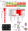
Extended Data Fig. 1 |
Extended Data Fig. 1 |. Antigenic screening and HRV 3 C purification through capture of co-expression enriches for minority population of trimeric Env.
a, Sequence homology between HIV-1 and several SIV strains is shown. b, The 96-well screening results are presented as red-white heatmap with the top scoring constructs enlarged on the right panel showing their specific values for HIV-1 and SIV antibodies. The bottom-right table summarized the screening antibody epitopes. The ITS92 epitope was unknown during the screening and the accompanying structures revealed it to be a gp41 conformationally dependent epitope. c, Negative stain 2D images show a small population of trimers, highlighted here in the green squares. d, Schematic of the purification strategy is shown with on column binding of the supernatant to ITS92.02 followed by HRV 3 C cleavage, allowing the avoidance of harsh purification buffers. A similar strategy was used for PGT145 but included co-expression with the HRV 3 C IgG.
Extended Data Fig. 2 |
Extended Data Fig. 2 |. Cryo-EM analysis of SIVE660.CR54 SOS-2P in complex with ITS92.02 reveals a trimer with high flexibility at the apex.
a, Representative micrograph and CTF of the micrograph are shown. 8,259 micrographs were collected in total. b, Representative 2D class averages are shown. c, The gold-standard Fourier shell correlation resulted in a resolution of 4.32 Å using non-uniform refinement with C3 symmetry. d, The orientations of all particles used in the final refinement are shown as a heatmap. e, The local resolution of the full map is shown generated through cryoSPARC using an FSC cutoff of 0.5. f, A wild-type sequence alignment near the ITS92.02 epitope is shown highlighting residues within a 5 Å footprint in red. We note that non-conserved residue E613 forms a salt bridge with R94 of the ITS92.02 heavy chain. g, The prefusion (magenta) and postfusion (gray, PDB ID 1QBZ) conformations of gp41 protomers are aligned with the prefusion footprint residues colored red and the corresponding residues in the postfusion conformation are shown in blue. Footprint residues between positions 638 and 652 (HXB2 numbering) align well, residues from 608–614 extend away from the helical region of the epitope.
Extended Data Fig. 3 |
Extended Data Fig. 3 |. Cryo-EM analysis of SIVmac239 SOS-2P in complex with PGT145 contains a stabilized apex region.
a, Representative micrograph and CTF of the micrograph are shown. 6,255 micrographs total were collected. b, Representative 2D class averages are shown. c, The gold-standard Fourier shell correlation resulted in a resolution of 4.12 Å using non-uniform refinement with C1 symmetry. d, The orientations of all particles used in the final refinement are shown as a heatmap. e, The local resolution of the full map is shown in two contours. Maps were generated through cryoSPARC using an FSC cutoff of 0.5.
Extended Data Fig. 4 |
Extended Data Fig. 4 |. SIVmac239 sequence and structural comparisons.
a, The SIVmac239 trimer aligns well to the CD4-bound SIVmac239 core (PDB: ID 6TYB). The glycan shield shows strong similarity for most glycans available in the core with notable differences at positions 47 and 464 where sequons were not glycosylated in the core and position 88 where the gp41 glycan at position N625 overlaps in space with N88 of the core. b, A sequence alignment of HIV-1 (BG505) and SIV (mac239) are shown with secondary structure shown below. Notable positions are indicated for SOSIP mutations. c, The Cα residue-by-residue distances are shown for HIV-1 and SIVcpz compared to residues of SIVmac239 indicating areas of close structural alignment and highlighting regions that diverge significantly.
Extended Data Fig. 5 |
Extended Data Fig. 5 |. CD4 binding details in SIVmac239 and HIV-1.
a, (left) SIVmac239 gp120 from the trimer structure (magenta) is shown aligned to the CD4-bound SIVmac239 gp120 core (gray). (right) SIVmac239 gp120 from the trimer structure (magenta) is shown aligned to an HIV-1 BG505 gp120 from a trimer (light blue). b, (left) The CD4 (yellow) binding site is shown corresponding to the structural alignments in panel a. W427 adjusts out of the pocket that the F43 of CD4 inserts into. (right) Residue 375, used to confer rhCD4 binding to SHIV, is shown in red. Relative positions of W427 in SIV and HIV-1 are shown proximal to the F43 pocket. c, Conformations of the region from residues 50–80 are shown. The region adopts various conformations in SIV and HIV-1 and undergoes a switch upon CD4 binding.
Extended Data Fig. 6 |
Extended Data Fig. 6 |. Details of the primate immunodeficiency viruses conserved features.
a, An additional disulfide was observed in one branch which placed cys at residues 165 and 168, stabilizing the turn between the B and C strands of V1V2. b, Disulfides observed in the hypervariable V1 loop are highlighted in purple. Due to poor alignment confidence in this region the sequences are shown from residue cys157 conserved in all viruses in the tree. c, The HIV-2 glycan shield is modeled as in Fig. 5e with minor deviations from SIVmac239. d, The fusion peptide region of SIVmac239 adopts a helical structure and abuts the core of the Env contrary to the flexible conformations observed in HIV-1 Env (BG505, PDB ID: 5FYL). We note that the FP is one residue longer in the viruses more proximal to HIV-1.
Extended Data Fig. 7 |
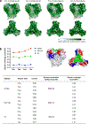
Extended Data Fig. 7 |. Impact of O-linked saccharides on glycan shielding.
a, Glycan shielding overlaid on SIVmac239 surfaces viewing from side (top panel) and top (bottom panel). b, Glycan coverage of epitope CD4bs (CD4 binding site), PGT145 and V3 regions of SIVmac239. The epitope regions are shown (top middle and top right panels) with the same color code as the plot (top left).
Extended Data Fig. 8 |
Extended Data Fig. 8 |. N-linked glycan conservation across diverse primate immunodeficiency viruses.
(top) Bar graph depicting the percent conservation of N-linked glycan sequon positions (HXB2 numbering from 1–683) for the 34 viruses depicted in the phylogenetic tree of Fig. 4. Hypervariable regions extending beyond HXB2 numbering are excluded due to high variability and low confidence alignments. * at positions 143 and 185 denotes artificially high bars due to multiple glycans in inserts. Dotted horizontal lines are shown at 50% and 75% conservation and locations above these thresholds are labeled. (bottom) A detailed plot of sequon locations for all 34 sequences in the phylogenetic tree of Fig. 4. Locations are colored as in Fig. 4b.
Extended Data Fig. 9 |
Extended Data Fig. 9 |. Cryo-ET density fit to the model with MPER region extended from the membrane.
a, A slice through a representative tomogram shows multiple SIV virions with Env spikes, black scale bar in the bottom right represents 50 nm. 55 tomograms of SIVmac239 and 105 tomograms of SIVmac239 complex with ITS90.03 were collected. b, FSC curves of the ITS90.03-bound (blue) and unliganded SIV (black) sub-tomogram averages. c, Superposition of the ITS90.03 bound crystal structure with that of the SIV trimer based on the gp120 alignment. d, Glycan N625 of the SIVmac239 trimer must re-orient to accommodate the binding of ITS90.03. e, The V1 insertion region is observed in the ITS90.03-bound and unliganded cryo-ET density. f, Side view of the gp41 density with the gp41 built from the soluble map fit to the density. g, Side view to panel d is shown. h, The pedestal at the base of the gp41 region in HIV-1 Bal is shown in yellow, segmented from density of EMDB map EMD-21412. Coordinates are shown for BG505 SOSIP.664. i, The side of gp41 is shown with the MPER region density shown in gold. j, The fit of the MPER region in relation to the membrane surface density. k, The pedestal at the base of the gp41 region in HIV is shown in yellow with membrane density shown.
Fig. 1 |
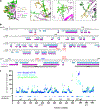
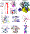
Fig. 1 |. Stabilization of SIVE660.CR54 Env through ITS92.02 screening-enabled cryo-EM structure determination.
a, Left: designs of stabilized soluble SIV trimers based on HIV-1 structures were assessed through antigenic screening of a 96-well plate. Middle: antigenicity for the designs, shown as a heatmap, with each designed construct as a row and each antibody as a column, with ITS92.02 on the rightmost column (Extended Data Fig. 1b). Right: introduction of a proline mutation at 569, in addition to the proline mutation of SOSIP at 559, showed the best binding to ITS92.02. b, SIVCR54 SOS-2P with ITS92.02 cryo-EM density. Density at the apex is less well-defined than the core and gp41 regions. c, The atomic model of the structure is shown in cartoon format. d, Density at the ITS92.02 epitope, shown with the model. e, The ITS92.02 epitope is highlighted in red and shown in the bound-prefusion conformation (looking up from the membrane) of SIVE660.CR54 and the postfusion conformation (side view; PDB 1QBZ) of SIVmac239. f, The density of the SIV trimer (left) did not allow for unambiguous model-building of the apex region, including the V1V2 and V3 domains, which are highlighted in HIV-1 (right; PDB 4TVP) in blue and orange, respectively.
Fig. 2 |
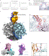
Fig. 2 |. Structure of stabilized SIVmac239 Env trimer in a prefusion conformation bound to PGT145.
a, PGT145 neutralization curves are shown for three SIV viruses. WT SIVmac251.30, which has a native Thr at position 169, is neutralized. The error bars indicate the s.d. of duplicate measurements. b, The 169S/T mutation at the trimer apex required for PGT145 to bind and neutralize SIV is shown in red. c, Cryo-EM density at 4.1 Å, shown for SIVmac239 SOS-2P trimer with the K169T mutation, in complex with PGT145. d, Density at the apex allowed unambiguous model-building for the trimer and PGT145 CDR H3. e, The overall structure of the SIV Env trimer looking down the three-fold axis shows the three-blade propeller. gp120 and gp41 for each protomer are colored individually and labeled. f, Cα alignment of SIVmac239 protomer with HIV-1 (PDB 4TVP) and SIVcpz (PDB 6OHY). g, Modeling the native Lys at position 169 shows clashes with residues R100a and F100d of the PGT145 CDR H3, which are not seen in the K169T mutant structure.
Fig. 3 |
Fig. 3 |. SIV antigenic evasion through extended variable loops.
a, Sequence alignment of HIV-1 and SIVmac239 V1V2 region, marking an extended SIV V1 loop with predicted O-linked glycosylation sites (POGs) in red. N-linked glycans are shown in gold and green for HIV-1 and SIV, respectively. b, Surface representation of HIV-1 (gray) with cartoon representation of SIVmac239 (magenta), highlighting the extended surface coverage by the SIV V1 and V4 loops, POGs are colored in red and V4 in blue. c, The surface region of the HIV-1 N332 (gold) V3-glycan epitope (cyan) is occluded by the SIV V1 loop. This region is marked by a cyan box in b. d, The SIVmac239 V1V2 region, highlighting the N-linked (green) and O-linked (red) glycosylation sites. The two disulfides not found in HIV-1 are shown in dark purple, with the resulting loops shown in thicker tube radius. e, The V4 loop of SIVmac239 is oriented towards the apex covering the region protected by glycans N363, N386 and N392 in HIV-1. The HIV-1 V4 is oriented away from the apex (gray, PDB 2B4C).
Fig. 4 |
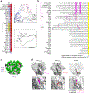
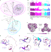
Fig. 4 |. Conserved sequence features in primate immunodeficiency viruses observed in the SIVmac239 structure.
a, A phylogenetic tree of representative Env sequences from HIV-1, HIV-2 and SIV. Branches that conserve features such as the a0 insert and the V1 and V2 disulfides are highlighted. All branches except HIV-1 and SIVcpz are predicted to contain O-linked glycans. b, Amino acid lengths of the V1 (C131 and C157) and V2 (C183–C191) hypervariable regions and the number of N-linked sequons for the full Env. c, SIV sequence entropy, mapped to the surface of SIVmac239 with high sequence conservation in white and low sequence conservation in purple. d, The V1 hypervariable loops of HIV-1, SIVcpz and SIVmac239, overlayed with residues between the conserved disulfide bond at positions C131 and C157 following strand B and preceding strand C. The disulfide in SIVmac239 is shown in purple. e, The V2 regions from 179 to 191 are shown with an additional disulfide in SIVmac239 at positions 183 and 191, which are Gln and Tyr in HIV-1BG505 or Ala and Tyr in SIVcpzMT145K, respectively. BG505 structures are typically disordered in parts of this loop. f, The region proximal to α0 contains a seven-residue insert in HIV-1 and SIVcpz that is not observed in the majority of SIVs.
Fig. 5 |
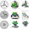
Fig. 5 |. The SIV glycan shield contains N- and O-linked glycans.
a, Cryo-EM density, with green regions corresponding to N-linked glycans and red regions O-linked glycans. b, Density, shown as mesh, surrounding V1 Thr residues predicted to have O-linked glycosylation. The resolution of the density in this region was not high enough to build with atomic-level certainty, but the level of excess density strongly suggested there were heterogeneously glycosylated residues in the loop. c, Full glycan shield for HIV-1 BG505 (gold; PDB 5FYL) and SIV mac239 (green and red), with some direct overlap (that is, N160, N197), some neighboring overlap (that is, SIV N229 with HIV-1 N241) and non-overlapping coverage (that is, SIV N47). d, Cryo-EM density of the PGT145-bound SIVmac239 trimer in complex with the O-linked glycan-binding jacalin is shown as transparent, with the atomic model fit and V1 loops colored in red. e, Modeled glycan shields based on SIV-HIV templates. Glycans are colored green if present in SIVmac239 and otherwise colored as in a and b by their initial appearance. Glycans that arise in hypervariable regions of V1, V2 or V4 or those shifted within two residues or less of a previously present glycan are not recolored. Sequence identity to SIVmac239 of the Env (31 to 664 by HXB2 numbering) is shown below, with other features of the related viruses, including alternate V1 disulfides (Extended Data Fig. 6) and POGS calculated from NetOGlyc-4.0 with a conservative cutoff score of over 0.75.
Fig. 6 |
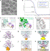
Fig. 6 |. In situ cryo-ET of SIVmac239 Env trimer on virions confirms the prefusion-stabilized trimer as the conformation targeted by neutralizing antibodies.
a, A slice of the sub-tomogram average of the ITS90.03-bound SIVmac239 Env on an AT-2-inactivated virion is shown from a view at the apex of the Env looking down the three-fold axis. b, A sub-tomogram average slice is shown, as in a, but rotated 90° to show the side view. c, Cryo-ET volume density, shown from the same view, with each protomer colored as gray, pink and blue. N-linked glycans are colored green, and potential O-linked glycan sites are in red. The neutralizing antibody ITS90.03 is colored purple and tan for the light and heavy chain, respectively. d, As in c, but rotated 90° to a side view to show the angle of approach of ITS90.03. e, The cryo-ET density is shown as a transparent surface with the trimeric atomic model fit. The ITS90.03 antibody is shown from the structure PDB 6TYB after alignment with the trimer by the gp120 core region. f, View of the ITS90.03 crystal structure PDB 6TYB docked into the cryo-ET density. g, A slice of a sub-tomogram average of the SIV ligand-free trimer, displayed in the same orientation as a. h, Cryo-ET volume of the ligand-free trimer as in e. i, The model from the soluble trimer is fit to the density. Extra density is observed for the MPER region, displacing the Env from the membrane by over 10 Å. Little density is observed for residues 512–569.

References

    1. Daniel MD et al. Isolation of T-cell tropic HTLV-III-like retrovirus from macaques. Science 228, 1201–1204 (1985). - PubMed
    1. Kanki PJ et al. Serologic identification and characterization of a macaque T-lymphotropic retrovirus closely related to HTLV-III. Science 228, 1199–1201 (1985). - PubMed
    1. Aghokeng AF et al. Extensive survey on the prevalence and genetic diversity of SIVs in primate bushmeat provides insights into risks for potential new cross-species transmissions. Infect. Genet. Evol. 10, 386–396 (2010). - PMC - PubMed
    1. Peeters M, D’Arc M & Delaporte E Origin and diversity of human retroviruses. AIDS Rev. 16, 23–34 (2014). - PMC - PubMed
    1. Bell SM & Bedford T Modern-day SIV viral diversity generated by extensive recombination and cross-species transmission. PLoS Pathog. 13, e1006466 (2017). - PMC - PubMed

Publication types

MeSH terms

Substances