Review
. 2022 Nov 7;29(1):93.
doi: 10.1186/s12929-022-00878-z.

Cancer-derived extracellular succinate: a driver of cancer metastasis

Affiliations
Review

Cancer-derived extracellular succinate: a driver of cancer metastasis

Cheng-Chin Kuo et al. J Biomed Sci. .

Abstract

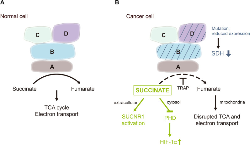
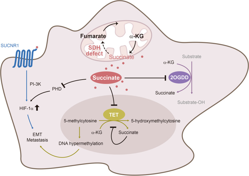
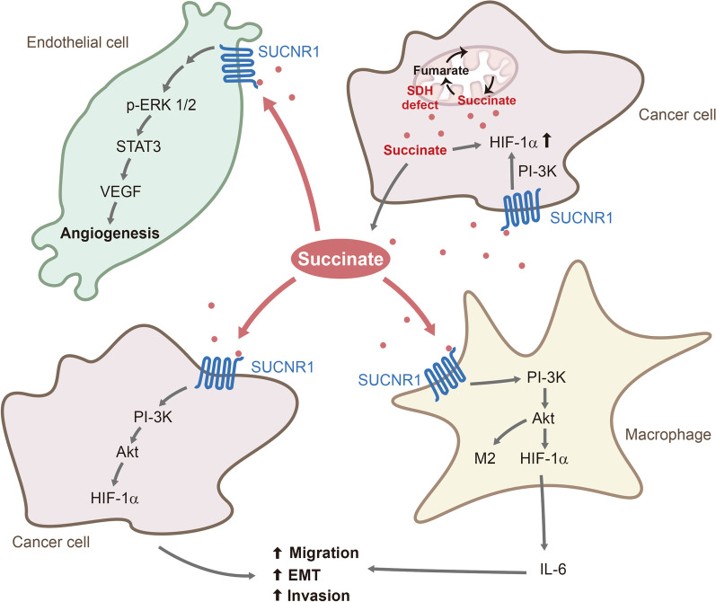
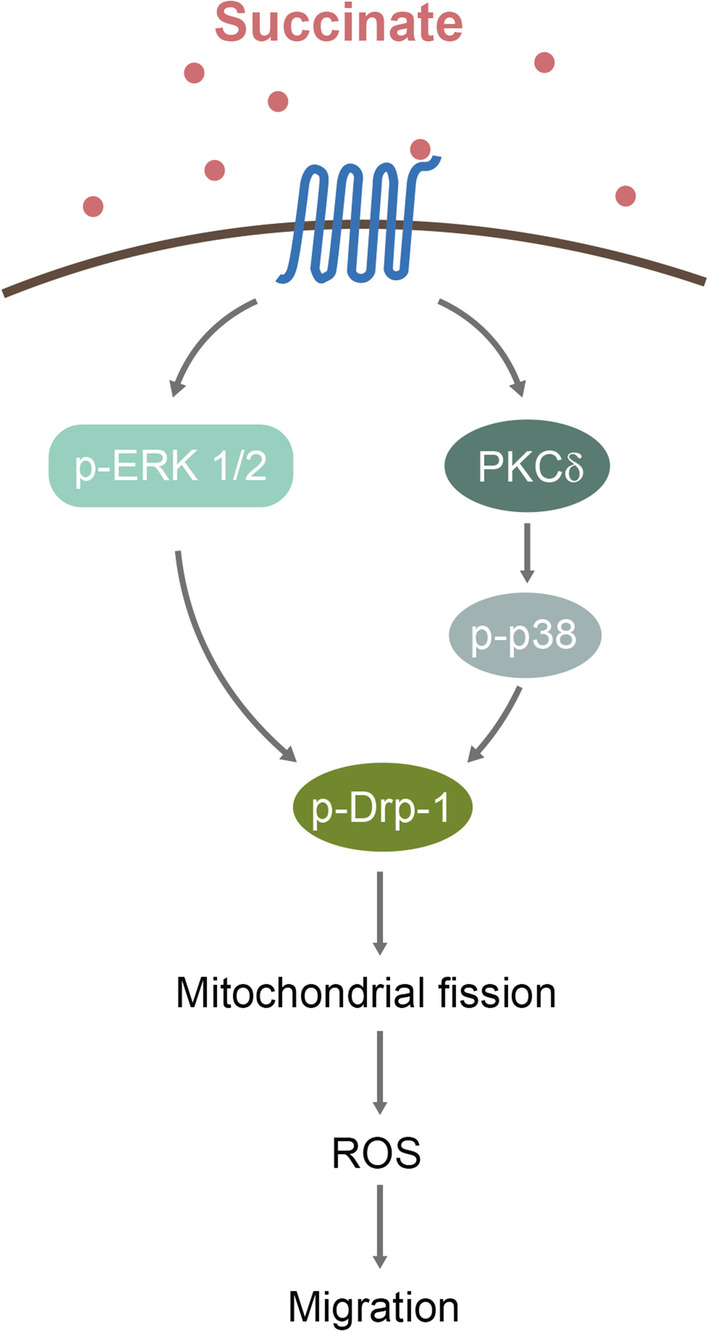
Succinate is a tricarboxylic acid (TCA) cycle intermediate normally confined to the mitochondrial matrix. It is a substrate of succinate dehydrogenase (SDH). Mutation of SDH subunits (SDHD and SDHB) in hereditary tumors such as paraganglioma or reduction of SDHB expression in cancer results in matrix succinate accumulation which is transported to cytoplasma and secreted into the extracellular milieu. Excessive cytosolic succinate is known to stabilize hypoxia inducible factor-1α (HIF-1α) by inhibiting prolyl hydroxylase. Recent reports indicate that cancer-secreted succinate enhances cancer cell migration and promotes cancer metastasis by activating succinate receptor-1 (SUCNR-1)-mediated signaling and transcription pathways. Cancer-derived extracellular succinate enhances cancer cell and macrophage migration through SUCNR-1 → PI-3 K → HIF-1α pathway. Extracellular succinate induces tumor angiogenesis through SUCNR-1-mediated ERK1/2 and STAT3 activation resulting in upregulation of vascular endothelial growth factor (VEGF) expression. Succinate increases SUCNR-1 expression in cancer cells which is considered as a target for developing new anti-metastasis drugs. Furthermore, serum succinate which is elevated in cancer patients may be a theranostic biomarker for selecting patients for SUCNR-1 antagonist therapy.

Keywords: Cancer metastasis; G-protein-coupled receptor 91 (GPR91); Hereditary paraganglioma; Succinate; Succinate dehydrogenase; Succinate receptor-1.


Conflict of interest statement

The authors declare no competing interests.

Figures

Fig. 1
Fig. 1
Succinate dehydrogenase (SDH) in normal and cancer cell. A. Schematic illustration of SDH subunits. It catalyzes conversion of succinate to fumarate in TCA cycle and electron transport in ETC. B Mutation or expression defect of subunit B or D results in reduced SDH activity and accumulation of succinate. TRAP inhibits SDH catalytic activity also resulting in succinate accumulation. Cytosolic succinate stabilizes HIF-1α through inhibition of PHD while extracellular succinate promotes cancer metastasis via SUCNR-1
Fig. 2
Fig. 2
Succinate promotes cancer cell growth by inhibiting 2OGDD and through ligating SUCNR-1. Succinate accumulation due to SDH defect leads to increased cytosolic succinate and secretion of succinate. Cytosolic succinate exerts its effect by inhibiting a large number of 2-oxoglutarate-dependent dioxygenases (2OGDD or α-keto glutarate-dependent enzymes) such as prolyl hydroxylase (PHD) and TET (Ten to Eleven Translocation). Inhibition of PHD results in HIF-1α stabilization while inhibition of TET leads to DNA hypermethylation. Extracellular succinate interacts with succinate receptor-1 (SUCNR-1) which signals via PI-3 K to increase HIF-1α expression
Fig. 3
Fig. 3
Cancer cell-derived succinate acts on macrophages, endothelial cells and cancer cells to drive cancer metastasis
Fig. 4
Fig. 4
Schematic illustration of the signaling pathway via which succinate phosphorylate Drp-1 and induces mitochondrial fission. ROS generated drive cancer cell migration

References

    1. Martínez-Reyes I, Chandel NS. Mitochondrial TCA cycle metabolites control physiology and disease. Nat Commun. 2020;11:102. - PMC - PubMed
    1. Chouchani ET, Pell VR, Gaude E, Aksentijević D, Sundier SY, Robb EL, et al. Ischaemic accumulation of succinate controls reperfusion injury through mitochondrial ROS. Nature. 2014;515:431–435. - PMC - PubMed
    1. Zhang J, Wang YT, Miller JH, Day MM, Munger JC, Brookes PS. Accumulation of succinate in cardiac ischemia primarily occurs via canonical Krebs cycle activity. Cell Rep. 2018;23:2617–2628. - PMC - PubMed
    1. Kohlhauer M, Dawkins S, Costa ASH, Lee R, Young T, Pell VR, et al. Metabolomic profiling in acute ST-segment-elevation myocardial infarction identifies succinate as an early marker of human ischemia-reperfusion injury. J Am Heart Assoc. 2018;7:e007546. - PMC - PubMed
    1. Nair S, Sobotka KS, Joshi P, Gressens P, Fleiss B, Thornton C, et al. Lipopolysaccharide-induced alteration of mitochondrial morphology induces a metabolic shift in microglia modulating the inflammatory response in vitro and in vivo. Glia. 2019;67:1047–1061. - PubMed