Comparison of the radiological parameters between dynamic-referencing tactile guidance robotic system and Microplasty® instrumentation in unicompartmental knee arthroplasty
- PMID: 36345186
- PMCID: PMC9647677
- DOI: 10.52312/jdrs.2022.742
Comparison of the radiological parameters between dynamic-referencing tactile guidance robotic system and Microplasty® instrumentation in unicompartmental knee arthroplasty
Abstract
Objectives: This study aims to compare the radiological outcomes of unicompartmental knee arthroplasty (UKA) performed by a navigation-based robotic system versus Microplasty® instrumentation.
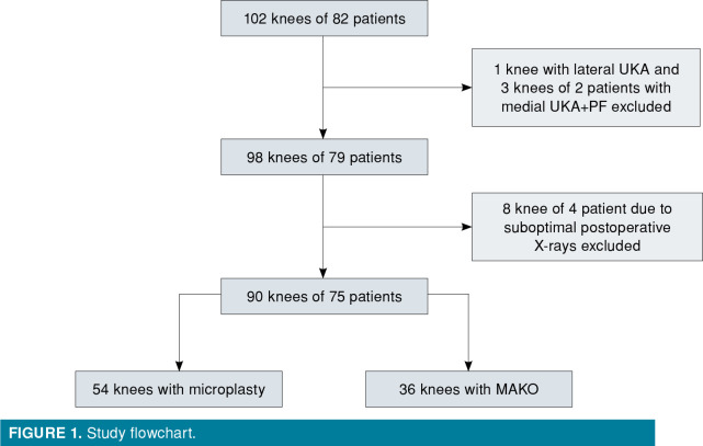
Patients and methods: Between January 2018 and January 2019, a total of 90 knees of 75 patients (65 males, 10 females; mean age: 62.0±9.4 years; range, 50 to 73 years) were included. Among these, 54 knees underwent Oxford mobile-bearing UKA with an Microplasty® instrumentation set and 36 knees were operated with the aid of a Restoris® MCK with MAKO navigation-based robotic system. Postoperative anteroposterior and lateral X-rays of all patients were evaluated according to nine different parameters. On the femoral side, femoral varus-valgus angle, flexion-extension angle, femoral condyle posterior fit; on tibial side, tibial component varus/valgus, tibial posterior slope, medial, anterior, posterior and lateral fit of tibial component assessed.
Results: There was no significant difference between groups in terms of age, sex, and affected side. On the femoral side, no significant difference was observed in the component position between groups. On the tibial side, tibial component medial fit (p=0.032) and anterior fit (p=0.007) were better in navigation-based robotic system group.
Conclusion: Microplasty® instrumentation may lead to comparable implant positioning compared to a tactile-based navigated robotic instrumentation.
Conflict of interest statement
Figures


References
-
- Brown NM, Sheth NP, Davis K, Berend ME, Lombardi AV, Berend KR, et al. Total knee arthroplasty has higher postoperative morbidity than unicompartmental knee arthroplasty: A multicenter analysis. J Arthroplasty. 2012;27(8 Suppl):86–90. - PubMed
-
- Hopper GP, Leach WJ. Participation in sporting activities following knee replacement: Total versus unicompartmental. Knee Surg Sports Traumatol Arthrosc. 2008;16:973–979. - PubMed
-
- Campi S, Pandit HG, Dodd CAF, Murray DW. Cementless fixation in medial unicompartmental knee arthroplasty: A systematic review. Knee Surg Sports Traumatol Arthrosc. 2017;25:736–745. - PubMed
-
- Saenz CL, McGrath MS, Marker DR, Seyler TM, Mont MA, Bonutti PM. Early failure of a unicompartmental knee arthroplasty design with an all-polyethylene tibial component. Knee. 2010;17:53–56. - PubMed
MeSH terms
LinkOut - more resources
Full Text Sources
Medical