Review
. 2022 Oct 26:13:1044143.
doi: 10.3389/fmicb.2022.1044143. eCollection 2022.

Bacteriophage-encoded lethal membrane disruptors: Advances in understanding and potential applications

Affiliations
Review

Bacteriophage-encoded lethal membrane disruptors: Advances in understanding and potential applications

Gayan S Abeysekera et al. Front Microbiol. .

Abstract

Holins and spanins are bacteriophage-encoded membrane proteins that control bacterial cell lysis in the final stage of the bacteriophage reproductive cycle. Due to their efficient mechanisms for lethal membrane disruption, these proteins are gaining interest in many fields, including the medical, food, biotechnological, and pharmaceutical fields. However, investigating these lethal proteins is challenging due to their toxicity in bacterial expression systems and the resultant low protein yields have hindered their analysis compared to other cell lytic proteins. Therefore, the structural and dynamic properties of holins and spanins in their native environment are not well-understood. In this article we describe recent advances in the classification, purification, and analysis of holin and spanin proteins, which are beginning to overcome the technical barriers to understanding these lethal membrane disrupting proteins, and through this, unlock many potential biotechnological applications.

Keywords: alphaFold structure; bacteriophage; holins spanins; holins spanins applications; holins spanins future potential potential; membrane protein.


Conflict of interest statement

The authors declare that the research was conducted in the absence of any commercial or financial relationships that could be construed as a potential conflict of interest.

Figures
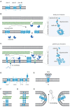
Figure 1
Figure 1
(A) Transmembrane α-helical segment topology of the most studied holin classes (I-III). (B) Schematic representation of canonical pathway: Following the late gene expression initiated in the bacteriophage lytic cycle, canonical holins and endolysins accumulate in the inner membrane and cytoplasm, respectively. At an allele-specific time, holins make micron-scale holes in the inner membrane and endolysins escape to the periplasm, degrading the peptidoglycan. (C) Schematic representation of pinholin pathway: When late gene expression is initiated, pinholins and inactive SAR endolysins accumulated in the inner membrane. At an allele-specific time, pinholins make heptameric channels with a lumen of ~2 nm which destabilised the proton motive pump. SAR endolysins transform into their active form as they are sensitive to the proton motive pump and finally degrade the peptidoglycan. (D) Topology of lambda and lambdoid bacteriophage ϕ21 holins (S105, S2168) and its antiholins (S107, S2171). (E) T4 holin complex can make a complex with its RI, RIII antiholins, and DNA to respond to superinfections. (F) Two-component spanins (G) Unimolecular spanins (H) Fusion inner and outer membrane.
Figure 2
Figure 2
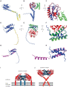
(A) AlphaFold Colab prediction of complete holin T structure. The colour of the predicted holin T structure represents the score of the predicted local distance difference test (pLDDT). (B) Structure comparisons of AlphaFold Colab predicted holin T (blue) and 6PSK(soluble domain of the resolved holin T and RI antiholin complex; RMSD = 0.688 angstroms). (C) An enlarged version of structure comparisons of AlphaFold Colab predicted holin T (blue) and 6PSK (red; soluble domain of holin T and green; soluble domain of RI antiholin). (D) AlphaFold Colab prediction of RI antiholin. The colour of the predicted RI antiholin structure represents the score of the predicted local distance difference test (pLDDT). (E) Structure comparisons of AlphaFold Colab predicted RI antiholin (blue) and 6PSK (soluble domain of the resolved holin T and RI antiholin complex; RMSD = 1.537 angstroms). (F) An enlarged version of structure comparisons of AlphaFold Colab predicted RI antiholin (blue) and 6PSK (red; soluble domain of holin T and green; soluble domain of RI antiholin). (G) Structure comparisons of AlphaFold Colab predicted RI antiholin (blue) and 6PSH (soluble domain of the resolved RI antiholin monomer; RMSD = 21.077 angstroms). (H) An enlarged version of structure comparisons of AlphaFold Colab predicted RI antiholin (blue) and 6PSH (purple). (I) RI–T complex model with RI SAR domain by Krieger et al. (2020). (J) RI–T complex model with cleavable signal peptide (new model) by Mehner-Breitfeld et al. (2021).

References

    1. Agu C. A., Klein R., Schwab S., König-Schuster M., Kodajova P., Ausserlechner M., et al. (2006). The cytotoxic activity of the bacteriophage λ-holin protein reduces tumour growth rates in mammary cancer cell xenograft models. J. Gene Med. 8, 229–241. doi: 10.1002/jgm.833, PMID: - DOI - PubMed
    1. Ahammad T., Drew D. L., Sahu I. D., Serafin R. A., Clowes K. R., Lorigan G. A. (2019). Continuous wave electron paramagnetic resonance spectroscopy reveals the structural topology and dynamic properties of active pinholin S2168 in a lipid bilayer. J. Phys. Chem. B 123, 8048–8056. doi: 10.1021/acs.jpcb.9b06480, PMID: - DOI - PMC - PubMed
    1. Barenboim M., Chang C. Y., Dib Hajj F., Young R. (1999). Characterization of the dual start motif of a class II holin gene. Mol. Microbiol. 32, 715–727. doi: 10.1046/j.1365-2958.1999.01385.x, PMID: - DOI - PubMed
    1. Basit A., Qadir S., Qureshi S., Rehman S. U. (2021). Cloning and expression analysis of fused holin-endolysin from RL bacteriophage; exhibits broad activity against multi drug resistant pathogens. Enzym. Microb. Technol. 149:109846. doi: 10.1016/j.enzmictec.2021.109846, PMID: - DOI - PubMed
    1. Berman H. M., Westbrook J., Feng Z., Gilliland G., Bhat T. N., Weissig H., et al. (2000). The protein data bank. Nucleic Acids Res. 28, 235–242. doi: 10.1093/nar/28.1.235, PMID: - DOI - PMC - PubMed

LinkOut - more resources