Case Reports
. 2022 Oct;11(10):1717-1725.
doi: 10.21037/tp-22-15.

Novel pathogenic variant combination in LPL causing familial chylomicronemia syndrome in an Asian family and experimental validation in vitro: a case report

Affiliations
Case Reports

Novel pathogenic variant combination in LPL causing familial chylomicronemia syndrome in an Asian family and experimental validation in vitro: a case report

Huiping Shi et al. Transl Pediatr. 2022 Oct.

Abstract

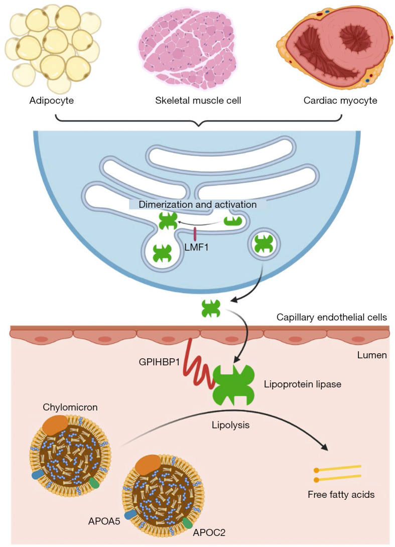
Background: Familial chylomicronemia syndrome (FCS) is a rare autosomal recessive disorder, typically caused by biallelic pathogenic variants in the lipoprotein lipase (LPL) gene. Lipoprotein lipase, encoded by the LPL gene, catalyzes the hydrolysis of triglycerides, and its deficiency or dysfunction can lead to chylomicronemia and potentially fatal recurrent acute pancreatitis.

Case description: Here, we report an Asian child with FCS due to compound heterozygous LPL variants. The 4-year-old patient presented with splenomegaly and severe hypertriglyceridemia, specifically chylomicronemia which resulted in abnormal coagulation measured by a turbidity-based assay. Based on the clinical features and family history, the diagnosis of FCS was suspected, and confirmed by the identification of compound heterozygous variants in the LPL gene (c.461A>G; p.His154Arg and c.788T>A; p.Leu263Gln) in the patient, inheriting one from each parent. According to the clinical and genetic findings, the patient was diagnosed with FCS. In vitro experimental validation found that the LPL p.H154R variant reduced the expression of lipoprotein lipase and decreased its lipolytic activity, while the LPL p.L263Q variant mainly impaired its lipolytic activity.

Conclusions: FCS was molecularly diagnosed using whole exome sequencing in the case presented. When interpreting abnormal coagulation profiles measured by turbidity-based assay, the possibility of lipemic blood (or chylomicronemia) should be considered and the presence of this phenomenon might indicate the diagnosis of FCS. In vitro experiments showed that the two LPL variants impaired lipoprotein lipase expression and/or function making them likely to be pathogenic.

Keywords: LPL gene; case report; familial chylomicronemia syndrome; lipoprotein lipase; novel pathogenic variant.


Conflict of interest statement

Conflicts of Interest: Both authors have completed the ICMJE uniform disclosure form (available at https://tp.amegroups.com/article/view/10.21037/tp-22-15/coif). The authors report that this study was funded by Jiangsu Provincial Special Program of Medical Science (BL2012005); Jiangsu Province’s Key Medical Center (ZX201102); The Priority Academic Program Development of Jiangsu Higher Education Institutions (PAPD). The authors have no other conflicts of interest to declare.

Figures

Figure 1
Figure 1
Lipoprotein lipase catalyzes the hydrolysis of triglycerides. Lipoprotein lipase is synthesized in adipocytes, skeletal muscle cells and cardiac myocytes. After dimerization and activation with the help of lipase maturation factor 1 (LMF1), lipoprotein lipase is released extracellularly and binds to glycosylphosphatidylinositol-anchored high-density lipoprotein-binding protein 1 (GPIHBP1) expressed on capillary endothelial cells, which shuttles lipoprotein lipase into the capillary lumen. There, lipoprotein lipase catalyzes the hydrolysis of triglycerides, with the help of two regulators: apolipoprotein C-II and apolipoprotein A-V. ApoC-II serves as a cofactor for lipoprotein lipase, and apoA-V affects plasma triglyceride levels possibly by stabilizing the lipolytic machinery via binding to lipoproteins, endothelial proteoglycans, and lipoprotein lipase. Figure is created with BioRender.
Figure 2
Figure 2
Characterization of an Asian family of familial chylomicronemia syndrome caused by compound heterozygous variants in LPL. (A) The triglyceride levels in the patient. (B) Pedigree presentation of the studied family. The LPL p.H154R variant was colored in black while the LPL p.L263Q variant was colored in grey. Arrow indicated the patient. The triglyceride levels were labelled below the corresponding individuals. TG: triglyceride. (C) Sequencing analysis of the LPL gene showing the p.H154R variant in patient and father, and the p.L263Q variant in patient and mother. Amino acid abbreviations: V, valine; H, histidine; R, arginine; L, leucine; Q, glutamine. Nucleotide abbreviations: G, guanine; T, thymine; C, cytosine; A, adenine. (D) Visualization of the p.H154R and p.L263Q variants in the lipoprotein lipase protein structure using pymol. The residues were colored according to the following color scheme: acidic residue: red; basic residue: marine; nonpolar: orange; polar: green; cysteine: yellow. In the mutant site, both the original and mutant residues were shown. (E) Homologous protein alignment using MutationTaster2 showing the conservation of His-154 and Leu-263 residues among different species. Seq, sequencing; LPL, lipoprotein lipase.
Figure 3
Figure 3
Functional validation of the LPL pathogenic variants in vitro. (A) Quantification of the expression levels of wild-type and mutant lipoprotein lipase proteins using ELISA. The LPL p.H154R variant resulted in reduced level of lipoprotein lipase whereas the LPL p.L263Q variant did not alter lipoprotein lipase level when compared to wild type (WT) control. (B) Comparison of the lipolytic activity of wild-type and mutant lipoprotein lipase proteins. One-way ANOVA, *, P<0.05; ns, non-significant. LPL, lipoprotein lipase; AU, arbitrary unit; ANOVA, analysis of variance.

Comment in

References

    1. Goldberg RB, Chait A. A Comprehensive Update on the Chylomicronemia Syndrome. Front Endocrinol (Lausanne) 2020;11:593931. 10.3389/fendo.2020.593931 - DOI - PMC - PubMed
    1. Baass A, Paquette M, Bernard S, et al. Familial chylomicronemia syndrome: an under-recognized cause of severe hypertriglyceridaemia. J Intern Med 2020;287:340-8. 10.1111/joim.13016 - DOI - PubMed
    1. Brahm AJ, Hegele RA. Chylomicronaemia--current diagnosis and future therapies. Nat Rev Endocrinol 2015;11:352-62. 10.1038/nrendo.2015.26 - DOI - PubMed
    1. Marogi EP, Ohiomoba RO, Stone NJ. Eruptive Xanthomas: Importance of Recognition to Reduce Delay of Effective Triglyceride Reduction. Am J Med 2022;135:444-7. 10.1016/j.amjmed.2021.09.026 - DOI - PubMed
    1. Cabodevilla AG, Tang S, Lee S, et al. Eruptive xanthoma model reveals endothelial cells internalize and metabolize chylomicrons, leading to extravascular triglyceride accumulation. J Clin Invest 2021;131:145800. 10.1172/JCI145800 - DOI - PMC - PubMed

Publication types

LinkOut - more resources