. 2023 Mar 8;211(1):31-45.
doi: 10.1093/cei/uxac096.

A novel selective spleen tyrosine kinase inhibitor SKI-O-703 (cevidoplenib) ameliorates lupus nephritis and serum-induced arthritis in murine models

Affiliations

A novel selective spleen tyrosine kinase inhibitor SKI-O-703 (cevidoplenib) ameliorates lupus nephritis and serum-induced arthritis in murine models

Somi Cho et al. Clin Exp Immunol. .

Abstract

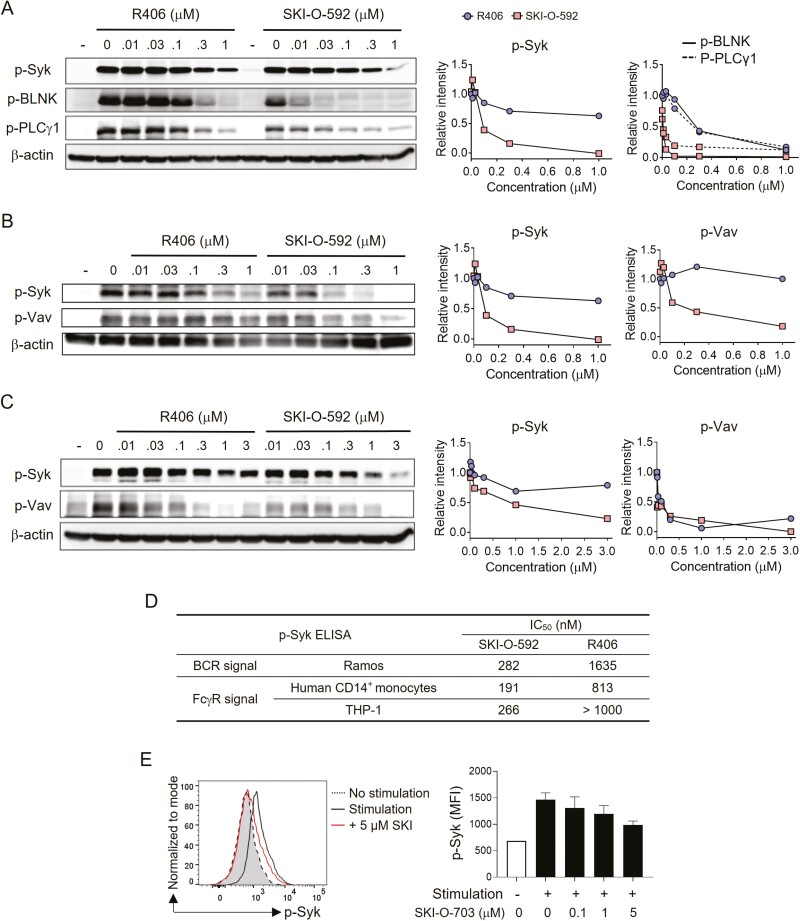
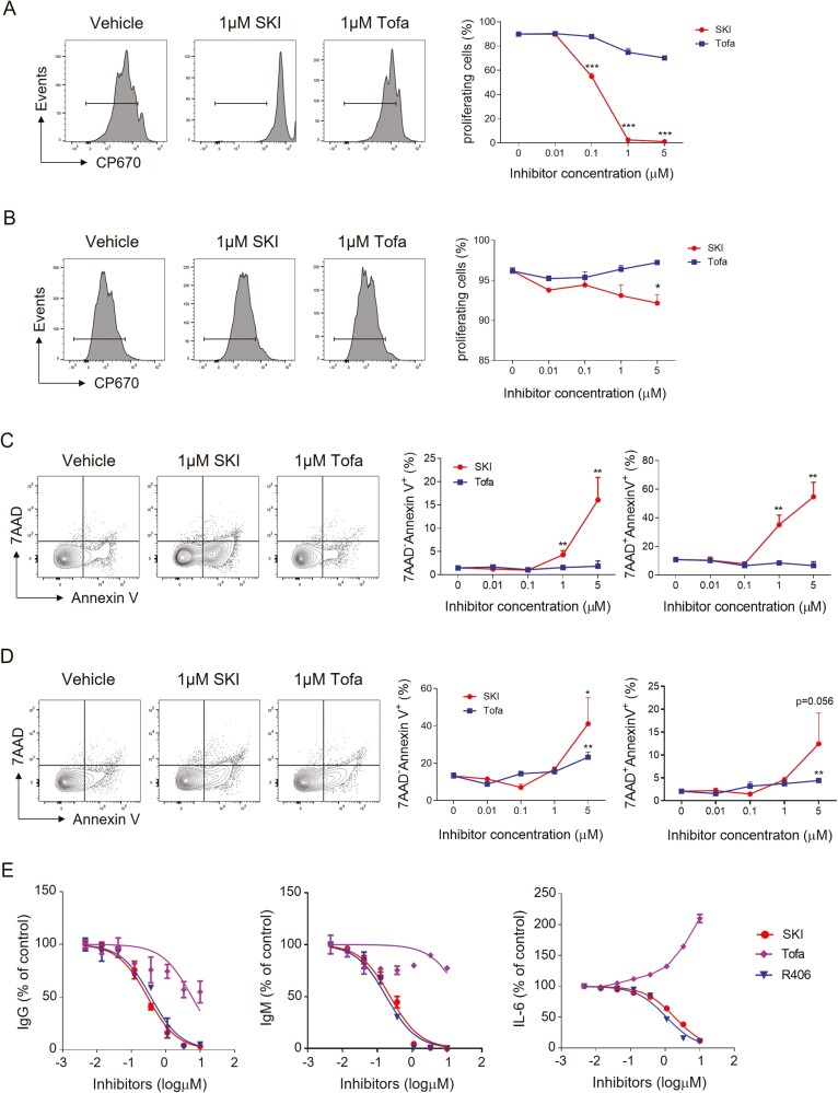
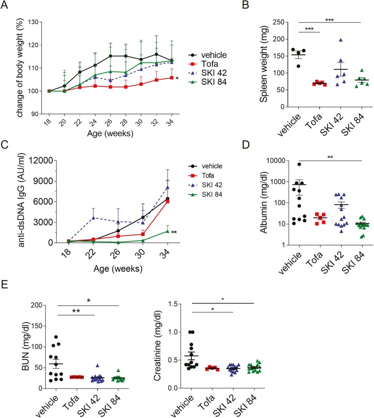
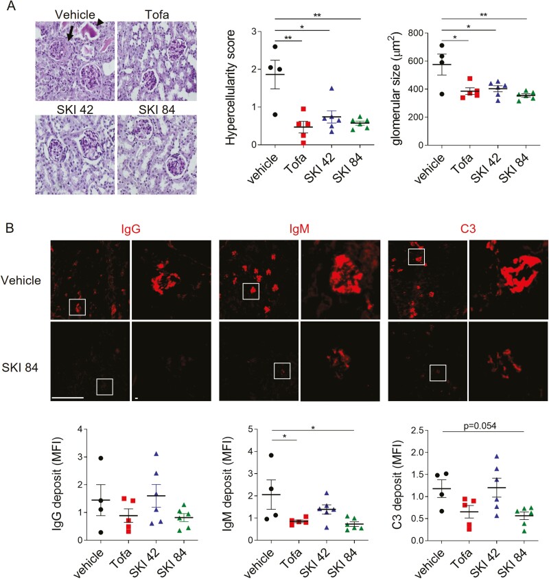
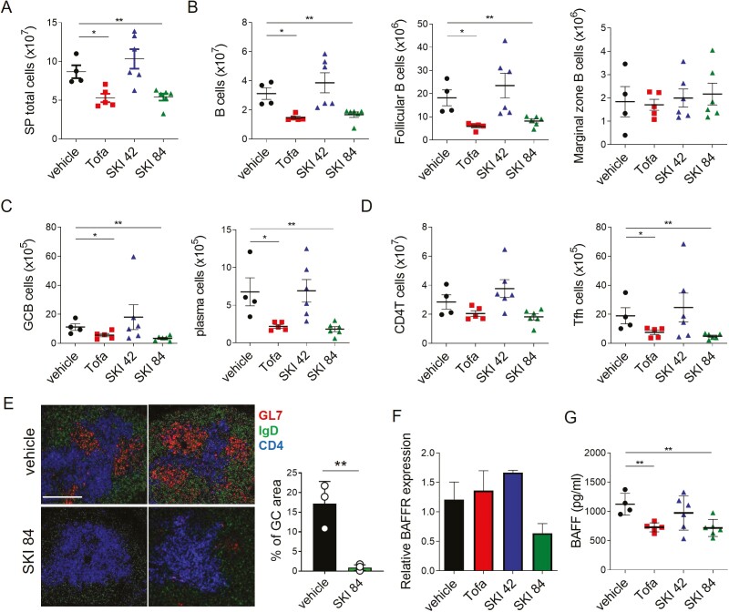
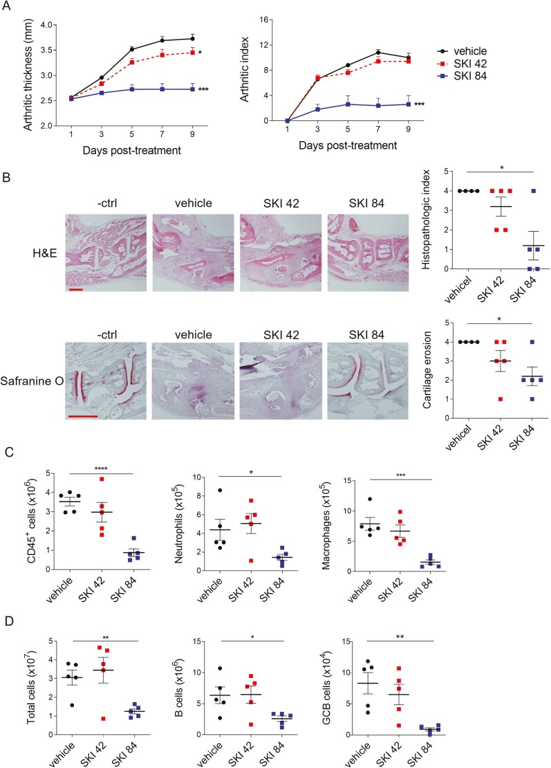
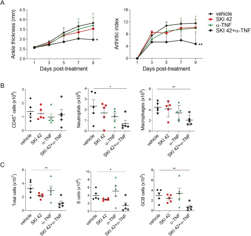
Spleen tyrosine kinase (Syk) plays a pivotal role in the activation of B cells and innate inflammatory cells by transducing immune receptor-triggered signals. Dysregulated activity of Syk is implicated in the development of antibody-mediated autoimmune diseases including systemic lupus erythematosus (SLE) and rheumatoid arthritis, but the effect of Syk inhibition on such diseases remains to be fully evaluated. We have developed a novel selective Syk inhibitor, SKI-O-592, and its orally bioavailable salt form, SKI-O-703 (cevidoplenib). To examine the efficacy of SKI-O-703 on the progression of SLE, New Zealand black/white mice at the autoimmunity-established phase were administrated orally with SKI-O-703 for 16 weeks. Levels of IgG autoantibody, proteinuria, and glomerulonephritis fell significantly, and this was associated with hypoactivation of follicular B cells via the germinal center. In a model of serum-transferred arthritis, SKI-O-703 significantly ameliorated synovitis, with fewer neutrophils and macrophages infiltrated into the synovial tissue. This effect was recapitulated when mice otherwise refractory to anti-TNF therapy were treated by TNF blockade combined with a suboptimal dose of SKI-O-703. These results demonstrate that the novel selective Syk inhibitor SKI-O-703 attenuates the progression of autoantibody-mediated autoimmune diseases by inhibiting both autoantibody-producing and autoantibody-sensing cells.

Keywords: Syk inhibitor SKI-O-703; autoimmune disease; spleen tyrosine kinase; systemic lupus erythematosus.


Figures

Graphical Abstract
Graphical Abstract
Figure 1:
Figure 1:
in vitro inhibitory effects of SKI-O-592 and -703 on Syk in B cells and monocytes. Ramos cells (A), human primary monocytes (B), and THP cells (C) were pretreated with inhibitors at the indicated concentrations and stimulated with anti-human IgM (A) and human IgG (B and C). The cells were then lysed and assayed by immunoblotting methods. The relative intensities of protein phosphorylation levels were quantitated with reference to each lane of β-actin control bands. (D) Ramos cells, human primary monocytes, and THP cells were treated as in A–C for 24 h and levels of phosphorylated Syk (p-Syk) were measured by ELISA. (E) Mouse primary B cells were stimulated with anti-IgM mAb for 60 min in the presence or absence of SKI-O-703 and assayed for p-Syk by FACS. A representative FACS profile and data on the dose-response of mean fluorescence intensity (MFI) are shown. The data are representative of two independent experiments.
Figure 2:
Figure 2:
in vitro effects of SKI-O -703 on the proliferation, survival, and differentiation of B cells. Mouse primary B cells were labeled with CP670, and stimulated with either anti-IgM mAb, CD40L and IL-4 (A) or LPS (B) for 72 h in the presence or absence of SKI-O-703 and tofacitinib at the indicated concentrations; they were then assayed by FACS. (C and D) Mouse primary B cells were cultured as in A and B for 24 h, stained with Annexin V and 7-AAD, and assayed by FACS. Representative FACS profiles and graphs displaying mean Annexin V-positivity are shown. (E) Human primary B cells were stimulated with CpG ODN plus IL-2 (to detect IgM and IgG) or CpG ODN 2006 plus IFN-α (to detect IL-6) in the presence or absence of inhibitors. The culture supernatants were assayed by ELISA. Graphs are expressed as % positivity relative to the controls (without inhibitor). The data are representative of 2–3 independent experiments. *P < 0.05, **P < 0.01, and ***P < 0.001, compared with vehicle control by two-tailed unpaired Students t-tests.
Figure 3:
Figure 3:
in vivo effect of oral SKI-O-703 on lupus-like signs in NZB/W mice. Female NZB/W F1 mice were administrated orally with 42 mpk of SKI-O-703 (SKI 42), 84 mpk of SKI-O-703 (SKI 84), or tofacitinib (Tofa) from 18 to 34 weeks of age. (A) Body weights were measured every 2 weeks and are displayed as mean percent change of body weight, with SEM. (B) Spleen weights measured at the end of the experiment. (C) Serum was collected every 4 weeks and assayed by ELISA to measure titers of anti-dsDNA IgG. Mean titers are shown. AU, arbitrary unit. (D) Urine collected at 34 weeks of age and urinary albumin was assayed by ELISA. (E) Concentrations of blood urine nitrogen (BUN) and creatinine at 34 weeks of age. Graphs display means ± SEMs, and symbols represent values of individual mice (B, D, and E). *P < 0.05, **P < 0.01, and ***P < 0.001, compared with the vehicle control group by two-way ANOVA (A and C) and two-tailed unpaired Students t-test (B, D, and E).
Figure 4:
Figure 4:
histopathological alteration of kidney tissues by SKI-O-703 in NZB/W mice. Female NZB/W F1 mice were administrated orally with 42 mpk SKI-O-703 (SKI 42), 84 mpk SKI-O-703 (SKI 84), or tofacitinib (Tofa) from 18 to 34 weeks of age, and kidneys were examined at 34 weeks by histopathological methods. (A) Paraffin sections were stained with PAS and hematoxylin. An eosinophilic protein cast and crescent are indicated by the arrowhead and arrow, respectively. The images are representative of each group. Graphs show means ± SEMs with symbols representing values of individual mice. (B) Cryosections were stained with anti-IgG, anti-IgM, and anti-C3 Abs and observed by fluorescence confocal microscopy. Boxes in the left images are magnified in the right images. Representative images with mean fluorescence intensities are shown. Bar scale, 512 μm. *P < 0.05 and **P < 0.01 by two-tailed unpaired Students t-test.
Figure 5:
Figure 5:
reduced numbers of spleen cells in SKI-O-703-treated NZB/W mice. Female NZB/W F1 mice were administrated orally with 42 mpk SKI-O-703 (SKI 42), 84 mpk SKI-O-703 (SKI 84), or tofacitinib (Tofa) from 18 to 34 weeks of age. Spleens and sera were collected at 34 weeks of age. (AD) Spleens were assayed by FACS. The gating strategy is displayed in Supplementary Fig. S2. (E) Spleens were cryosectioned, stained with Abs to GL-7, IgD, and CD4, and analyzed by confocal microscopy. Two representative images per group are shown. Bar scale, 80 μm. Percentage of GC area (area of GL7+/area of total area) from 3 to 4 images/group are displayed as mean ± SD. (F) Splenic B cells were assayed by quantitative RT-PCR. Levels of BAFFR mRNA were normalized to the level of β-actin mRNA. (G) Concentrations of BAFF measured by ELISA in sera. The data are representative of at least two independent experiments. Graphs show means + SEMs, and symbols represent values of individual mice. *P < 0.05 and **P < 0.01 by two-tailed unpaired Students t-test.
Figure 6:
Figure 6:
SKI-O-703 attenuates KSTA. BALB/c mice were infused with K/BxN serum and orally dosed with 42 mpk SKI-O-703 (SKI 42) or 84 mpk SKI-O-703 (SKI 84) twice a day for 9 days. (A) Ankle thickness and arthritic indexes are displayed as means + SEMs. (B) On Day 9, hind paws were examined by histopathologic methods after staining with H&E and Safranine O. Representative images and graphs displaying means ± SEMs with symbols representing each individual are shown. (C) Spleen cells and joint-draining LN cells were assayed by FACS. Graphs display means ± SEMs, with symbols representing values of individual mice. The data are representative of four independent experiments. *P < 0.05, **P < 0.01, and ***P < 0.001, compared with the vehicle control group by two-way ANOVA (A) and two-tailed unpaired Student’s t-test (B-D).
Figure 7:
Figure 7:
effects of SKI-O-703 combined with TNF blockade on KSTA. BALB/c mice were infused with K/BxN serum and treated with 42 mpk SKI-O-703 (SKI 42) alone, etanercept (a-TNF) alone, or both, for 9 days. (A) Ankle thickness and arthritic indexes on a given day are shown as means + SEM. (B and C) Spleen and dLN cells were analyzed by FACS. The data are representative of four independent experiments. *P < 0.05, **P < 0.01, and ***P < 0.001, compared with the vehicle control group by two-way ANOVA (A) and two-tailed unpaired Student’s t-test (B and C).

Similar articles

Cited by

References

    1. Theofilopoulos AN, Kono DH, Baccala R.. The multiple pathways to autoimmunity. Nat Immunol 2017, 18, 716–24. doi:10.1038/ni.3731. - DOI - PMC - PubMed
    1. Suurmond J, Diamond B.. Autoantibodies in systemic autoimmune diseases: specificity and pathogenicity. J Clin Invest 2015, 125, 2194–202. doi:10.1172/JCI78084. - DOI - PMC - PubMed
    1. Isenberg DA, Manson JJ, Ehrenstein MR, Rahman A.. Fifty years of anti-ds DNA antibodies: are we approaching journey’s end?. Rheumatology (Oxford) 2007, 46, 1052–6. doi:10.1093/rheumatology/kem112. - DOI - PubMed
    1. Rahman A, Isenberg DA.. Systemic lupus erythematosus. N Engl J Med 2008, 358, 929–39. doi:10.1056/NEJMra071297. - DOI - PubMed
    1. Tsokos GC. Autoimmunity and organ damage in systemic lupus erythematosus. Nat Immunol 2020, 21, 605–14. doi:10.1038/s41590-020-0677-6. - DOI - PMC - PubMed