. 2023 May 2;146(5):1932-1949.
doi: 10.1093/brain/awac418.

Anti-pan-neurofascin antibodies induce subclass-related complement activation and nodo-paranodal damage

Affiliations

Anti-pan-neurofascin antibodies induce subclass-related complement activation and nodo-paranodal damage

Luise Appeltshauser et al. Brain. .

Abstract

Autoimmune neuropathy associated with antibodies against pan-neurofascin is a new subtype of nodo-paranodopathy. It is relevant because it is associated with high morbidity and mortality. Affected patients often require intensive care unit treatment for several months, and data on the reversibility and long-term prognosis are limited. The pathogenicity including IgG subclass-associated mechanisms has not been unravelled, nor directly compared to anti-neurofascin-155 IgG4-related pathology. Understanding the underlying pathology might have a direct impact on treatment of these severely affected patients. By a multicentre combined prospective and retrospective approach, we provide clinical data of a large cohort of patients with anti-neurofascin-associated neuropathy (n = 18) including longitudinal titre and neurofilament light chain assessment via Ella® and relate clinical data to in vitro pathogenicity studies of anti-neurofascin antibodies. We assessed antibody binding characteristics and the pathogenic effects of anti-pan-neurofascin versus neurofascin-155 antibodies on living myelinating dorsal root ganglia co-cultures. Additionally, we analysed the IgG subclass profile and the complement binding capacity and effector functions considering the effects of intravenous immunoglobulin preparations via enzyme-linked immunosorbent and cell-based assays. In contrast to chronic neurofascin-155 IgG4-associated neuropathy, anti-pan-neurofascin-associated disease presented with a high morbidity and mortality, but as a monophasic and potentially reversible disorder. During follow-up, antibodies were no longer detectable in 8 of 11 patients. Anti-pan-neurofascin had direct access to the nodes of Ranvier in myelinating cultures titre-dependently, most probably inducing this severe phenotype. Antibody preincubation led to impaired paranode formation, destruction of paranodal architecture and alterations on paranodal myelin and sensory neurons in the cultures, with more severe effects than neurofascin-155 antibodies. Besides IgG4, subclass IgG3 was detected and associated with complement binding and cytotoxic effects in vitro. As a possible correlate of axonal damage in vivo, we detected highly increased serum neurofilament light chain levels (sNF-L), correlating to serum C3a. Still, sNF-L was not identified as a marker for poor prognosis, but rather as an intra- and interindividual marker for acuteness, severity and course, with a strong decrease during recovery. Our data provide evidence that anti-pan-neurofascin antibodies directly attack the node and induce severe and acute, but potentially reversible, nodo-paranodal pathology, possibly involving complement-mediated mechanisms. Screening for autoantibodies thus is crucial to identify this subset of patients who benefit from early antibody-depleting therapy. Titre and sNF-L might serve as valuable follow-up parameters. The prospect of a favourable outcome has high relevance for physicians, patients and relatives during months of critical care.

Keywords: autoantibodies; complement; neurofascin; neurofilament light chain; nodo-paranodopathy.


Conflict of interest statement

L.A., F.L., C.S. and K.D. work for an academic institution offering commercial antibody diagnostics. I.A. has received travel grants from Biogen Idec and Guthy-Jackson Charitable Foundation, served on scientific advisory boards for Roche, Alexion, Merck and received research support from Diamed, none related to this manuscript. A.L.F. received research funding from Georgius Agricola Stiftung Ruhr, Ruhr University Bochum (FoRUM-program) and GBS CIDP Foundation International. She owns shares of Fresenius SE & Co., Gilead Sciences, Medtronic PLC and Novartis AG. F.L. serves or has served on advisory boards for Biogen, Roche and Alexion and received speaker honoraria from Roche, Biogen, Grifols, Alexion, Desitin and Novartis. He serves as editorial board member for Neurology N2. J.M. received travel grants from Biogen idec. His research is funded by Klaus Tschira Foundation, Hertie Foundation, Novartis, Biogen and Ruhr-University, Bochum (FoRUM-Program), none related to this work. K.P. received travel grants and speaker honoraria from Biogen Idec and Bayer Schering, Novartis, Celgene and Grifols and has participated in advisory board meeting for Celgene, none related to this manuscript. C.S. has served on scientific advisory boards for Akcea, Algiax, Air Liquide, Bayer, Grifols, Ipsen, LFB, Immunic, Merz, Pfizer, Roche and Takeda. She received speaker honoraria from Akcea, Alnylam Amicus, Grifols, Pfizer and Teva. She serves or has served as a journal editor, associate editor or editorial advisory board member for the European Journal of Neurology, PLoS One and PAIN Reports. The other authors report no competing interests.

Figures

Figure 1
Figure 1
ELISA subclass profile and C1q IgG binding. (A) Mean OD450nm in ELISA including standard deviations (error bars) and individual values. We used titre-adjusted sera of nine anti-pan-neurofascin and five anti-neurofascin-155 seropositive patients and secondary antibodies against human IgG (‘total IgG’) and titrated subclass-specific IgG1, IgG2, IgG3 and IgG4 secondary antibodies. Medians were compared using Mann–Whitney U-test, significant differences are highlighted by asterisks (see below). (B) Box-and-whisker plot showing median (line), first and third quartiles (box) and min/max values (whiskers) of OD450nm of C1q binding of single patients’ means. We compared the neurofascin-155 cohort versus the pan-neurofascin using two-sided t-test, significant differences are highlighted by asterisks (see below). (C) The scatter dot chart including simple linear regression (line) with 95% confidence intervals (dotted lines) illustrates a significant correlation of the OD450nm of C1q binding with the OD450nm of the IgG3 subclass ELISA in patients with anti-pan-neurofascin and anti-neurofascin-155. Significance level: *P < 0.05, **P < 0.01.
Figure 2
Figure 2
Effects of IVIg in ELISA and complement binding assay. (A and B) schematic display of the (A) ELISA and ELISA-based C1q complement binding assay, (B) generation of F(ab′)2 fragments in patient 12 and ELISA with preincubation of IVIg with F(ab′)2. All patients’ mean relative OD to an IVIg/BSA/milk powder concentration of 0 mg/ml are displayed with standard deviations in bar graphs using a IVIg/BSA/milk powder dilution series from 0.78 to 25 mg/ml. (C) No reduction of antibody binding was detected when preincubating the bound antigen with IVIg. (D) No reduction of antibody binding was detected when preincubating soluble antibodies with IVIg. (E) A slight but not significant reduction of mean C1q OD levels was noted when preincubating IVIg with antigen-bound IgG antibodies in the C1q binding assay. (F) IVIg incubation has C1q neutralizing effects, with a significant reduction of relative ODs in a dose-dependent manner, when preincubating IVIg with C1q before addition to the assay. Statistical testing was performed using paired t-test and Wilcoxon signed rank test, depending on normal distribution of the data. Significance level: *P < 0.05, **P < 0.01, ***P < 0.001, ****P < 0.0001. Schematic display created with BioRender.com.
Figure 3
Figure 3
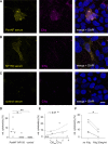
Complement binding and effects in the cell-based assay. (AC) Photomicrographs of neurofascin-155 (NF155) transfected HEK293 cells incubated with serum and complement factor C1q and triple immunostaining with anti-human IgG (yellow), anti-C1q (magenta) and DAPI (blue). Incubation with serum containing anti-pan-neurofascin (PanNF) antibodies leads to complement C1q deposition on transfected cells (A), whereas no complement deposition was observed using serum with NF155 antibodies (B) or serum of a healthy control (C). Scale bar = 10 µm. (D) The relative cytotoxicity measured by LDH assay and calculated according to instructions of the manufacturer is displayed in percent (%) for preincubation of five sera with anti-PanNF, NF155 antibodies and healthy control sera. Kruskal–Wallis test with Dunn’s correction for multiple testing was used for statistical testing. (E) The scatter dot chart illustrated the Spearman correlation of the relative cytotoxicity (%, y-axis) with the OD450nm of the C1q binding assay in seropositive patients. (F) The mean relative cytotoxicity (%) is displayed in two patients with anti-PanNF antibodies without and with IVIg co-incubation with complement serum. Two-sided paired t-test was used for comparison. Significance level: *P < 0.05, **P < 0.01. rel. = relative.
Figure 4
Figure 4
Nodo-paranodal accessibility in a living cell culture model. Photomicrographs including high magnification of myelinated fibres and nodes of Ranvier in dorsal root ganglia/Schwann cell explant co-cultures are shown after serum preincubation of a healthy control serum (A), serum of a patient with neurofascin-155 (NF155) antibodies (B and C) and with pan-neurofascin (PanNF) antibodies (D). Myelin associated glycoprotein (MAG)-staining is shown in cyan, commercial pan-neurofascin in magenta and serum IgG in yellow. (A) After preincubation with a control serum, no specific IgG deposition at the paranodes was observed. (B and C) After preincubation with NF155 serum, we did not observe a perfect colocalization of commercial antibody against neurofascin and IgG at the paranodes, but just flanking paranodal IgG deposits as indicated by asterisk. Furthermore, we observed specific IgG deposits at the outer myelin sheath (small arrowheads) and at Schwann cell bodies of myelinating Schwann cells (big arrowheads). (D) After preincubation with anti-PanNF-serum, strong IgG deposition was found at the node, co-localizing with the commercial nodal fraction of PanNF and thus neurofascin-186 isoform. No deposits were observed at the paranodes (arrows). Scale bar = 5 µm.
Figure 5
Figure 5
Effects of serum containing anti-neurofascin antibodies on living myelinating co-cultures. (A and B) Triple immunostaining [Caspr-1: green; MAG: cyan; anti-pan-neurofascin (Pan NF): magenta] after 5-day serum preincubation during node of Ranvier (NOR) formation. A shows intact NOR formation (arrowheads) and heminodes (asterisk) after control preincubation. B shows incomplete NOR formation with Caspr-1 hemistaining despite complete myelination and elongated nodes (arrow) after anti-PanNF preincubation. Scale bar = 100 µm, inlet = 5 µm. (C) High magnification of preincubation during myelination with overlay photomicrographs (first row) and single-channel photomicrographs (Caspr-1, PanNF) shows healthy NOR formation after control preincubation (left), partial loss and dispersion of Caspr-1 (arrowheads) after neurofascin-155 (NF155) preincubation and a complete loss of Caspr-1 and paranodal neurofascin and nodal elongation (double arrow) after anti-PanNF preincubation. Scale bar = 5 µm. (D) Double immunofluorescence after serum preincubation on already formed NOR. Anti-NF155 preincubation (middle) leads to a destruction of already formed paranodes with partial loss of paranodal NF155 and Caspr-1 (simple arrows) and a nodal elongation. Preincubation with anti-PanNF serum (right) leads to destruction of both paranodal and nodal neurofascin and nodal elongation. Scale bar = 2 µm. (E) Myelin alterations (MAG, cyan) after anti-pan-neurofascin preincubation with paranodal swelling (asterisk) and myelin balls (arrow). Scale bar = 5 µm. (F) Neurofascin-186 overexpression at the sensory neuron membrane after anti-PanNF preincubation (curved arrowheads). Scale bar = 10 µm. (G) Count of intact NOR per segmented MAG area in mm2 shows reduction of intact nodes after anti-PanNF preincubation. (H) Nodo-paranodal length (Caspr-1 staining) after serum preincubation during and after NOR formation. (I and J) The total neurofascin length after preincubation during NOR (I) and after NOR formation (J) gives evidence of (partial) degradation of neurofascin after serum anti-neurofascin preincubation. (K and L) Nodal gap (Caspr-1 gap) length was significantly elongated after serum preincubation during (K) and after (L) NOR formation. Statistical testing was performed using Kruskal–Wallis test with Dunn’s correction for multiple testing.
Figure 6
Figure 6
Neurofilament light chain levels, clinical scores and titre including follow-up. (A) Serum NF-L levels are shown at the first recruitment in patients with pan-neurofascin (PanNF, blue), neurofascin-155 (NF155, grey) and controls (white) and compared using Kruskal–Wallis test with Dunn’s correction for multiple testing. (B) The Spearman correlation of sNF-L and the overall disability scale at the first recruitment is shown as scatter dot chart including simple linear regression (line) and 95% confidence intervals (dotted lines), with PanNF-patient marked in blue and neurofascin-155 patients marked in grey. (C) Line blots show a significant decrease of sNF-L and the autoantibody titre between the nadir of the disease and the last follow-up, as calculated by Wilcoxon signed rank test. (D) Correlation matrices show the correlation of absolute sNF-L with clinical scores in follow-up sera and the correlation of the relative titre reduction with reduction of relative clinical scores in follow-up sera. Numbers show Spearman’s coefficient with colour coding as indicated in the legend. Significant correlations are highlighted in bold letters and by asterisk. (E and F) Absolute antibody titres and ODSS are shown at follow-up intervals between 0 and 24 months in patients with anti-PanNF (E) and NF155 antibodies (F). In contrast to NF155, antibody titres decrease completely in PanNF-associated disease. Significance level: *P < 0.05, **P < 0.01, ***P < 0.001, ****P < 0.00001.

References

    1. van den Berg B, Walgaard C, Drenthen J, Fokke C, Jacobs BC, van Doorn PA. Guillain–Barré syndrome: Pathogenesis, diagnosis, treatment and prognosis. Nat Rev Neurol. 2014;10:469–482. - PubMed
    1. Leonhard SE, Mandarakas MR, Gondim FAA, et al. . Diagnosis and management of Guillain–Barré syndrome in ten steps. Nat Rev Neurol. 2019;15:671–683. - PMC - PubMed
    1. Ng JK, Malotka J, Kawakami N, et al. . Neurofascin as a target for autoantibodies in peripheral neuropathies. Neurology. 2012;79:2241–2248. - PMC - PubMed
    1. Devaux JJ, Odaka M, Yuki N. Nodal proteins are target antigens in Guillain–Barré syndrome. J Peripher Nerv Syst. 2012;17:62–71. - PubMed
    1. Miura Y, Shahrizaila N, Yuki N. Biomarkers of ‘acute-onset’ chronic inflammatory demyelinating polyneuropathy. Brain. 2015;138(Pt 3):e335. - PMC - PubMed

Publication types