. 2022 Dec 21;7(6):e0048522.
doi: 10.1128/msphere.00485-22. Epub 2022 Nov 8.

Characterization of MxiE- and H-NS-Dependent Expression of ipaH7.8, ospC1, yccE, and yfdF in Shigella flexneri

Affiliations

Characterization of MxiE- and H-NS-Dependent Expression of ipaH7.8, ospC1, yccE, and yfdF in Shigella flexneri

Chelsea P Hall et al. mSphere. .

Abstract

Shigella flexneri uses a type 3 secretion system (T3SS) apparatus to inject virulence effector proteins into the host cell cytosol. Upon host cell contact, MxiE, an S. flexneri AraC-like transcriptional regulator, is required for the expression of a subset of T3SS effector genes encoded on the large virulence plasmid. Here, we defined the MxiE regulon using RNA-seq. We identified virulence plasmid- and chromosome-encoded genes that are activated in response to type 3 secretion in a MxiE-dependent manner. Bioinformatic analysis revealed that similar to previously known MxiE-dependent genes, chromosome-encoded genes yccE and yfdF contain a regulatory element known as the MxiE box, which is required for their MxiE-dependent expression. The significant AT enrichment of MxiE-dependent genes suggested the involvement of H-NS. Using a dominant negative H-NS system, we demonstrate that H-NS silences the expression of MxiE-dependent genes located on the virulence plasmid (ipaH7.8 and ospC1) and the chromosome (yccE and yfdF). Furthermore, we show that MxiE is no longer required for the expression of ipaH7.8, ospC1, yccE, and yfdF when H-NS silencing is relieved. Finally, we show that the H-NS anti-silencer VirB is not required for ipaH7.8 and yccE expression upon MxiE/IpgC overexpression. Based on these genetic studies, we propose a model of MxiE-dependent gene regulation in which MxiE counteracts H-NS-mediated silencing. IMPORTANCE The expression of horizontally acquired genes, including virulence genes, is subject to complex regulation involving xenogeneic silencing proteins, and counter-silencing mechanisms. The pathogenic properties of Shigella flexneri mainly rely on the acquisition of the type 3 secretion system (T3SS) and cognate effector proteins, whose expression is repressed by the xenogeneic silencing protein H-NS. Based on previous studies, releasing H-NS-mediated silencing mainly relies on two mechanisms involving (i) a temperature shift leading to the release of H-NS at the virF promoter, and (ii) the virulence factor VirB, which dislodges H-NS upon binding to specific motifs upstream of virulence genes, including those encoding the T3SS. In this study, we provide genetic evidence supporting the notion that, in addition to VirB, the AraC family member MxiE also contributes to releasing H-NS-mediated silencing in S. flexneri.

Keywords: H-NS; MxiE; Shigella; T3SS; anti-silencing; silencing.


Conflict of interest statement

The authors declare no conflict of interest.

Figures

FIG 1
FIG 1
Design and validation of the mxiEΔDBD strain. Schematic of the MxiE protein domains, including the DNA-binding domain (DBD), which is comprised of two helix-turn-helix (HTH) motifs. (B) qPCR of ipgD in mxiEΔDBD relative to WT. (C) Invasion assay where the number of foci formed in HT-29 cells was quantified and compared relative to WT. Representative images of infection foci for WT and mxiEΔDBD at 8 h postinfection. The scale bar is 100 μm. WT, mxiEΔDBD, and mxiEΔDBD complemented with pBAD mxiE (pmxiE) strains were grown at 37°C without and with Congo red dye (CR) and qPCR was performed for (D) ipaH7.8 and (E) ospC1 mRNA expression relative to WT without CR. Data shown are the averages of three independent biological experiments and the error bars represent standard deviation. Student’s t test (B and C) or a one-way analysis of variance (ANOVA) (D and E) with Tukey’s multiple-comparison test was performed; ns, not significant; ****, P < 0.0001.
FIG 2
FIG 2
RNA-seq identified MxiE-dependent chromosomal genes. Gene expression peaks from the RNA-seq data comparison of mxiEΔDBD versus wild-type S. flexneri, both with the addition of Congo red dye, which revealed chromosome-encoded genes (A) yccE and (B) yfdF that were downregulated in the mxiEΔDBD mutant compared to wild-type. MxiE box motifs found using FIMO bioinformatic analysis are indicated with the arrowheads. GC content for (A) yccE and (B) yfdF and the flanking regions are depicted below the gene expression peaks.
FIG 3
FIG 3
MxiE is necessary for yccE and yfdF expression. WT, mxiEΔDBD, and mxiEΔDBD complemented with pBAD mxiE (pmxiE) strains were grown at 37°C without and with Congo red dye (CR) and qPCR was performed for (A) yccE and (B) yfdF mRNA expression relative to WT. Data shown are the averages of three independent biological experiments and the error bars represent standard deviation. One-way ANOVA with Tukey’s multiple-comparison test was performed; ns, not significant; ****, P < 0.0001.
FIG 4
FIG 4
yccE and yfdF have functional MxiE box cis-regulatory elements. Alignment of the MxiE box sequences of MxiE-dependent genes with mutation sites underlined. WT, mxiEΔDBD, and mxiEΔDBD complemented with pBAD mxiE (pmxiE) strain harboring the CFP reporter constructs, promoter-less control (prmless), yccE/yfdF promoter (yccE/yfdF prm), and yccE/yfdF mutated MxiE box promoter (mut box prm), were grown at 37°C with Congo red dye (CR) to induce T3SS secretion and qPCR was performed for CFP mRNA expression as a proxy for (B) yccE and (C) yfdF promoter activation relative to WT promoter-less. Data shown are the averages of three independent biological experiments and the error bars represent standard deviation. One-way ANOVA with Tukey’s multiple-comparison test was performed; ns, not significant; ****, P < 0.0001.
FIG 5
FIG 5
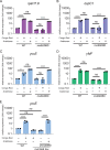
Sequestration of H-NS in S. flexneri leads to ipaH7.8, ospC1, yccE, and yfdF expression in nonpermissive conditions. Schematic of the H-NS dominant negative overexpression system where a truncated H-NS (ΔDBD) was induced by arabinose and oligomerizes with endogenous H-NS preventing DNA-binding and transcriptional repression. The S. flexneri WT strain with the dominant negative H-NS construct was grown at 30°C or 37°C with either no arabinose (−Ara), with arabinose (+Ara), or no arabinose but with Congo red (−Ara +CR) and qPCR was performed for mRNA expression levels of (B) ipaH7.8, (C) ospC1, (D) yccE, and (E) yfdF. Data shown are the averages of three independent biological experiments and the error bars represent standard deviation. One-way ANOVA with Tukey’s multiple-comparison test was performed; ns, not significant; ***, P = 0.0002; ****, P < 0.0001.
FIG 6
FIG 6
MxiE is not necessary for the expression of ipaH7.8, ospC1, yccE, or yfdF when H-NS is depleted. The S. flexneri WT and mxiEΔDBD strains with the dominant negative H-NS construct (phnsΔDBD) were grown at 37°C with or without Congo red and/or Arabinose to induce expression of phnsΔDBD and qPCR was performed for mRNA expression levels of (A) ipaH7.8, (B) ospC1, (C) yccE, and (D) yfdF relative to WT. An S. flexneri strain with a mutated MxiE box upstream of yccE without and with the dominant negative H-NS construct was grown at 37°C with or without Congo red and/or Arabinose to induce expression of phnsΔDBD and qPCR was performed for the mRNA expression level of (E) yccE compared to WT. Data shown are the averages of three independent biological experiments and the error bars represent standard deviation. One-way ANOVA with Tukey’s multiple-comparison test was performed; ns, not significant; **, P = 0.008 (ospC1); P = 0.0013 (SF1005); ***, P = 0.0003; ****, P < 0.0001.
FIG 7
FIG 7
VirB is not required for the expression of ipaH7.8 or yccE upon MxiE/IpgC overexpression. The S. flexneri WT, ΔvirB deletion strain, and ΔvirB with pMMB mxiE and pBAD ipgC were grown at 37°C with Congo red and with either no induction (X) or induction of both ipgC and mxiE (+Ara +IPTG) and qPCR was performed for mRNA expression levels of (A) ipaH7.8, (B) yccE, and (C) ipgD relative to WT. Data shown are the averages of three independent biological experiments and the error bars represent standard deviation. One-way ANOVA with Tukey’s multiple-comparison test was performed; ns, not significant; **, P = 0.0013; ***, P = 0.0001 (ΔvirB); ***, P = 0.0002 (+Ara+IPTG); ****, P < 0.0001.
FIG 8
FIG 8
Proposed model of MxiE- and H-NS-dependent regulation in S. flexneri. Before T3SS activation at 37°C, MxiE is bound by the antiactivator, OspD1, whose chaperone is Spa15, and IpgC is functioning as a chaperone for the translocon proteins, IpaB and IpaC. H-NS is repressing the transcription of MxiE-dependent genes. Upon T3SS activation, IpaB and IpaC form the translocon pore and OspD1 is secreted. Transcription of MxiE-dependent genes is activated when MxiE and IpgC are both free to interact and MxiE binds to the box sequence upstream, which results in the counter-silencing of H-NS.

References

    1. Musher DM, Musher BL. 2004. Contagious acute gastrointestinal infections. N Engl J Med 351:2417–2427. doi:10.1056/NEJMra041837. - DOI - PubMed
    1. DuPont HL, Levine MM, Hornick RB, Formal SB. 1989. Inoculum size in shigellosis and implications for expected mode of transmission. J Infect Dis 159:1126–1128. doi:10.1093/infdis/159.6.1126. - DOI - PubMed
    1. Sansonetti PJ. 1998. Molecular and cellular mechanisms of invasion of the intestinal barrier by enteric pathogens. The paradigm of Shigella. Folia Microbiol (Praha) 43:239–246. doi:10.1007/BF02818608. - DOI - PubMed
    1. Yum LK, Byndloss MX, Feldman SH, Agaisse H. 2019. Critical role of bacterial dissemination in an infant rabbit model of bacillary dysentery. Nat Commun 10:1826. doi:10.1038/s41467-019-09808-4. - DOI - PMC - PubMed
    1. Galan JE, Lara-Tejero M, Marlovits TC, Wagner S. 2014. Bacterial type III secretion systems: specialized nanomachines for protein delivery into target cells. Annu Rev Microbiol 68:415–438. doi:10.1146/annurev-micro-092412-155725. - DOI - PMC - PubMed

Publication types

MeSH terms

LinkOut - more resources