. 2023 Jan 2;155(1):e202213249.
doi: 10.1085/jgp.202213249. Epub 2022 Nov 8.

Variants of the myosin interacting-heads motif

Affiliations

Variants of the myosin interacting-heads motif

Raúl Padrón et al. J Gen Physiol. .

Abstract

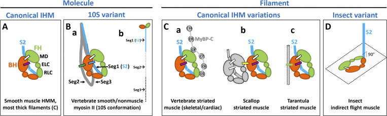
Under relaxing conditions, the two heads of myosin II interact with each other and with the proximal part (S2) of the myosin tail, establishing the interacting-heads motif (IHM), found in myosin molecules and thick filaments of muscle and nonmuscle cells. The IHM is normally thought of as a single, unique structure, but there are several variants. In the simplest ("canonical") IHM, occurring in most relaxed thick filaments and in heavy meromyosin, the interacting heads bend back and interact with S2, and the motif lies parallel to the filament surface. In one variant, occurring in insect indirect flight muscle, there is no S2-head interaction and the motif is perpendicular to the filament. In a second variant, found in smooth and nonmuscle single myosin molecules in their inhibited (10S) conformation, S2 is shifted ∼20 Å from the canonical form and the tail folds twice and wraps around the interacting heads. These molecule and filament IHM variants have important energetic and pathophysiological consequences. (1) The canonical motif, with S2-head interaction, correlates with the super-relaxed (SRX) state of myosin. The absence of S2-head interaction in insects may account for the lower stability of this IHM and apparent absence of SRX in indirect flight muscle, contributing to the quick initiation of flight in insects. (2) The ∼20 Å shift of S2 in 10S myosin molecules means that S2-head interactions are different from those in the canonical IHM. This variant therefore cannot be used to analyze the impact of myosin mutations on S2-head interactions that occur in filaments, as has been proposed. It can be used, instead, to analyze the structural impact of mutations in smooth and nonmuscle myosin.


Figures

Figure 1.
Figure 1.
The canonical myosin IHM and its variants. (A) Canonical IHM present in smooth muscle HMM and most thick filaments (see C), showing the blocked and free heads (BH, orange; FH, green), with BH–FH interaction (white bar) and BH–S2 interaction (purple bar). MD, myosin II motor domain; RLC, myosin II regulatory light chain; ELC, myosin II essential light chain. (B) 10S variant present in isolated vertebrate smooth and nonmuscle myosin II molecules in their inhibited, folded conformation, with interactions between BH and FH (white bar), BH and tail segments 1 and 3 (blue bar), and the BH and tail segment 2 (green bar). Inset b shows myosin molecule with tail extended. (C) IHM variations in thick filaments, where the canonical IHM placed in a thick filament environment may undergo additional (intermolecular) interactions: (a) vertebrate striated, showing hypothetical interaction with MyBP-C; (b) scallop striated, with additional interactions between other IHMs in the same crown (yellow bar); and (c) tarantula striated, with additional interaction (green bar) between BH and S2 from IHM in the axially adjacent crown. IHMs in all these variations are parallel to the filament surface. (D) IHM variant in Lethocerus indirect flight muscle thick filaments (insect variant), where IHM is perpendicular to S2 and the filament surface, showing BH–FH interactions (white bar) but no interaction with S2 (blue).
Figure 2.
Figure 2.
20 Å difference in position of segment 1 of the 10S IHM variant compared with S2 in the canonical IHM. (A) PDB structures for the 10S molecule variant (6XE9 [yellow], 6Z47 [purple], 7MF3 [green]) show similar locations for the three tail segments (Segs1–3). (B) Top: Comparison of 10S variant (PDB accession no. 6XE9 [yellow]) with tarantula canonical IHM (PDB accession no. 3JBH [blue]) with light chains removed for simplicity, showing Seg1 of the molecule shifted ∼20 Å closer to the tip of the BH than S2 in the filament (red double arrow). Bottom: Horizontal slice of top view, rotated 90° to show Seg1 of the molecule ∼10 Å above the BH (yellow), implying little or no interaction, while S2 in the tarantula canonical IHM (blue) is within 3 Å of the BH mesa and could interact. (C) Top: Superposition of the cryo-EM densities of the 10S variant (EMD accesison no. EMD-22145; low-pass filtered to 10 Å resolution, yellow) and the ∼12 Å resolution tarantula filament canonical IHM (blue), showing a ∼22 Å S2–Seg1 shift (red double arrow). This view is rotated 180° around a vertical axis compared with A and B to best reveal S2/Seg1, on the rear of the IHM. Bottom: Individual 3-D map densities showing that Seg1 (red arrow) does not interact with the BH in the molecule variant (yellow), while S2 (red arrow) of the tarantula canonical IHM (blue) docks on the BH mesa. A and B were made with UCSF Chimera, C was made with UCSF ChimeraX.

References

    1. Abrams, J., Einhorn Z., Seiler C., Zong A.B., Sweeney H.L., and Pack M.. 2016. Graded effects of unregulated smooth muscle myosin on intestinal architecture, intestinal motility and vascular function in zebrafish. Dis. Model. Mech. 9:529–540. 10.1242/dmm.023309 - DOI - PMC - PubMed
    1. Al-Khayat, H.A., Kensler R.W., Squire J.M., Marston S.B., and Morris E.P.. 2013. Atomic model of the human cardiac muscle myosin filament. Proc. Natl. Acad. Sci. U.S.A. 110:318–323. 10.1073/pnas.1212708110 - DOI - PMC - PubMed
    1. Alamo, L., Ware J.S., Pinto A., Gillilan R.E., Seidman J.G., Seidman C.E., and Padron R.. 2017. b. Effects of myosin variants on interacting-heads motif explain distinct hypertrophic and dilated cardiomyopathy phenotypes. Elife. 6. e24634. 10.7554/eLife.24634 - DOI - PMC - PubMed
    1. Alamo, L., Pinto A., Sulbaran G., Mavarez J., and Padron R.. 2018. Lessons from a tarantula: New insights into myosin interacting-heads motif evolution and its implications on disease. Biophys. Rev. 10:1465–1477. 10.1007/s12551-017-0292-4 - DOI - PMC - PubMed
    1. Alamo, L., Qi D., Wriggers W., Pinto A., Zhu J., Bilbao A., Gillilan R.E., Hu S., and Padron R.. 2016. Conserved intramolecular interactions maintain myosin interacting-heads motifs explaining tarantula muscle super-relaxed state structural basis. J. Mol. Biol. 428:1142–1164. 10.1016/j.jmb.2016.01.027 - DOI - PMC - PubMed

Publication types