. 2022 Nov 8;10(1):47.
doi: 10.1186/s40635-022-00474-3.

Effect of noradrenaline on propofol-induced mitochondrial dysfunction in human skeletal muscle cells

Affiliations

Effect of noradrenaline on propofol-induced mitochondrial dysfunction in human skeletal muscle cells

Adéla Krajčová et al. Intensive Care Med Exp. .

Abstract

Background: Mitochondrial dysfunction is a hallmark of both critical illness and propofol infusion syndrome and its severity seems to be proportional to the doses of noradrenaline, which patients are receiving. We comprehensively studied the effects of noradrenaline on cellular bioenergetics and mitochondrial biology in human skeletal muscle cells with and without propofol-induced mitochondrial dysfunction.

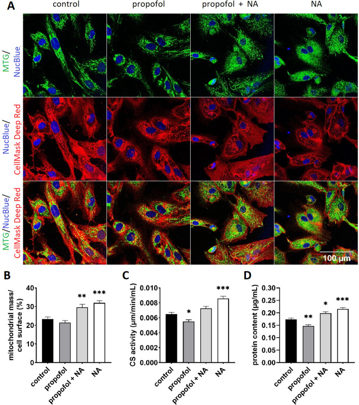
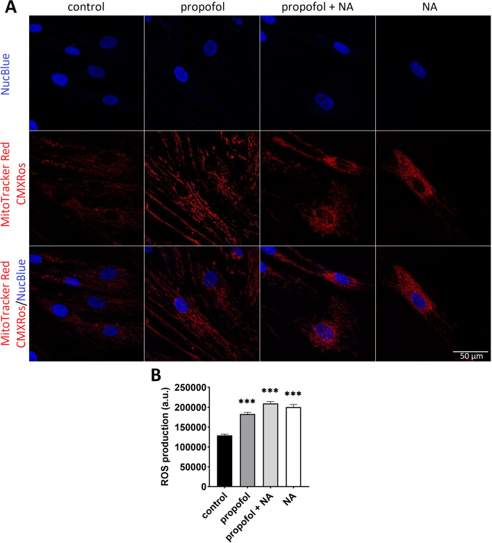
Methods: Human skeletal muscle cells were isolated from vastus lateralis biopsies from patients undergoing elective hip replacement surgery (n = 14) or healthy volunteers (n = 4). After long-term (96 h) exposure to propofol (10 µg/mL), noradrenaline (100 µM), or both, energy metabolism was assessed by extracellular flux analysis and substrate oxidation assays using [14C] palmitic and [14C(U)] lactic acid. Mitochondrial membrane potential, morphology and reactive oxygen species production were analysed by confocal laser scanning microscopy. Mitochondrial mass was assessed both spectrophotometrically and by confocal laser scanning microscopy.

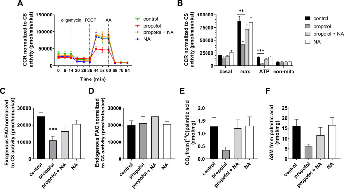
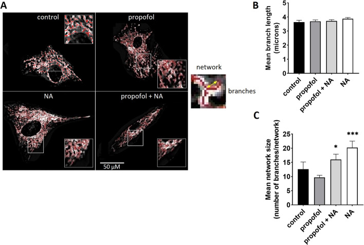
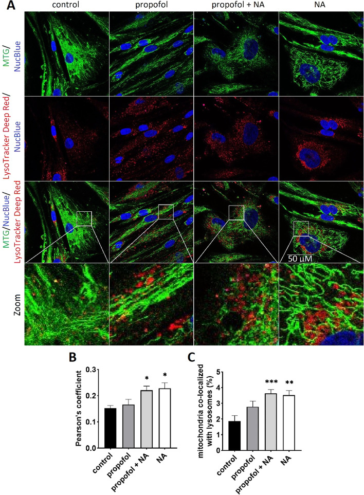
Results: Propofol moderately reduced mitochondrial mass and induced bioenergetic dysfunction, such as a reduction of maximum electron transfer chain capacity, ATP synthesis and profound inhibition of exogenous fatty acid oxidation. Noradrenaline exposure increased mitochondrial network size and turnover in both propofol treated and untreated cells as apparent from increased co-localization with lysosomes. After adjustment to mitochondrial mass, noradrenaline did not affect mitochondrial functional parameters in naïve cells, but it significantly reduced the degree of mitochondrial dysfunction induced by propofol co-exposure. The fatty acid oxidation capacity was restored almost completely by noradrenaline co-exposure, most likely due to restoration of the capacity to transfer long-chain fatty acid to mitochondria. Both propofol and noradrenaline reduced mitochondrial membrane potential and increased reactive oxygen species production, but their effects were not additive.

Conclusions: Noradrenaline prevents rather than aggravates propofol-induced impairment of mitochondrial functions in human skeletal muscle cells. Its effects on bioenergetic dysfunctions of other origins, such as sepsis, remain to be demonstrated.

Keywords: Critical illness; Mitochondrial dysfunction; Noradrenaline; Propofol infusion syndrome; Skeletal muscle.


Conflict of interest statement

The authors declare that they have no competing interests.

Figures

Fig. 1
Fig. 1
Extracellular flux analysis and substrate oxidation assays. A Real-time measurement of OCR at baseline and after sequential injection of oligomycin, FCCP and antimycin A. Each data-point represents the mean of 7 subjects measured in tri- or tetraplicates. Values are normalized to mitochondrial content (CS activity). B Global mitochondrial parameters. Basal respiration, maximal respiratory capacity, ATP production and non-mitochondrial respiration determined from OCRs shown in part A. C Exogenous oxidation of fatty acids after palmitate addition to the medium during measurement. D Endogenous oxidation of fatty acids in palmitate-free medium. E CO2 production from [14C]palmitic acid (complete oxidation). F Oxidation of [14C]palmitic acid to ASM (incomplete oxidation). Error bars in each graph indicate standard error of the mean. **p < 0.01, *** p < 0.001 vs. control group. AA antimycin A, FCCP  carbonyl cyanide-4- (trifluoromethoxy)phenylhydrazone, FAO  fatty acid oxidation, NA  noradrenaline, OCR  oxygen consumption rate
Fig. 2
Fig. 2
A and B Analysis of mitochondrial mass by confocal imaging of exposed myoblasts. A Representative confocal images of each channel after dual staining with Mitotracker Green™ FM (accumulating in mitochondria) and CellMask™ Deep Red (binding into the cell membrane). Additionally, all cells were stained with nuclear blue-fluorescent probe NucBlue. Experiments were performed at least at 50 cells per each condition from 3 independent measurements (= cells established from 3 individual subjects). B Mitochondrial mass calculated as a fraction (%) of a cell surface area in 2D cross-sectional images. Mitochondrial mass (mitochondrial footprint) was analysed as a sum of positive pixels (binary image) per cell representing mitochondrial area using ImageJ™ tool “MINA”. C Activity of CS enzyme measured spectrophotometrically. D Total protein content from frozen cell pellets was determined using Bradford assay as described elsewhere [53]. For both CS activity and protein content measurement, experiments were performed in n = 7 replicates in tri- or tetraplicates. Error bars indicate standard error of the mean. *p < 0.05, **p < 0.01, ***p < 0.001 vs. control group. CS citrate synthase, MTG MitoTracker Green™ FM, NA noradrenaline
Fig. 3
Fig. 3
Mitochondrial morphology. Confocal images of myoblasts stained with MitoTracker™ Green FM analysed with the ImageJ™ plugin “MINA”. A Representative skeletonized images show mitochondrial network in different groups (left). Measurements were performed at least at 50 cells per each group from 3 independent experiments (= 3 individual subjects). Yellow arrow (right) shows one of the three branches in an example of mitochondrial network. B and C Mean branch length and mean network size (= number of branches per network) were analysed in each cell separately. Error bars indicate standard error of the mean. *p < 0.05, ***p < 0.001 vs. control group. NA noradrenaline
Fig. 4
Fig. 4
Co-localization of mitochondria with lysosomes. A Representative confocal images of each channel after dual staining with Mitotracker Green™ FM (accumulating in mitochondria) and LysoTracker™ Deep Red (binding to acidic lysosomes). Additionally, all cells were stained with blue-fluorescent probe NucBlue to label nuclei. Experiments were performed at least at 40 cells per each condition from 3 independent measurements (= 3 individual subjects). B Pearson’s coefficient, characterizing a degree of overlap between labelled mitochondria and lysosomes. The quantification of the co-localization was performed using ImageJ™ plugin “JACoP”. C Percent co-localization was calculated by total area of co-localized lysosomes (red channel) over total area of mitochondria (green channel). Error bars indicate standard error of the mean. *p < 0.05, **p < 0.01, ***p < 0.001 vs. control group. NA noradrenaline
Fig. 5
Fig. 5
ROS production. A Representative confocal images from myoblasts stained with MitoTracker Red CM-H2XRos to detect accumulation of mitochondrial specific-reactive oxygen species. Experiments were performed on at least 90 cells per group from n = 3 independent experiments (with cells from 3 individual subjects). B Determination of ROS production in different groups. Error bars indicate standard error of the mean. ***p < 0.001 vs. control group. NA noradrenaline

References

    1. Brealey D, Brand M, Hargreaves I, Heales S, Land J, Smolenski R, Davies NA, Cooper CE, Singer M. Association between mitochondrial dysfunction and severity and outcome of septic shock. Lancet. 2002;360(9328):219–223. doi: 10.1016/S0140-6736(02)09459-X. - DOI - PubMed
    1. Supinski GS, Schroder EA, Callahan LA. Mitochondria and critical illness. Chest. 2020;157(2):310–322. doi: 10.1016/j.chest.2019.08.2182. - DOI - PMC - PubMed
    1. Branca D, Vincenti E, Scutari G. Influence of the anesthetic 2,6-diisopropylphenol (propofol) on isolated rat heart mitochondria. Comp Biochem Physiol C Pharmacol Toxicol Endocrinol. 1995;110(1):41–45. doi: 10.1016/0742-8413(94)00078-o. - DOI - PubMed
    1. Branca D, Roberti MS, Lorenzin P, Vincenti E, Scutari G. Influence of the anesthetic 2,6-diisopropylphenol on the oxidative phosphorylation of isolated rat liver mitochondria. Biochem Pharmacol. 1991;42(1):87–90. doi: 10.1016/0006-2952(91)90684-w. - DOI - PubMed
    1. Branca D, Roberti MS, Vincenti E, Scutari G. Uncoupling effect of the general anesthetic 2,6-diisopropylphenol in isolated rat liver mitochondria. Arch Biochem Biophys. 1991;290(2):517–521. doi: 10.1016/0003-9861(91)90575-4. - DOI - PubMed

LinkOut - more resources