. 2022 Nov 8;20(11):e3001851.
doi: 10.1371/journal.pbio.3001851. eCollection 2022 Nov.

Hippo signaling pathway activation during SARS-CoV-2 infection contributes to host antiviral response

Affiliations

Hippo signaling pathway activation during SARS-CoV-2 infection contributes to host antiviral response

Gustavo Garcia Jr et al. PLoS Biol. .

Abstract

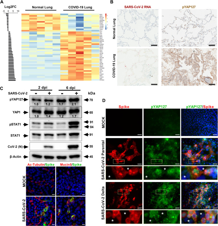
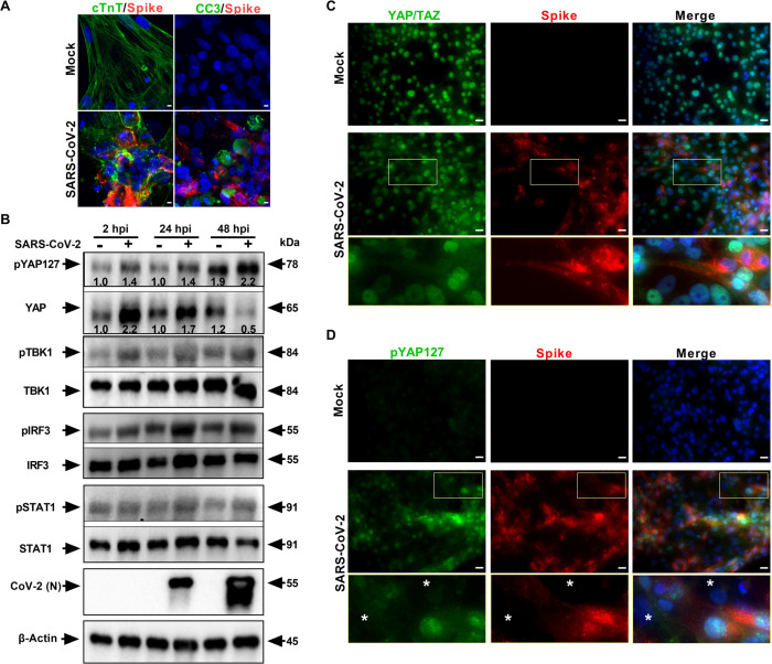
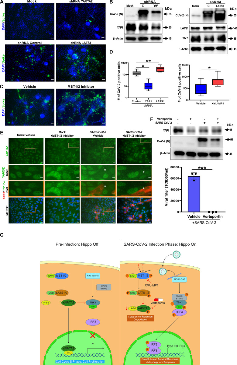
Severe Acute Respiratory Syndrome Coronavirus 2 (SARS-CoV-2), responsible for the Coronavirus Disease 2019 (COVID-19) pandemic, causes respiratory failure and damage to multiple organ systems. The emergence of viral variants poses a risk of vaccine failures and prolongation of the pandemic. However, our understanding of the molecular basis of SARS-CoV-2 infection and subsequent COVID-19 pathophysiology is limited. In this study, we have uncovered a critical role for the evolutionarily conserved Hippo signaling pathway in COVID-19 pathogenesis. Given the complexity of COVID-19-associated cell injury and immunopathogenesis processes, we investigated Hippo pathway dynamics in SARS-CoV-2 infection by utilizing COVID-19 lung samples and human cell models based on pluripotent stem cell-derived cardiomyocytes (PSC-CMs) and human primary lung air-liquid interface (ALI) cultures. SARS-CoV-2 infection caused activation of the Hippo signaling pathway in COVID-19 lung and in vitro cultures. Both parental and Delta variant of concern (VOC) strains induced Hippo pathway. The chemical inhibition and gene knockdown of upstream kinases MST1/2 and LATS1 resulted in significantly enhanced SARS-CoV-2 replication, indicating antiviral roles. Verteporfin, a pharmacological inhibitor of the Hippo pathway downstream transactivator, YAP, significantly reduced virus replication. These results delineate a direct antiviral role for Hippo signaling in SARS-CoV-2 infection and the potential for this pathway to be pharmacologically targeted to treat COVID-19.


Conflict of interest statement

The authors have declared that no competing interests exist.

Figures

Fig 1
Fig 1. Hippo signaling pathway is activated in lung tissues of COVID-19 and infected human lung in vitro cell cultures.
(A) Transcriptome analysis of normal and COVID-19 autopsy lung samples (n = 5) is presented. Bar graph shows Log2FC (FC >1) of 45 Hippo signaling pathway genes that were differentially expressed in COVID-19 lung samples (p < 0.01). The adjacent heatmap depicts Z scores as expression levels of these 45 DEGs. Blue and red colors represent 12 down-regulated and 33 up-regulated genes, respectively. The KEGG pathway database was used to examine the Hippo signaling pathway genes in the differentially expressed gene list. Individual quantitative observations that underlie the data summarized can be located under the Supporting Information File as S1 Data. (B) IHC of COVID-19 lung autopsy tissue shows high level of phospho-YAP Ser127 protein (dark brown). SARS-CoV-2 RNA (red) presence in COVID-19 lung was confirmed by RNA-FISH (RNAscope) (scale bar: 100 μm). (C) Western blot analyses of lung ALI cells show activation of Hippo-STAT1 pathways during SARS-CoV-2 infection (MOI 1). Density of bands compared to that of 2 dpi mock is provided for pYAP127 and YAP. Proximal lung air interface cultures are susceptible to SARS-CoV-2 infection. Antibody targeting Spike protein (green) was used for probing infection by immunohistochemistry. Cell-specific markers, such as Ac-tubulin (ciliated cells) and Mucin5 (mucus cells) were detected by antibody probes (red) to define infected cell types (6 dpi). Scale bar: 10 μm. (D) Human airway epithelial cells (Calu-3) were analyzed with IHC for pYAP127 and SARS-CoV-2 Spike protein (red) in uninfected (Mock) and infected cells at 48 hours post-infection. Parental and Delta strains were used for infection studies (MOI 0.1). Scale bar: 50 μm. Insets highlight phospho-YAP S127 in infected cells, and white asterisks denote uninfected cells. Representative data from 3 independent studies are provided. ALI, air–liquid interface; COVID-19, Coronavirus Disease 2019; DEG, differentially expressed gene; IHC, immunohistochemistry; KEGG, Kyoto Encyclopedia of Genes and Genomes; MOI, multiplicity of infection; SARS-CoV-2, Severe Acute Respiratory Syndrome Coronavirus 2.
Fig 2
Fig 2. SARS-CoV-2 infection activates Hippo and antiviral RLR-STAT pathways in hPSC-CMs.
(A) Confocal image analysis of SARS-CoV-2 Spike protein (red) in infected cardiomyocytes shows extensive damage to cTNT positive (green) cells, which undergo apoptotic cell death (green; cleaved caspase 3). Scale bar: 5 μm; n = 6 independent experiments. (B) Western blot analyzes show activation of Hippo, RLR, and STAT1 pathways. Phospho-YAP127 level is increased upon SARS-CoV-2 infection (MOI 0.01); n = 3 independent experiments. Density of bands compared to that of 2 hpi mock is provided for pYAP127 and YAP. (C) Immunohistochemistry analysis of SARS-CoV-2 Spike protein in infected hPSC-CMs at 48 hpi reveals cytoplasmic localization and depletion of YAP/TAZ and (D) increase in pYAP127 level. Scale bar: 25 μm. Insets denote total YAP or pYAP127 in infected cells, and white asterisks highlight uninfected cells. hPSC-CM, human pluripotent stem cell-derived cardiomyocyte; MOI, multiplicity of infection; SARS-CoV-2, Severe Acute Respiratory Syndrome Coronavirus 2.
Fig 3
Fig 3. ShRNA-mediated knockdown and pharmacological modulation of SARS-CoV-2 replication.
(A) IHC analysis of shRNA-mediated knockdown of YAP1 and LATS1-specific shRNAs showed efficiently reduced or increased SARS-CoV-2 replication (green) relative to shRNA control, respectively, in hPSC-CMs. Scale bar: 50 μm. (B) Western blot analysis of shRNA-mediated knockdown of YAP1 and LATS1 respective protein expression. (Con: Control shRNA). (C) IHC images of XMU-MP-1 (MST1/2 inhibitor) and vehicle-treated hPSC-CMs are shown. Note: XMU-MP-1 increased SARS-CoV-2 replication (green) in hPSC-CM. (D) Graphs depict quantification of SARS-CoV-2 positive cells in infected hPSC-CM respective to panels A and C. Data represented as the average of technical replicates and SEM. Student t test. **P > 0.001; n = 2 independent experiments. (E) IHC Images show YAP/TAZ protein (green) and SARS-CoV-2 Spike antigen (red) in Calu-3 cells. Note, MST1/2 inhibitor-treated Calu-3 cells have higher number of infected cells. Inset and white asterisk hovers infected cells showing depletion of YAP/TAZ. Scale bar: 25 μm. Inset scale bar: 10 μm. (F) Western blot analysis of Calu-3 cells treated with Verteporfin (1 μm) and SARS-CoV-2 infection. Drug treatment resulted in reduction in SARS-CoV-2 infection. Graph shows the viral titer (TCID50/ml) measurement of infected as well as treated Calu-3 culture supernatant (representative data from 2 independent experiments). (G) Schematic diagram of our hypothetical model integrating Hippo and TBK1 signaling pathways during preinfection (Hippo off) and SARS-CoV-2 infection states (Hippo on). c-GAS, cyclic GMP-AMP synthase; IKKe, inhibitor of nuclear factor kappa B kinase subunit epsilon; MAVS, mitochondrial antiviral-signaling protein; RIG-I, retinoic acid inducible gene I protein; STING, stimulator of interferon response cGAMP interactor 1; TEAD, TEA domain transcription factors. Individual quantitative observations that underlie the data summarized in Fig 3D and 3F can be located under the Supporting Information File as S2 and S3 Data files, respectively. hPSC-CM, human pluripotent stem cell-derived cardiomyocyte; SARS‑CoV‑2, Severe Acute Respiratory Syndrome Coronavirus 2.

Update of

Similar articles

Cited by

References

    1. Ramaiah A, Arumugaswami V. Insights into Cross-species Evolution of Novel Human Coronavirus 2019-nCoV and Defining Immune Determinants for Vaccine Development. bioRxiv. 2020:2020.01.29.925867.
    1. Wu F, Zhao S, Yu B, Chen Y-M, Wang W, Song Z-G, et al.. A new coronavirus associated with human respiratory disease in China. Nature. 2020;579(7798):265–269. doi: 10.1038/s41586-020-2008-3 - DOI - PMC - PubMed
    1. Zhou P, Yang X-L, Wang X-G, Hu B, Zhang L, Zhang W, et al.. A pneumonia outbreak associated with a new coronavirus of probable bat origin. Nature. 2020;579(7798):270–273. doi: 10.1038/s41586-020-2012-7 - DOI - PMC - PubMed
    1. Xu H, Zhong L, Deng J, Peng J, Dan H, Zeng X, et al.. High expression of ACE2 receptor of 2019-nCoV on the epithelial cells of oral mucosa. Int J Oral Sci. 2020;12(1):8. doi: 10.1038/s41368-020-0074-x - DOI - PMC - PubMed
    1. Wang K, Chen W, Zhang Z, Deng Y, Lian JQ, Du P, et al.. CD147-spike protein is a novel route for SARS-CoV-2 infection to host cells. Signal Transduct Target Ther. 2020;5(1):283. doi: 10.1038/s41392-020-00426-x - DOI - PMC - PubMed

Publication types

Substances

Supplementary concepts