Review
. 2022 Dec 1;276(6):943-956.
doi: 10.1097/SLA.0000000000005434. Epub 2022 Mar 9.

Hepatic Artery Infusion Pumps: A Surgical Toolkit for Intraoperative Decision-Making and Management of Hepatic Artery Infusion-Specific Complications

Affiliations
Review

Hepatic Artery Infusion Pumps: A Surgical Toolkit for Intraoperative Decision-Making and Management of Hepatic Artery Infusion-Specific Complications

Jeremy M Sharib et al. Ann Surg. .

Abstract

Background: Hepatic artery infusion (HAI) is a liver-directed therapy that delivers high-dose chemotherapy to the liver through the hepatic arterial system for colorectal liver metastases and intrahepatic cholangiocarcinoma. Utilization of HAI is rapidly expanding worldwide.

Objective and methods: This review describes the conduct of HAI pump implantation, with focus on common technical pitfalls and their associated solutions. Perioperative identification and management of common postoperative complications is also described.

Results: HAI therapy is most commonly performed with the surgical implantation of a subcutaneous pump, and placement of its catheter into the hepatic arterial system for inline flow of pump chemotherapy directly to the liver. Intraoperative challenges and abnormal hepatic perfusion can arise due to aberrant anatomy, vascular disease, technical or patient factors. However, solutions to prevent or overcome technical pitfalls are present for the majority of cases. Postoperative HAI-specific complications arise in 22% to 28% of patients in the form of pump pocket (8%-18%), catheter (10%-26%), vascular (5%-10%), or biliary (2%-8%) complications. The majority of patients can be rescued from these complications with early identification and aggressive intervention to continue to deliver safe and effective HAI therapy.

Conclusions: This HAI toolkit provides the HAI team a reference to manage commonly encountered HAI-specific perioperative obstacles and complications. Overcoming these challenges is critical to ensure safe and effective pump implantation and delivery of HAI therapy, and key to successful implementation of new programs and expansion of HAI to patients who may benefit from such a highly specialized treatment strategy.


Conflict of interest statement

The authors report no conflict of interests.

Figures

FIGURE 1.
FIGURE 1.
Hepatic artery infusion (HAI) pump placement in normal anatomy and common variants. A, HAI pump placement with conventional arterial anatomy. B, Replaced left hepatic artery (originating from the left gastric) in situ and following HAI pump placement (C). D, Replaced right hepatic artery (originating from the SMA) in situ and following HAI pump placement after ligation of the aberrant artery lateral to the common bile duct (E). F, Intraoperative photos of uncommon arterial variant: common hepatic artery (long white arrow) branches into the right (asterisk) and left (triangle) hepatic arteries and the GDA (short white arrow) emanates from the right hepatic artery. An accessory right hepatic artery (looped) is also present. G, Following ligation and division of the accessory right hepatic artery and native left hepatic artery (clipped), the GDA (short white arrow) is cannulated which facilitated single vessel in-line flow to the liver via the right hepatic artery (asterisk).
FIGURE 2.
FIGURE 2.
Introperative and postoperative perfusion studies. A, Intraoperative blue dye and (B) indocyanine green (ICG) injection confirming normal bilobar perfusion. C, Incomplete perfusion with blue dye only perfusing left hemiliver due to competitive flow from accessory right hepatic artery (yellow vessel loop). Note demarcation along Cantlie’s line. Bilobar flow via proper hepatic artery in-line from HAI pump was achieved after ligation of accessory vessel (not shown). D, Extrahepatic perfusion (EHP) to duodenal bulb (black arrow), which is resolved following further dissection along GDA and periportal lymphatics, confirmed with ICG (E). F, Planar images from a normal postoperative perfusion study of the anterior (left) and posterior (right) abdomen with perfusion confined to the liver. Note heterogeneous perfusion in the liver reflects known liver metastases. G, In contrast, EHP is identified with Technetium-99 uptake in duodenal bulb (white arrows). H, This was further confirmed on axial SPECT/CT to reflect EHP to the pancreatic uncinate and duodenum (white arrows). I, Confirmation of EHP by celiac arteriography demonstrates an anomalous origin of the posterior superior pancreaticoduodenal artery (white arrows) from an accessory segment 6 artery that arises from the distal proper hepatic artery as the source of nontarget perfusion. J, After coil embolization of the pancreaticoduodenal and accessory segment 6 artery (white arrow), there is resolution of nontarget artery opacification on angiography. Subsequent scintigraphy confirmed absence of radiotracer uptake in the pancreatic head and duodenum (inset). Note, HAI catheter remains in GDA and was functional following embolization.
FIGURE 3.
FIGURE 3.
Pump-and pocket-related complications. A, Erythema and blistering over pump pocket which was warm on physical exam with (B) corresponding CT findings of pump pocket seroma, which resolved after aspiration of serous fluid and empiric antibiotics. C, Coronal maximum intensity projection following pump relocation to right abdomen using catheter extension tunneled through the subcutaneous tissue (white arrow). D, Pump erosion presenting at blister (left) and full thickness ulcer with exposed device (right). E, Fluoroscopy shows evidence of a flipped pump: note the orientation of the access port and catheter connector (black arrow) is superior-lateral rather than inferior indicating the opening faces inward and accounts for the inability to access the pump. In addition, the catheter has disconnected from the intraabdominal portion and only a short length remains attached to the pump (white arrow), whereas the intraabdominal portion terminates inferiorly (out of view). F, Catheter migration through pump pocket incision following pump removal. This was managed by ligation and trimming of catheter at the level of the fascia and closure of the anterior rectus sheath defect.
FIGURE 4.
FIGURE 4.
Catheter-related complications. (A–C) Catheter disloged from GDA. A, Axial CT of a patient with fluid incidentally noted adjacent to GDA (white arrow) and pump catheter tubing that dislodged from the GDA insertion. B, 3D-rendering of same patient shows the catheter tip (black arrow) paralleling the course of the common hepatic artery (white arrow), and is no longer sited within the vessel. Optimal positioning of the catheter will mimic the position of the GDA and is commonly more perpen-dicular to the common hepatic artery. C, Contrast injection through the pump demonstrates opacification from the pump catheter tip (black arrow) into an amorphous collection (white arrows) that was confirmed to be extraluminal on cone-beam CT scan (not shown). D, Coronal maximum intensity projection in a patient with an HAI pump in the left abdomen. The catheter is correctly positioned distally at the hepatic hilum (white arrow), whereas the tubing is inappropriately disconnected proximally, residing freely in the pelvis (long black arrow). The tubing disconnected close to the pump; a short segment of tubing is seen exiting the pump proximal to the tubing fracture (short black arrow). E, CT image of a catheter which appears intraluminal (white arrow) at the gastrojejunostomy of a patient with a prior gastric bypass. F, Intraluminal catheter erosion (white arrow) was confirmed on upper endoscopy adjacent to an Axios stent placed for access to the gastric remnant, which likely created friction leading to catheter erosion. Jejunal ulcer healed following catheter removal and laparoscopic repair.
FIGURE 5.
FIGURE 5.
Vascular complications. (A–C) Arterial dissection. A, Axial CT angiography of GDA dissection of the common hepatic artery which has propagated beyond the bifurcation of the right and left hepatic arteries. B, In the same patient, contrast injection through the pump demonstrates opacification from the pump catheter tip (black arrow) into the false lumen of an extensive dissection (white arrow) that extends from the common hepatic artery into the right hepatic artery, with opacification of right hepatic artery branches (black asterisk) via distal right hepatic artery fenestrations into the true lumen, but does not communicate with the left hepatic artery (nonopacified). C, Celiac arteriogram confirms a narrow common hepatic artery (true lumen, black arrow) with filling only of the left hepatic artery (white arrow). (D–F) Pseudoaneurysm. D, Abdominal CT shows dense layered fluid (white arrows) surrounding the catheter insertion into the GDA, concerning for pseudoaneurysm. E, Celiac arteriogram of the same patient demonstrates active filling of a pseudoaneurysm (white arrow) originating at the site of the pump catheter tip (black arrow). F, Rescue was achieved by deployment of a self-expanding covered stent graft positioned to traverse the gastroduodenal artery origin (margins denoted by bracket), to prevent active filling of the pseudoaneurysm. Note the residual contrast within the now thrombosed pseudoaneurysm (white arrow).
FIGURE 6.
FIGURE 6.
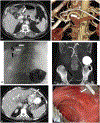
Biliary complications. A, Short segment right hepatic duct stricture and significant dilation of left hepatic ductal system on MRCP. B, Transhepatic cholangiogram demonstrates a central stricture involving the origins of both the right and left hepatic ducts (white arrow) with otherwise normal common bile duct and peripheral right ducts. A long segment stricture of the left hepatic duct is also present. C, The central stricture was managed with percutaneous biliary drainage. D, Long segment right hepatic duct stricture identified as biliary dilatation on CT (white arrow) obtained for hyperbilirubinemia. E, Stricture at origin of right hepatic duct (black arrow) was confirmed by endoscopic retrograde cholangiography and managed by (F) balloon dilation (black arrow) and (G) deployment of an indwelling plastic biliary stent (black arrow). Note, A–C from patient 1 and D–G from patient 2.

Comment in

  • Hepatic Arterial Infusion Pump: Quo Vadis?
    Qadan M, Ryan DP. Qadan M, et al. Ann Surg. 2022 Dec 1;276(6):957-958. doi: 10.1097/SLA.0000000000005580. Epub 2022 Jul 15. Ann Surg. 2022. PMID: 35837950 No abstract available.

References

    1. Kemeny NE, Niedzwiecki D, Hollis DR, et al. Hepatic arterial infusion versus systemic therapy for hepatic metastases from colorectal cancer: a randomized trial of efficacy, quality of life, and molecular markers (CALGB 9481). J Clin Oncol. 2006;24:1395–1403. - PubMed
    1. D’Angelica MI, Correa-Gallego C, Paty PB, et al. Phase II trial of hepatic artery infusional and systemic chemotherapy for patients with unresectable hepatic metastases from colorectal cancer: conversion to resection and longterm outcomes. Ann Surg. 2015;261:353–360. - PMC - PubMed
    1. Pak LM, Kemeny NE, Capanu M, et al. Prospective phase II trial of combination hepatic artery infusion and systemic chemotherapy for unresectable colorectal liver metastases: long term results and curative potential. J Surg Oncol. 2018;117:634–643. - PMC - PubMed
    1. Dhir M, Jones HL, Shuai Y, et al. Hepatic arterial infusion in combination with modern systemic chemotherapy is associated with improved survival compared with modern systemic chemotherapy alone in patients with isolated unresectable colorectal liver metastases: a case-control study. Ann Surg Oncol. 2017;24:150–158. - PubMed
    1. Jarnagin WR, Schwartz LH, Gultekin DH, et al. Regional chemotherapy for unresectable primary liver cancer: results of a phase II clinical trial and assessment of DCE-MRI as a biomarker of survival. Ann Oncol. 2009;20:1589–1595. - PMC - PubMed

MeSH terms