. 2022 Dec 5;32(23):5057-5068.e5.
doi: 10.1016/j.cub.2022.10.028. Epub 2022 Nov 7.

Reduced mitochondria provide an essential function for the cytosolic methionine cycle

Affiliations

Reduced mitochondria provide an essential function for the cytosolic methionine cycle

Justyna Zítek et al. Curr Biol. .

Abstract

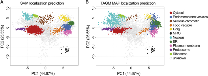
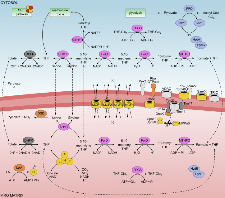
The loss of mitochondria in oxymonad protists has been associated with the redirection of the essential Fe-S cluster assembly to the cytosol. Yet as our knowledge of diverse free-living protists broadens, the list of functions of their mitochondrial-related organelles (MROs) expands. We revealed another such function in the closest oxymonad relative, Paratrimastix pyriformis, after we solved the proteome of its MRO with high accuracy, using localization of organelle proteins by isotope tagging (LOPIT). The newly assigned enzymes connect to the glycine cleavage system (GCS) and produce folate derivatives with one-carbon units and formate. These are likely to be used by the cytosolic methionine cycle involved in S-adenosyl methionine recycling. The data provide consistency with the presence of the GCS in MROs of free-living species and its absence in most endobionts, which typically lose the methionine cycle and, in the case of oxymonads, the mitochondria.

Keywords: LOPIT; Paratrimastix; glycine cleavage system; methionine cycle; mitochondrion-related organelle; one-carbon metabolism; proteome; spatial proteomics.


Conflict of interest statement

Declaration of interests The authors declare no competing interests.

Figures

None
Graphical abstract
Figure 1
Figure 1
Application of LOPIT-DC on P. pyriformis cells (A) Schematic representation of the fractionation workflow. (B) Venn diagram displaying common and unique proteins identified in four replicates. The total number of quantified proteins for each replicate is shown in brackets. (C) Distribution profile of marker proteins across ten labeled fractions. (D) Principal-component analysis (PCA) plot mapping 226 marker proteins for 11 compartments. See also Figures S1 and S2.
Figure 2
Figure 2
PCA plots with proteins assigned to compartments labeled (A) PCA projection after SVM classification and (B) PCA projection after TAGM MAP classification. See also Figure S3.
Figure 3
Figure 3
Metabolic map of P. pyriformis MRO based on LOPIT-DC analysis Proteins used as markers for classification are colored yellow, proteins involved in folate-mediated 1-carbon metabolism are colored pink, proteins involved in extended glycolysis and hydrogen production are colored blue, previously unknown-function proteins are colored light gray, and proteins present in the genome but missing in LOPIT dataset are colored dark gray with a dashed outline. H, L, P, and T, components of glycine cleavage system; SHMT, serine hydroxymethyltransferase; FolD, methylenetetrahydrofolate dehydrogenase/methenyl tetrahydrofolate cyclohydrolase; MTHFD, formate-tetrahydrofolate ligase; FPGS, folylpolyglutamate synthase; MTHFR, methylenetetrahydrofolate reductase; DHFR, dihydrofolate reductase; LplA, lipoate ligase; DSD, D-serine dehydratase; PFO, pyruvate:ferredoxin oxidoreductase; Fdx, ferredoxin; HydA, [FeFe]hydrogenase; HydE, HydF, and HydG, hydrogenase maturases; MPPαβ, mitochondrial processing peptidase subunits α and β; VDAC, voltage-dependent anion channel; TRIC, trimeric intracellular ion channel; Pex3, peroxisomal biogenesis factor 3; Cpn10, mitochondrial chaperonin 10; Cpn60, mitochondrial chaperonin 60; DnaK, chaperone protein; MCF1, MCF2, MCF3, and MCF4, mitochondrial carrier family proteins; THF, tetrahydrofolate; LA, lipoic acid. See also Data S1 and S2.
Figure 4
Figure 4
Schematic phylogeny of the Tim17/22/23 family with the positions of P. pyriformis homologs Employing a previously compiled dataset, the ML tree was inferred by IQ-TREE using the posterior mean site frequency empirical model with the Shimodaira-Hasegawa approximate likelihood ratio test/ultrafast bootstrapping strategy both supporting their phylogenetic position. See also STAR Methods.

Comment in

References

    1. Karnkowska A., Vacek V., Zubáčová Z., Treitli S.C., Petrželková R., Eme L., Novák L., Žárský V., Barlow L.D., Herman E.K., et al. A eukaryote without a mitochondrial organelle. Curr. Biol. 2016;26:1274–1284. - PubMed
    1. Roger A.J., Muñoz-Gómez S.A., Kamikawa R. The origin and diversification of mitochondria. Curr. Biol. 2017;27:R1177–R1192. - PubMed
    1. Santos H.J., Makiuchi T., Nozaki T. Reinventing an organelle: the reduced mitochondrion in parasitic protists. Trends Parasitol. 2018;34:1038–1055. - PubMed
    1. Leger M.M., Kolisko M., Kamikawa R., Stairs C.W., Kume K., Čepička I., Silberman J.D., Andersson J.O., Xu F., Yabuki A., et al. Organelles that illuminate the origins of Trichomonas hydrogenosomes and Giardia mitosomes. Nat. Ecol. Evol. 2017;1:0092. - PMC - PubMed
    1. Mi-ichi F., Abu Yousuf M.A., Nakada-Tsukui K., Nozaki T. Mitosomes in Entamoeba histolytica contain a sulfate activation pathway. Proc. Natl. Acad. Sci. USA. 2009;106:21731–21736. - PMC - PubMed

Publication types