. 2022 Nov 8;17(6):061003.
doi: 10.1116/6.0002183.

Shape-dependent gold nanoparticle interactions with a model cell membrane

Affiliations

Shape-dependent gold nanoparticle interactions with a model cell membrane

Thaddeus W Golbek et al. Biointerphases. .

Abstract

Customizable gold nanoparticle platforms are motivating innovations in drug discovery with massive therapeutic potential due to their biocompatibility, stability, and imaging capabilities. Further development requires the understanding of how discrete differences in shape, charge, or surface chemistry affect the drug delivery process of the nanoparticle. The nanoparticle shape can have a significant impact on nanoparticle function as this can, for example, drastically change the surface area available for modifications, such as surface ligand density. In order to investigate the effects of nanoparticle shape on the structure of cell membranes, we directly probed nanoparticle-lipid interactions with an interface sensitive technique termed sum frequency generation (SFG) vibrational spectroscopy. Both gold nanostars and gold nanospheres with positively charged ligands were allowed to interact with a model cell membrane and changes in the membrane structure were directly observed by specific SFG vibrational modes related to molecular bonds within the lipids. The SFG results demonstrate that the +Au nanostars both penetrated and impacted the ordering of the lipids that made up the membrane, while very little structural changes to the model membrane were observed by SFG for the +Au nanospheres interacting with the model membrane. This suggests that the +Au nanostars, compared to the +Au nanospheres, are more disruptive to a cell membrane. Our findings indicate the importance of shape in nanomaterial design and provide strong evidence that shape does play a role in defining nanomaterial-biological interactions.


Figures

FIG. 1.
FIG. 1.
Experimental schematic showing a PTFE (a) trough filled with MilliQ water, nanoparticles, and a dDPPS lipid monolayer at the surface (not to scale). The injection of the nanoparticles is done by syringe (shown but not to scale). The visible and IR beams are overlapped in space and time at the aqueous-air interface. Representative TEM images of (b) Au nanostars and (c) Au nanospheres provided by nanoCmposix.
FIG. 2.
FIG. 2.
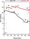
Graph shows surface pressure vs time of different nanoparticles of +Au star NP (red) and +Au sphere NP (black) interacting with the dDPPS lipid monolayer. A lipid monolayer is spread at the surface (traces I and IV) by the addition of lipids in solvent until a desired amount is added to the surface (∼5 ul of 0.7 mg/ml dDPPS lipids). After injection of the NPs into the subphase, the surface pressure is allowed to go to equilibrium (II and V). This took approximately 50 min (III and VI).
FIG. 3.
FIG. 3.
SFG spectrum of (a) the lipid monolayer membrane and (b) the calculated percent change in the ordering ratio of the same lipid monolayer after NP interaction is depicted. The CD region ssp polarization combination SFG spectrum of the dDPPS lipid monolayer before NP interaction (black), after +Au nanospheres interact, and after +Au nanostars interact (blue) shows an increase in the peak amplitudes relating to the CD3 and Fermi resonance peaks and a decrease in the CD2 peak amplitudes. Upon quantifying the ratio of the CD3 and CD2 symmetric peak amplitudes, the +Au star NPs cause a 773 ± 112% order increase while the +Au sphere NPs only cause a 73 ± 24%. This suggests that the +Au star NPs have more impact on the dDPPS lipid monolayer compared to the +Au sphere NPs. The representative SFG spectra are offset for clarity. The error bars are the standard deviation of the measured SFG ordering ratio.
FIG. 4.
FIG. 4.
Schematic for +Au nanostar (left) and +Au nanosphere (right) NPs interacting with a dDPPS lipid monolayer. The schematic of the +Au star NP depicts the large increase in lipid order measured by SFG in the form of insertion of the +Au star NP into the lipid membrane, and the decrease in surface pressure shown by the mass of the lipids and the +Au star NPs leaving the surface. For the +Au sphere NP, we show the dimpling of the membrane supported by the small lipid order increase compared to the +Au star NPs measured by SFG and the pressure decrease measured by tensiometry.

References

    1. Bathe M. and Rothemund P. W. K., MRS Bull. 42, 882 (2017). 10.1557/mrs.2017.279 - DOI
    1. Zhang Z., Shen W., Xue J., Liu Y., Liu Y., Yan P., Liu J., and Tang J., Nanoscale Res. Lett. 13, 1 (2018). 10.1186/s11671-017-2411-3 - DOI - PMC - PubMed
    1. George A., Shah P. A., and Shrivastav P. S., Int. J. Pharm. 561, 244 (2019). 10.1016/j.ijpharm.2019.03.011 - DOI - PubMed
    1. Albanese A., Tang P. S., and Chan W. C., Annu. Rev. Biomed. Eng. 14, 1 (2012). 10.1146/annurev-bioeng-071811-150124 - DOI - PubMed
    1. Parveen S., Misra R., and Sahoo S. K., Nanomed. Nanotechnol. Biol. Med. 8, 147 (2012). 10.1016/j.nano.2011.05.016 - DOI - PubMed

Publication types