. 2022 Nov 8;13(1):6742.
doi: 10.1038/s41467-022-34582-1.

Discovery of a small molecule that selectively destabilizes Cryptochrome 1 and enhances life span in p53 knockout mice

Affiliations

Discovery of a small molecule that selectively destabilizes Cryptochrome 1 and enhances life span in p53 knockout mice

Seref Gul et al. Nat Commun. .

Abstract

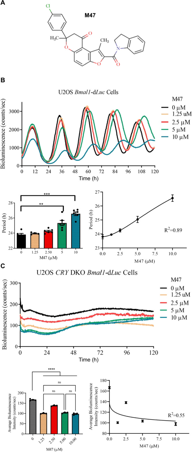
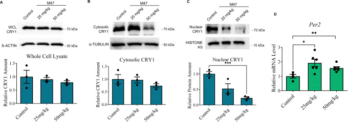
Cryptochromes are negative transcriptional regulators of the circadian clock in mammals. It is not clear how reducing the level of endogenous CRY1 in mammals will affect circadian rhythm and the relation of such a decrease with apoptosis. Here, we discovered a molecule (M47) that destabilizes Cryptochrome 1 (CRY1) both in vitro and in vivo. The M47 selectively enhanced the degradation rate of CRY1 by increasing its ubiquitination and resulted in increasing the circadian period length of U2OS Bmal1-dLuc cells. In addition, subcellular fractionation studies from mice liver indicated that M47 increased degradation of the CRY1 in the nucleus. Furthermore, M47-mediated CRY1 reduction enhanced oxaliplatin-induced apoptosis in Ras-transformed p53 null fibroblast cells. Systemic repetitive administration of M47 increased the median lifespan of p53-/- mice by ~25%. Collectively our data suggest that M47 is a promising molecule to treat forms of cancer depending on the p53 mutation.


Conflict of interest statement

The M47 studied in this paper is awarded of the following patent: WO2021137771A1 (World Intellectual Property Organization, Patent Cooperation Treaty). S.G., M.T., and I.H.K. are inventors in these patent applications, while Koc University is the assignee. The title of the patent is “11-(4-chlorophenyl)−4-(2,3-dihydro-1h-indole-1-carbonyl)−3,11-dimethyl-5,10,dioxatricyclo[7.4.0.0,2,6,]trideca-1,3,6,8-tetraen-13-one and derivatives as destabilizer of cry1 for the treatment of circadian rhythm associated diseases and disorders”. The invention provided an anticancer molecule against cancers harboring p53 mutations. The other authors declare that they have no competing interests.

Figures

Fig. 1
Fig. 1. Dose-dependent effect of M47 on the circadian rhythm of U2OS cells.
A Structure of M47. B The representative figure for the effect of M47 on the bioluminescence rhythm of U2OS cells stably expressing Bmal1-dLuc. M47 lengthened the circadian rhythm dose-dependently (Data represent the mean ± SEM, n = 4 with duplicates ***p = 0.0002, *****p < 0.0001, versus DMSO control by one-way ANOVA with Dunnet’s post hoc test). C Effect of M47 on the CRY1/CRY2 double knockout (DKO) U2OS cell line (Data represent the mean ± SEM, n = 3 with duplicates ns=not significant versus DMSO control by one-way ANOVA with Dunnet’s multiple comparison test; ****p < 0.001 versus M47 treated cells by one-way ANOVA with Dunnet’s multiple comparison test).
Fig. 2
Fig. 2. The effect of M47 on the half-life of CRYs.
A M47 increased the degradation rate of CRY1::LUC dose-dependently. Normalized half-life is shown with ± SEM (n = 4 with triplicates). *p = 0.01, ***p = 0.0008 versus DMSO treated cells with one-way ANOVA with Dunnet’s multiple comparison test. B M47 did not affect the degradation rate of CRY2::LUC. Normalized half-life is shown with ± SEM (n = 5 with triplicates one-way ANOVA with Dunnet’s multiple comparison test). C The representative figure for the effect of M47 on the bioluminescence rhythm of U2OS CRY1 knockout (KO) Bmal1-dLuc. M47 did not affect on the circadian rhythm of the CRY1 knockout U2OS Bmal1-dLuc in dose-dependently (Data represent the mean ± SEM, n = 3 with duplicates **p = 0.002, wild-type U2OS Bmal1-dLuc versus CRY1 KO U2OS Bmal1-dLuc by two-tailed student’s t-test; ns = statistically not significant one-way ANOVA in CRY1 KO U2OS Bmal1-dLuc DMSO vs M47 treatment). D The representative figure for the effect of M47 on the bioluminescence rhythm of CRY2 knockout U2OS Bmal1-dLuc. M47 increased the period length of the circadian rhythm in U2OS CRY2 KO Bmal1-dLuc in dose-dependently (Data represent the mean ± SEM, n = 3 with duplicates **p = 0.0072 and ****p < 0.0001 versus DMSO control by one-way ANOVA with Dunnet’s post hoc test). E Binding pose of M47 on the simulated CRY1 structure predicted by AutodockVina with −11.2 kcal/mol binding energy. Protein structure is shown in surface. FBXL3 binding residues are colored as red. M47 interacting residues are shown in sticks. F M47 did not affect the half-life of mutant CRY1R293AW399L-LUC degradation Normalized half-life is shown with ± SEM (n = 4 with triplicates with one-way ANOVA test). G The ubiquitination of the CRY1 in the presence of M47. (Data represent the mean ± SEM, n = 3; *p = 0.0297 versus DMSO control with t-test with two-tailed).
Fig. 3
Fig. 3. Physical interaction between M47 and CRYs.
A, B HEK293T cells transfected with pcDNA4A-Cry1-His-Myc, pcDNA4A-Cry2-His-Myc, or pcDNA4A-Cry1-T5-His-Myc plasmids then lysates were subjected to pull-down assay. Lysates were treated with solvent (DMSO), 50 µM bM47, and 50 µM bM47 with 100 µM M47 (competitor). While M47 binds to the PHR of CRY1 (A, B) it does not bind to CRY2 (A) (n = 3). C χ1 dihedral angle distribution M47 interacting residues of CRY1 and CRY2 throughout 300 ns MD simulation. D Two superimpose images from two different angles of CRY1 and CRY2 structures. M47 binding residues (Trp399, Trp292, Arg293, and Ser396) in CRY1 and corresponding residues in CRY2 (Trp310, Ser414) are shown in sticks.
Fig. 4
Fig. 4. The effect of M47 on core clock genes and proteins in synchronized U2OS cells.
Confluent U2OS cells were synchronized by 2 h treatment with dexamethasone (0.1 µM) and medium replaced with fresh medium containing M47 or solvent (DMSO final 0.5%). Cells were harvested at indicated time points. A The protein level in lysed cells was analyzed by the protein immunoblot technique. M47 decreased the protein level of CRY1 between 24th−48th h. The level of proteins was normalized to DMSO at 24th h (mean ± SEM, n = 3). (*p = 0.0286, ***p = 0.0001 versus DMSO control by two-way ANOVA with Dunnet’s multiple comparison test) (B) Cells were subjected to a reverse-transcription-quantitative polymerase chain reaction (RT-qPCR). M47 treatment increased overall Dbp level and not affect the Cry1 and Cry2 levels. At 24 and 28th h M47 increased Per2 (**p = 0.0082, ***p = 0.0003 versus DMSO control with t-test with two-tailed). The expression level of genes was normalized relative to the expression level of that gene at 24th h. (Data represent the mean ± SEM, n = 3 with duplicates). *ns non-specific band in western blot.
Fig. 5
Fig. 5. Toxicity of M47 in mice.
A Body temperatures in C57BL/6 J mice (n > 4) treated with single i.p. doses of 5 mg/kg, 50 mg/kg, 300 mg/kg M47 or vehicle during 15-day observation period. Body temperatures (°C) were expressed as mean ± SEM. “↓” indicates the treatment days of M47. ***p < 0.001, control vs 300 mg/kg M47; #p < 0.05, ##p < 0.01 control vs 5 mg/kg M47 (Two-way ANOVA with Bonferroni post hoc test). B Body weight changes (%) in C57BL/6 J mice (n > 4) treated with single i.p. doses of 5 mg/kg, 50 mg/kg, 300 mg/kg M47 or vehicle during 15-day observation period. Body weight changes (%) were expressed as mean ± SEM. “↓” indicates the treatment days of M47. *p < 0.05, **p < 0.01, control vs 300 mg/kg M47; ###p < 0.001 5 mg/kg vs 300 mg/kg M47; φp < 0.05, φφp < 0.01 control vs 50 mg/kg; Ɛp < 0.05, 5 mg/kg vs 50 mg/kg M47 (Two-way ANOVA with Bonferroni post hoc test). C Body temperatures in C57BL/6 J mice (n > 4) treated with i.p. doses of 40 mg/kg, 80 mg/kg, 150 mg/kg M47 or vehicle for 5 days. Body temperatures (°C) were expressed as mean ± SEM. “↓” indicates the treatment days of M47. *p < 0.05, **p < 0.01, ***p < 0.001, control vs 150 mg/kg M47; #p < 0.05, ##p < 0.01, ###p < 0.001 control vs 80 mg/kg M47 (Two-way ANOVA with Bonferroni post hoc test). D Body weight changes (%) in C57BL/6 J mice (n > 4) treated with i.p. doses of 40 mg/kg, 80 mg/kg, 150 mg/kg M47 or vehicle for 5 days. Body weight changes (%) were expressed as mean ± SEM. “↓” indicates the treatment days of M47. **p < 0.01 control vs 150 mg/kg M47 (Two-way ANOVA with Bonferroni post hoc test). All the sample sizes (n) are given in the figures.
Fig. 6
Fig. 6. Subacute toxicity and pharmacokinetics of M47.
A Body temperatures in C57BL/6 J mice (n > 5) treated with i.p. dose 60 mg/kg M47 or vehicle for 14 days. Body temperatures (°C) were expressed as mean ± SEM. “↓” indicates the treatment days of M47. *p < 0.05, **p < 0.01, ***p < 0.001, control vs 60 mg/kg M47 (Two-way ANOVA with Bonferroni post hoc test). B Body weight changes (%) in C57BL/6 J mice (n > 5) treated with i.p. dose of 60 mg/kg M47 or vehicle for 14 days. Body weight changes (%) were expressed as mean ± SEM. “↓” indicates the treatment days of M47. *p < 0.05, ***p < 0.001, control vs 60 mg/kg M47 (Two-way ANOVA with Bonferroni post hoc test). C Mean plasma concentration-time curve of M47 administered at 100 mg/kg i.p. to C57BL/6 J mice. Data were expressed as mean ± SEM (n = 4 per time point). D M47 levels in brain tissue at 2 h (n = 3) and 4 h (n = 3) following M47 administration at a dose of 100 mg/kg i.p. to C57BL/6 J mice. Data were expressed as mean ± SEM. All the sample sizes (n) are given in the figures.
Fig. 7
Fig. 7. The effect of M47 on CRY1 and Per2 expression levels in mice liver.
M47 (25 mg/kg or 50 mg/kg) or vehicle i.p. administered to C57BL/6 J mice. 6 h after the treatment mice were sacrificed (n = 3). While M47 slightly decreased the CRY1 levels in whole cell lysate (WCL) and cytosolic fraction, decreased the nuclear CRY1 level significantly. Effect of M47 in (A) WCL (B) cytosolic fraction, and (C) nuclear fraction of mice liver. M47 treatment caused the reduction of the CRY1 level in the nucleus (Data represent the mean ± SEM, n = 3, **p = 0.0008 versus control by t-test two-tailed). D Liver samples subjected to RT-PCR. M47 treatment increased the mRNA level of Per2. (Data represent the mean ± SEM, n = 3 (with duplicates in RT-PCR) *p = 0.0211 and **p = 0.0082 versus control by t-test two-tailed).
Fig. 8
Fig. 8. The effect of M47 in p53 null mouse skin fibroblast (MSF) cells in the presence of oxaliplatin and on the survival of p53 mutant mice.
A Ras pT24 transformed p53 null MSF fibroblast cells were treated with DMSO or M47 (5 μM) for 24 h and then 0, 10, 20 μM oxaliplatin for 16 h. Cells were lysed, and protein levels were analyzed by Western blot. In each case, M47 increases the cleaved PARP protein level and decreases CRY1 levels. Bar graph was drawn normalizing to 10 µM dosage of oxaliplatin (Data represent the mean ± SEM, n = 4, **p = 0.0051 versus DMSO control by two-way ANOVA). B Measurement of Caspase-3 activity in WT or p53-null Ras-transformed MSF cells (p53-null) treated with oxaliplatin in the absence (DMSO) and presence of M47. Cells were treated with DMSO or M47 (5 μM) for 24 h and then 0, 10, 20 μM oxaliplatin for 16 h. Caspase-3 activity in total cell lysates was measured using the Caspase-3 substrate, Z-DEVD-R110. The activity was normalized to total protein concentration and presented as fold activity to 0 μM treated samples. The results are the average of 3 biological replicates ± SEM (**p = 0.0077 versus M47 treated cells with one-way ANOVA with Dunnet’s post hoc test). C M47 in p53−/− mice increases age-adjusted survival. Kaplan–Meier survival analysis (log-rank test) of the time of death with evidence of tumors showed significant differences between vehicle-treated p53−/− and M47-treated p53−/− (**p = 0.0068) (n = 9 for vehicle-treated and n = 7 for the M47 treated). Arrow indicates M47 administration (18th week).

References

    1. Lowrey PL, Takahashi JS. Genetics of circadian rhythms in Mammalian model organisms. Adv. Genet. 2011;74:175–230. - PMC - PubMed
    1. Takahashi JS, Hong HK, Ko CH, McDearmon EL. The genetics of mammalian circadian order and disorder: implications for physiology and disease. Nat. Rev. Genet. 2008;9:764–775. - PMC - PubMed
    1. Green CB, Takahashi JS, Bass J. The meter of metabolism. Cell. 2008;134:728–742. doi: 10.1016/j.cell.2008.08.022. - DOI - PMC - PubMed
    1. Marcheva, B. et al. Circadian clocks and metabolism. Handb Exp. Pharmacol. 127–155, 10.1007/978-3-642-25950-0_6 (2013). - PMC - PubMed
    1. Onat OE, et al. Human CRY1 variants associate with attention deficit/hyperactivity disorder. J. Clin. Invest. 2020;130:3885–3900. - PMC - PubMed

Publication types