. 2022 Nov 8;12(1):18986.
doi: 10.1038/s41598-022-21871-4.

Estrogen-induced immune changes within the normal mammary gland

Affiliations

Estrogen-induced immune changes within the normal mammary gland

Helen Tower et al. Sci Rep. .

Abstract

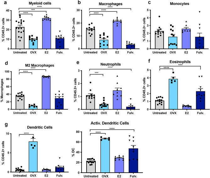
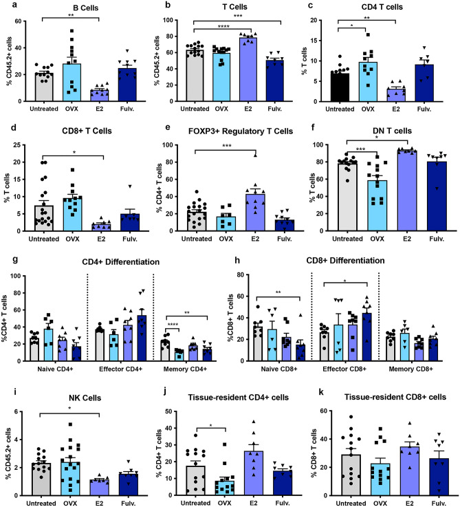
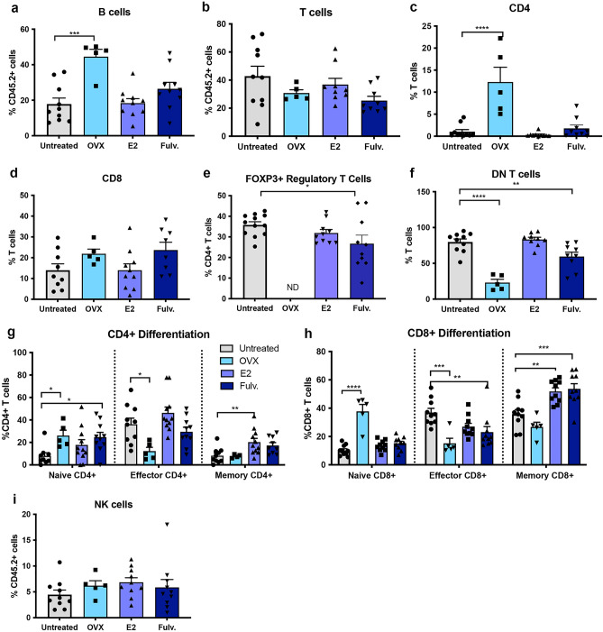
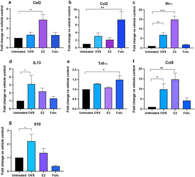
Breast cancer (BCa) incidence increases following aberrant hormone exposure, which has been linked to direct effects on estrogen receptor (ER)+ mammary epithelium. While estrogen exposure during mammary involution has been shown to drive tumour growth via neutrophils, the potential for the ER + immune microenvironment to mediate part (in addition to mammary epithelial cells) of hormonally controlled BCa risk during normal development has not been assessed. We collected mammary tissue, lymph nodes and blood from tumour naïve mice treated with, oophorectomy, estrogen (17β estradiol) or Fulvestrant. Flow cytometry was used to examine the impact on the frequency of innate and adaptive immune cells. Oophorectomy and fulvestrant decreased the proportion of macrophages, particularly pro-tumour polarized M2 macrophages and neutrophils. Conversely, dendritic cells were increased by these therapies, as were eosinophils. Estrogen increased the proportion of M2 macrophages and to a lesser extent CD4-CD8- double negative and FoxP3+ regulatory T cells but decreased CD8 + T cells and B cells. Excluding eosinophils, these changes were restricted to the mammary tissue. This suggests that inhibiting estrogen action lowers the immune suppressive myeloid cells, increases in antigen presentation and eosinophil-mediated direct or indirect cytotoxic effects. In contrast, estrogen exposure, which drives BCa risk, increases the suppressive myeloid cells and reduces anti-tumour cytotoxic T cells. The impact of hormonal exposure on BCa risk, may in part be linked to its immune modulatory activity.


Conflict of interest statement

PD and KB are on the editorial board for Scientific Reports. All other authors have no competing interests financial or non-financial to disclose.

Figures

Figure 1
Figure 1
Effects of estrogen, oophorectomy and fulvestrant on the innate immune cells within the non-neoplastic mouse mammary gland. Mice were either untreated, ovariectomised (Ooph) or treated for 6 weeks with estrogen (E2) or Fulvestrant (Fulv). (a) Myeloid cells, (b) macrophages, (c) monocytes, (d) M2 macrophages, (e) neutrophils, (f) eosinophils, (g) dendritic cells. Results are expressed percentage of immune cells (mean ± SEM). n = 11–17 mice/group. Data was analysed using a Turkey multiple comparisons one-way ANOVA if confirmed Gaussian distribution, or Kruskal–Wallis multiple comparisons 1-way ANOVA if non-Gaussian. Statistically significant results are denoted with *p < 0.05, **p < 0.01, ***p < 0.001, ****p < 0.0001.
Figure 2
Figure 2
Effects of estrogen, oophorectomy and fulvestrant on the adaptive immune cells within the non-neoplastic mouse mammary gland. Mice were either untreated, ovariectomised (Ooph) or treated for 6 weeks with estrogen (E2) or Fulvestrant (Fulv). (a) B cells, (b) T cells, (c) CD4 + T helper cells, (d) CD8 + cytotoxic T cells, (e) FoxP3 + regulatory T cells, (f) CD4- CD8- T cells, (g) CD4 + differentiation status, (h) CD8 + differentiation status, (i) NK cells, (j) tissue resident CD4 + T cells and (k) tissue resident CD8 + T cells. Results are expressed percentage of immune cells (mean ± SEM). n = 11–17 mice/group. Data was analysed using a Turkey multiple comparisons one-way ANOVA if confirmed Gaussian distribution, or Kruskal–Wallis multiple comparisons 1-way ANOVA if non-Gaussian. Statistically significant results are denoted with *p < 0.05, **p < 0.01, ***p < 0.001, ****p < 0.0001.
Figure 3
Figure 3
Effects of estrogen, oophorectomy and fulvestrant on the mammary lymph node immune cells. Mice were either untreated, ovariectomised (Ooph) or treated for 6 weeks with estrogen (E2) or Fulvestrant (Fulv). (a) macrophages, (b) CD206 + macrophages, (c) dendritic cells (d) activated (CD86 +) dendritic cells. Results are expressed percentage of immune cells (mean ± SEM). n = 7–21 mice/group. Data was analysed using a Turkey multiple comparisons one-way ANOVA if confirmed Gaussian distribution, or Kruskal–Wallis multiple comparisons 1-way ANOVA if non-Gaussian. Statistically significant results are denoted with *p < 0.05, **p < 0.01.
Figure 4
Figure 4
Effects of estrogen, oophorectomy and fulvestrant on the mammary lymph node immune cells. Mice were either untreated, ovariectomised (Ooph) or treated for 6 weeks with estrogen (E2) or Fulvestrant (Fulv). (a) T cells, (b) B cells, (c) CD4 + T helper cells, (d) CD8 + cytotoxic T cells, (e) Activated CD4 + T cells, (f) activated CD8 + T cells, (g) CD4 + differentiation status, (h) CD8 + differentiation status, (i) CD4- CD8- T cells, (j) CD4 + CD8 + T cells. Results are expressed percentage of immune cells (mean ± SEM). n = 7–21 mice/group. Data was analysed using a Turkey multiple comparisons one-way ANOVA if confirmed Gaussian distribution, or Kruskal–Wallis multiple comparisons 1-way ANOVA if non-Gaussian. Statistically significant results are denoted with *p < 0.05, **p < 0.01, ***p < 0.001.
Figure 5
Figure 5
Effects of estrogen, oophorectomy and fulvestrant on the blood innate immune cells. Mice were either untreated, ovariectomised (OVX) or treated for 6 weeks with estrogen (E2) or Fulvestrant (ICI). (a) Myeloid cells, (b) monocytes, (c) neutrophils, (d) eosinophils, (e) dendritic cells. Results are expressed percentage of immune cells (mean ± SEM). n = 5–17 mice/group. Data was analysed using a Turkey multiple comparisons one-way ANOVA if confirmed Gaussian distribution, or Kruskal–Wallis multiple comparisons 1-way ANOVA if non-Gaussian. Statistically significant results are denoted with *p < 0.05, **p < 0.01, ***p < 0.001, ****p < 0.0001.
Figure 6
Figure 6
Effects of estrogen, oophorectomy and fulvestrant on the blood adaptive immune cells. Mice were either untreated, ovariectomised (Ooph) or treated for 6 weeks with estrogen (E2) or Fulvestrant (Fulv). (a) B cells, (b) T cells, (c) CD4 + T helper cells, (d) CD8 + cytotoxic T cells, (e) FoxP3 + regulatory T cells, (f) CD4- CD8- T cells, (g) CD4 + differentiation status, (h) CD8 + differentiation status and (i) NK cells. Results are expressed percentage of immune cells (mean ± SEM). n = 5–17 mice/group. Data was analysed using a Turkey multiple comparisons one-way ANOVA if confirmed Gaussian distribution, or Kruskal–Wallis multiple comparisons 1-way ANOVA if non-Gaussian. Statistically significant results are denoted with *p < 0.05, **p < 0.01, ***p < 0.001, ****p < 0.0001.
Figure 7
Figure 7
Effects of estrogen, oophorectomy and fulvestrant on transcript expression of myeloid specific cytokines in the mouse mammary epithelial cells. Mice were either untreated, ovariectomised (Ooph) or treated for 6 weeks with estrogen (E2) or Fulvestrant (Fulv). (a) Csf-2, (b) Ccl2, (c) Ifn-α, (d) IL-13, (e) Tnf-α, (f) Ccl5, (g) Il-10. Results are expressed percentage of immune cells (mean ± SEM). n = 4 biological replicates of n = 2–10 mice/group. Data was analysed using a Turkey multiple comparisons one-way ANOVA if confirmed Gaussian distribution, or Kruskal–Wallis multiple comparisons 1-way ANOVA if non-Gaussian. Statistically significant results are denoted with *p < 0.05, **p < 0.01, ***p < 0.001, ****p < 0.0001.

Similar articles

Cited by

References

    1. Britt KL, Cuzick J, Phillips KA. Key steps for effective breast cancer prevention. Nat. Rev. Cancer. 2020;20:417–436. doi: 10.1038/s41568-020-0266-x. - DOI - PubMed
    1. Cuzick J, et al. Selective oestrogen receptor modulators in prevention of breast cancer: An updated meta-analysis of individual participant data. Lancet. 2013;381:1827–1834. doi: 10.1016/S0140-6736(13)60140-3. - DOI - PMC - PubMed
    1. Baek HJ, et al. Inhibition of estrogen signaling reduces the incidence of BRCA1-associated mammary tumor formation. Int. J. Biol. Sci. 2018;14:1755–1768. doi: 10.7150/ijbs.28142. - DOI - PMC - PubMed
    1. Chan SR, et al. STAT1-deficient mice spontaneously develop estrogen receptor alpha-positive luminal mammary carcinomas. Breast Cancer Res. 2012;14:R16. doi: 10.1186/bcr3100. - DOI - PMC - PubMed
    1. Torres-Arzayus MI, Zhao J, Bronson R, Brown M. Estrogen-dependent and estrogen-independent mechanisms contribute to AIB1-mediated tumor formation. Cancer Res. 2010;70:4102–4111. doi: 10.1158/0008-5472.CAN-09-4080. - DOI - PMC - PubMed

Publication types