. 2022 Nov 8;12(1):18985.
doi: 10.1038/s41598-022-22899-2.

Finite element modeling of effects of tissue property variation on human optic nerve tethering during adduction

Affiliations

Finite element modeling of effects of tissue property variation on human optic nerve tethering during adduction

Joseph Park et al. Sci Rep. .

Abstract

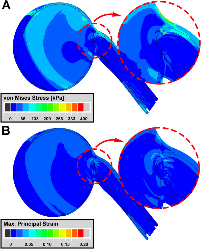
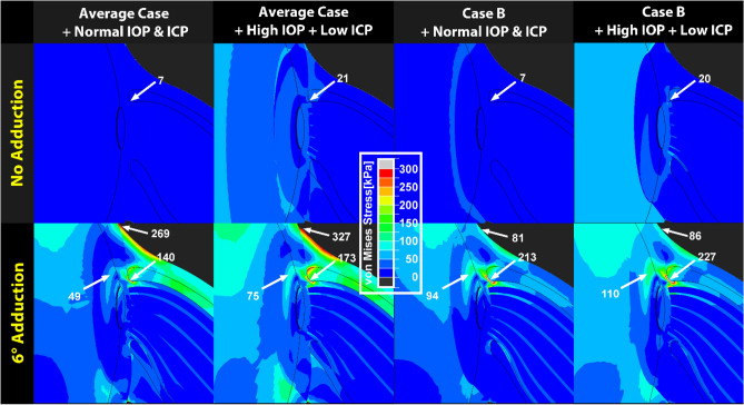
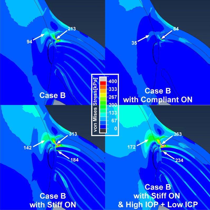
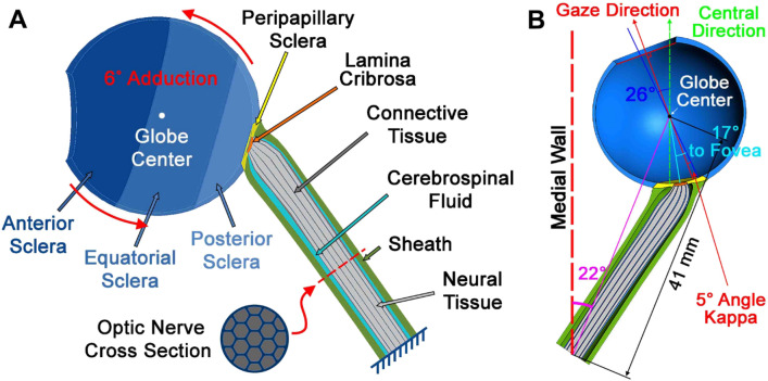
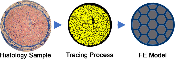
Tractional tethering by the optic nerve (ON) on the eye as it rotates towards the midline in adduction is a significant ocular mechanical load and has been suggested as a cause of ON damage induced by repetitive eye movements. We designed an ocular finite element model (FEM) simulating 6° incremental adduction beyond the initial configuration of 26° adduction that is the observed threshold for ON tethering. This FEM permitted sensitivity analysis of ON tethering using observed material property variations in measured hyperelasticity of the anterior, equatorial, posterior, and peripapillary sclera; and the ON and its sheath. The FEM predicted that adduction beyond the initiation of ON tethering concentrates stress and strain on the temporal side of the optic disc and peripapillary sclera, the ON sheath junction with the sclera, and retrolaminar ON neural tissue. However, some unfavorable combinations of tissue properties within the published ranges imposed higher stresses in these regions. With the least favorable combinations of tissue properties, adduction tethering was predicted to stress the ON junction and peripapillary sclera more than extreme conditions of intraocular and intracranial pressure. These simulations support the concept that ON tethering in adduction could induce mechanical stresses that might contribute to ON damage.


Conflict of interest statement

The authors declare no competing interests.

Figures

Figure 1
Figure 1
Simulation of adduction to 32° from initial ON tethering at 26°, employing average measured tissue hyperelastic functions. Heat maps of (A) von Mises stress and (B) principal strain. Stress–strain effects mainly occur in and around the ON head in the region enclosed by red dotted circles.
Figure 2
Figure 2
Sensitivity to variations in regional ocular material properties, as implemented by reduced polynomial functions for which the indicated qualities are shorthand, during adduction 6° beyond onset of tethering at 26°. Stiff and compliant material properties were set to 95th and 5th percentile values, respectively, of stress–strain functions measured after preconditioning (details in Table 1). Material properties for regions not noted here were set to average observed reduced polynomial functions.
Figure 3
Figure 3
Horizontal cross sections of finite element model superimposing initial configuration of 26° adduction at initiation of tethering (green) with final configuration of 32° adduction (red) for various combinations of observed tissue properties as defined in Fig. 2. Yellow region represents overlap. Blue inset shows 3 × magnified view of the optic disc region where arrows indicate locations of temporal edge of the lamina cribrosa, whose displacements are listed numerically for each case.
Figure 4
Figure 4
Finite element model of adduction 6° beyond onset of tethering at 26° demonstrating additional influences of intraocular pressure (IOP) and intracranial pressure (ICP) on stress distributions in the peripapillary and optic disc region. Material property cases are defined in Fig. 2. In upper panels without adduction tethering, note small stress due to 40 mmHg IOP (high) and 4 mmHg ICP (low), in comparison with lower panels showing larger effect of adduction. Comparison of lower right two panels for Case B demonstrates that during adduction, stress in the temporal peripapillary sclera and ON junction is 14 kPa higher when IOP is high and ICP is low, than when these pressures are normal.
Figure 5
Figure 5
Sensitivity to ON stiffness greater or less than average, assuming the least favorable combination of other tissue properties (case B). Simulations assumed normal IOP and ICP, except for the right bottom panel that assumes an extreme translaminar pressure gradient (40 mmHg IOP and 4 mmHg ICP).
Figure 6
Figure 6
Model geometry. (A) Sclera is parsed into anterior, equatorial, posterior (blue) and peripapillary regions (yellow). The lamina cribrosa (orange) abuts the peripapillary sclera. The posterior lamina cribrosa is joined to the ON (gray), and the ON sheath (green) is joined to the peripapillary sclera anteriorly, and to the orbital apex posteriorly as boundary conditions. Cerebrospinal fluid (CSF) is shown in light blue. (B) Dimensions of the model.
Figure 7
Figure 7
Model simplification of human optic nerve (ON). Transverse histological section of 57 year old human ON was chosen to calculate the proportion portion of connective versus neural tissue. In this 10 μm thick histological section stained with Mason trichrome at left, neural tissue (pink) was segmented from connective tissue (blue) as outlined as in the middle tracing. After assigning a 9:16 area ratio of connective to neural tissue, the ON was simplified as the honeycomb structure at right for modeling.
Figure 8
Figure 8
(A) Local mesh size was adjusted according to regional relevance, thus finest around the optic nerve head. (B) Mean and maximum values of von Mises stresses of all elements in the model were stable throughout all element numbers evaluated. Dotted lines indicate means.

Similar articles

Cited by

References

    1. Demer JL. Optic nerve sheath as a novel mechanical load on the globe in ocular duction. Investig. Ophthalmol. Vis. Sci. 2016;57:1826–1838. doi: 10.1167/iovs.15-18718. - DOI - PMC - PubMed
    1. Demer JL, et al. Magnetic resonance imaging of optic nerve traction during adduction in primary open-angle glaucoma with normal intraocular pressure. Investig. Ophthalmol. Vis. Sci. 2017;58:4114–4125. doi: 10.1167/iovs.17-22093. - DOI - PMC - PubMed
    1. Suh SY, Le A, Shin A, Park J, Demer JL. Progressive deformation of the optic nerve head and peripapillary structures by graded horizontal duction. Investig. Ophthalmol. Vis. Sci. 2017;58:5015–5021. doi: 10.1167/iovs.17-22596. - DOI - PMC - PubMed
    1. Epelboim J, et al. Gaze-shift dynamics in two kinds of sequential looking tasks. Vis. Res. 1997;37:2597–2607. doi: 10.1016/s0042-6989(97)00075-8. - DOI - PubMed
    1. Wu CC, Kowler E. Timing of saccadic eye movements during visual search for multiple targets. J. Vis. 2013 doi: 10.1167/13.11.11. - DOI - PMC - PubMed

Publication types