. 2022 Nov 8;12(1):18980.
doi: 10.1038/s41598-022-23238-1.

Ocular morphologic traits in the American Cocker Spaniel may confer primary angle closure glaucoma susceptibility

Affiliations

Ocular morphologic traits in the American Cocker Spaniel may confer primary angle closure glaucoma susceptibility

Sangwan Park et al. Sci Rep. .

Abstract

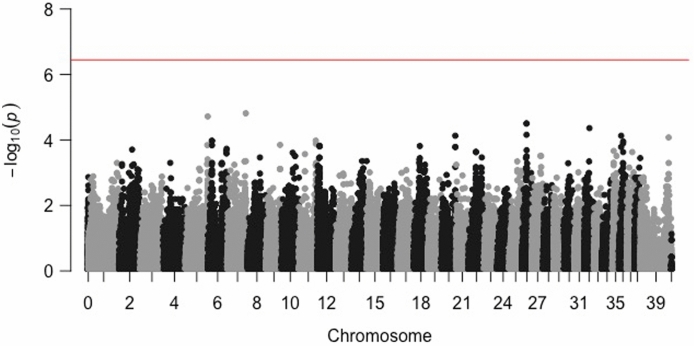
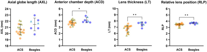
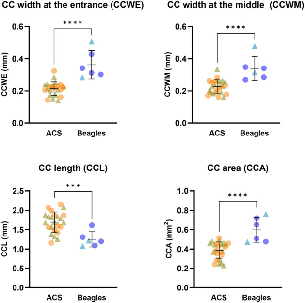
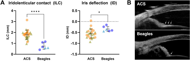
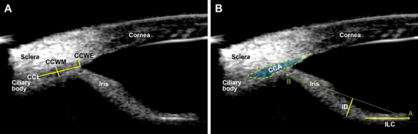
Acute primary angle closure glaucoma is a potentially blinding ophthalmic emergency requiring prompt treatment to lower the elevated intraocular pressure in humans and dogs. The PACG in most of canine breeds is epidemiologically similar to humans with older and female patients overrepresented with the condition. The American Cocker Spaniel (ACS) is among the most common breeds observed with PACG development in dogs. This study initially sought to identify genetic risk factors to explain the high prevalence of PACG in ACSs by using a case-control breed-matched genome-wide association study. However, the GWAS failed to identify candidate loci associated with PACG in this breed. This study then assessed intrinsic ocular morphologic traits that may relate to PACG susceptibility in this breed. Normal ACSs without glaucoma have a crowded anterior ocular segment and narrow iridocorneal angle and ciliary cleft, which is consistent with anatomical risk factors identified in humans. The ACSs showed unique features consisting of posterior bowing of iris and longer iridolenticular contact, which mirrors reverse pupillary block and pigment dispersion syndrome in humans. The ACS could hold potential to serve as an animal model of naturally occurring PACG in humans.


Conflict of interest statement

The authors declare no competing interests.

Figures

Figure 1
Figure 1
Manhattan plot for GWAS (Cases: 28 PACG-affected; Controls: 24 non-affected) in the ACS. The horizontal red line denotes the threshold for genome-wide statistical association and the black and grey colors alternate each other to delineate each chromosome easily. No statistically significant single nucleotide polymorphisms associated with PACG were identified.
Figure 2
Figure 2
The ACSs have significantly more crowded anterior ocular segment structures. While the overall globe size was not different between the two breeds, the anterior chamber was significantly shallower in ACS versus Beagles. Although the ACS have significantly thinner lenses, the lenses were more anteriorly located in ACS versus Beagles. An unpaired t-test was performed (n = 18 ACS and 7 Beagles; * = p value < 0.05; ** = P value < 0.01). The line and error bars indicate mean values and standard deviations. The circles and triangles indicate females and males, respectively.
Figure 3
Figure 3
The ciliary cleft (CC) of ACSs was significantly more narrow than that of Beagles. While the CC length (CCL) was significantly longer in ACS, the CC width at the entrance and the middle (CCWE and CCWM) were significantly shorter resulting in a significantly smaller CC area (CCA). An unpaired t-test was performed (n = 24 ACS and 6 Beagles; *** = p value < 0.001; **** = p value < 0.0001). The line and error bars indicate mean values and standard deviations. The circles and triangles indicate females and males, respectively.
Figure 4
Figure 4
The iris of the ACS was significantly more in contact with the anterior lens capsule and more concave (posteriorly deflected) in comparison to the Beagles. (A) The ACS have significantly longer iridolenticular contact (ILC) with a greater degree of posterior iris deflection (ID) versus Beagles. An unpaired t-test was performed (n = 24 ACS and 6 Beagles; * = p value < 0.05; **** = p value < 0.0001). The line and error bars indicate mean values and standard deviations. The circles and triangles indicate females and males, respectively. (B) Representative UBM images of ACS and Beagles are shown. Arrows indicate the iris in contact with the anterior lens capsule.
Figure 5
Figure 5
Measurements of UBM parameters (A: CCWE, CCWM, and CCL. B: CCA, ILC, and ID). CCWE, ciliary cleft width at the entrance; CCWM, ciliary cleft width at the middle; CCL, ciliary cleft length; CCA, ciliary cleft area; ILC, iridolenticular contact; ID, iris deflection.

References

    1. Zhang N, Wang J, Chen B, Li Y, Jiang B. Prevalence of primary angle closure glaucoma in the last 20 years: A meta-analysis and systematic review. Front. Med. 2021;7:1–10. doi: 10.3389/fmed.2020.624179. - DOI - PMC - PubMed
    1. Kondkar AA. Updates on genes and genetic mechanisms implicated in primary angle-closure glaucoma. Appl. Clin. Genet. 2021;14:89–112. doi: 10.2147/TACG.S274884. - DOI - PMC - PubMed
    1. Sun X, et al. Primary angle closure glaucoma: What we know and what we don’t know. Prog. Retin. Eye Res. 2017;57:26–45. doi: 10.1016/j.preteyeres.2016.12.003. - DOI - PubMed
    1. Miller PE, Bentley E. Clinical signs and diagnosis of the canine primary glaucomas. Vet. Clin. North Am. Small Anim. Pract. 2015;45:1183–1212. doi: 10.1016/j.cvsm.2015.06.006. - DOI - PMC - PubMed
    1. Plummer C, Regnier A, Gelatt K. The canine Glaucomas. In: Gelatt K, Gilger B, Kern T, editors. Veterinary Ophthalmology. Amsterdam: John Wiley & Sons Inc.; 2013. pp. 1050–1145.

Publication types