The internal structure of the infraspinatus muscle: a magnetic resonance study
- PMID: 36348046
- PMCID: PMC9674736
- DOI: 10.1007/s00276-022-03042-2
The internal structure of the infraspinatus muscle: a magnetic resonance study
Abstract
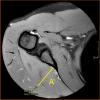
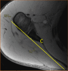
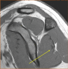
Purpose: This study aimed to describe the internal structure of the infraspinatus muscle. A secondary aim was to explore differences in internal structure between genders, sides, and correlations to demographic data.
Methods: In total, 106 shoulder MRI examinations of patients between 18 and 30 years of age seeking care in 2012-2020 at The Sahlgrenska University Hospital in Gothenburg, Sweden were re-reviewed.
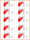
Results: The number of intramuscular tendons centrally in the infraspinatus muscle varied between 3 and 8 (median = 5). Laterally, the number of intramuscular tendons varied between 1 and 5 (median = 2). There was no difference in the median between the genders or sides. No correlations between the number of intramuscular tendons and demographic data were found. The muscle volume varied between 63 and 249 ml with a median of 188 ml for males and 122 ml for females. There was no significant difference in volume between the sides. The muscle volume correlated with body weight (Pearson's correlation coefficient, r = 0.72, p < 0.001) and height (r = 0.61, p < 0.001).
Conclusion: The anatomical variations of the infraspinatus muscle are widespread. In the medial part of the muscle belly, the number of intramuscular tendons varied between 3 and 8, while the number of intramuscular tendons laterally varied between 1 and 5. Results of our study may help to understand the internal structure of the infraspinatus muscle and its function in shoulder stabilization.
Keywords: Anatomy; Infraspinatus muscle; Magnetic resonance imaging; Rotator cuff.
© 2022. The Author(s).
Conflict of interest statement
The authors declare no competing interests.
Figures


References
-
- Campbell MJ. Medical statistics (electronic book): a textbook for the health sciences. 5. Hoboken: Wiley; 2020.
-
- Centralbyrån S. Undersökningarna av levnadsförhållanden (ULF) Stockholm: Statistikmyndigheten; 2020.
MeSH terms
LinkOut - more resources
Full Text Sources

