. 2023 Jun;30(3):1210-1218.
doi: 10.1007/s12350-022-03126-x. Epub 2022 Nov 8.

18F-FDG/PET-CT imaging findings after sternotomy

Affiliations

18F-FDG/PET-CT imaging findings after sternotomy

Maurits S H Blomjous et al. J Nucl Cardiol. 2023 Jun.

Abstract

Background: The clinical diagnosis of deep sternal wound infection (DSWI) is supported by imaging findings including 18F-fluorodeoxyglucose positron emission tomography/computed tomography (18F-FDG-PET/CT). To avoid misinterpretation due to normal post-surgery inflammation we assessed normal imaging findings in non-infected patients after sternotomy.

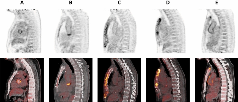
Methods: This is a prospective cohort study including non-infectious patients with sternotomy. All patients underwent 18F-FDG-PET/CT at either 5 weeks (group 1), 12 weeks (group 2) or 52 weeks (group 3) post-surgery. 18F-FDG uptake was scored visually in five categories and assessed quantitatively.

Results: A total of 44 patients were included. Sternal mean SUVmax was 7.34 (± 1.86), 5.22 (± 2.55) and 3.20 (± 1.80) in group 1, 2 and 3, respectively (p < 0.01). Sternal mean SUVmean was 3.84 (± 1.00), 2.69 (± 1.32) and 1.71 (± 0.98) in group 1, 2 and 3 (p < 0.01). All patients in group 1 had elevated uptake whereas group 2 and 3 showed 2/15 (13%) and 11/20 (55%) patients respectively with no elevated uptake. Group 3 still showed an elevated uptake pattern in in 9/20 (45%) and in 3/9 (33%) with a high-grade diffuse uptake pattern.

Conclusion: This study shows significant lower sternal 18F-FDG at 55 weeks compared to 5 weeks post-sternotomy however elevated uptake patterns may persist.


Figures

Figure 1
Figure 1
Boxplot of the SUVmean (A) and SUVmax (B) measurements distribution in each group. The dots indicated as “3”, “32”, “20”, “23”, (A, B) are outliers in the SUVmax and SUVmean measurements
Figure 2
Figure 2
Examples of different categories of sternal 18FDG uptake patterns post-sternotomy. (A) no elevated uptake; (B) low-grade diffuse uptake; (C) high-grade diffuse uptake (D); high-grade heterogeneous uptake; (E) focal uptake
Figure 3
Figure 3
Sternal 18F-FDG uptake on lateral view of attenuation-corrected (AC) maximum intensity projection (MIP) images in all patients divided by group and subdivided by visual uptake score. Group 1 (5 weeks post-sternomy); Group 2 (14 weeks post-sternomy); Group 3 (55 weeks post-sternomy). Category (A); no eleveated uptake; Category (B) low-grade diffuse uptake; Category (C) high-grade diffues uptake; Category (D) High-grade heterogeneous uptake; Category (E) focal uptake

References

    1. Filsoufi F, Castillo JG, Rahmanian PB, Broumand SR, Silvay G, Carpentier A, et al. Epidemiology of deep sternal wound infection in cardiac surgery. J Cardiothorac Vasc Anesth. 2009;23:488–494. doi: 10.1053/j.jvca.2009.02.007. - DOI - PubMed
    1. Mangram AJ, Horan TC, Pearson ML, Silver LC, Jarvis WR. Guideline for Prevention of Surgical Site Infection, 1999. Infect Cont Hosp Ep. 1999;20:250–278. doi: 10.1086/501620. - DOI - PubMed
    1. Lazar HL, Salm TV, Engelman R, Orgill D, Gordon S. Prevention and management of sternal wound infections. J Thorac Cardiovasc Surg. 2016;152:962–972. doi: 10.1016/j.jtcvs.2016.01.060. - DOI - PubMed
    1. Zhang R, Feng Z, Zhang Y, Tan H, Wang J, Qi F. Diagnostic value of fluorine-18 deoxyglucose positron emission tomography/computed tomography in deep sternal wound infection. J Plast Reconstr Aesthet Surg. 2018;71:1768–1776. doi: 10.1016/j.bjps.2018.07.017. - DOI - PubMed
    1. Hariri H, Tan S, Martineau P, Lamarche Y, Carrier M, Finnerty V, et al. Utility of FDG-PET/CT for the Detection and Characterization of Sternal Wound Infection Following Sternotomy. Nucl Med Mol Imaging. 2019;53:253–262. doi: 10.1007/s13139-019-00599-6. - DOI - PMC - PubMed

Substances

LinkOut - more resources