. 2022 Dec:47:101669.
doi: 10.1016/j.nantod.2022.101669. Epub 2022 Nov 3.

Multiplexed rapid antigen tests developed using multicolored nanoparticles and cross-reactive antibody pairs: Implications for pandemic preparedness

Affiliations

Multiplexed rapid antigen tests developed using multicolored nanoparticles and cross-reactive antibody pairs: Implications for pandemic preparedness

Helena de Puig et al. Nano Today. 2022 Dec.

Abstract

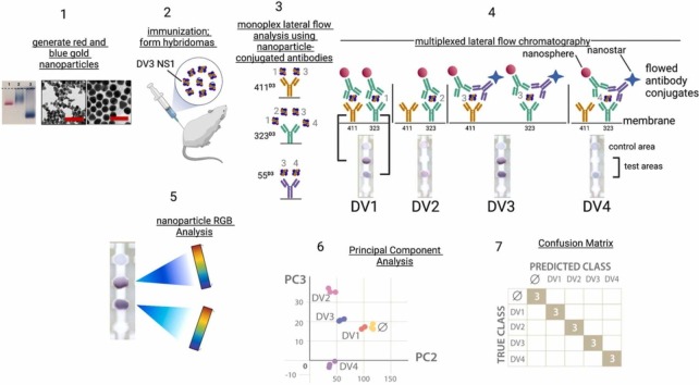
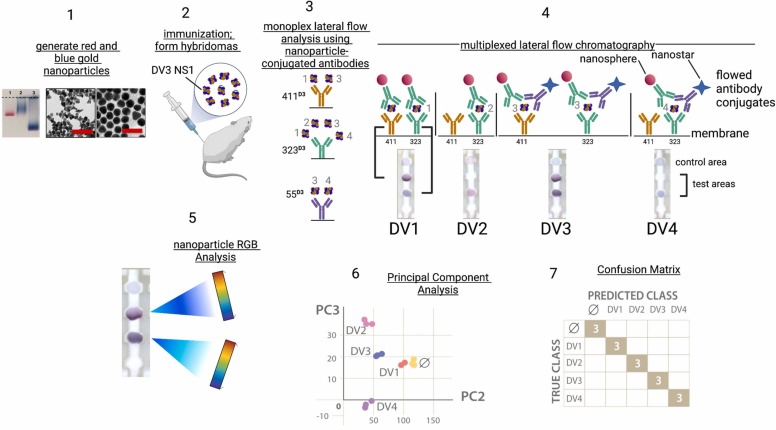
Global public health infrastructure is unprepared for emerging pathogen epidemics, in part because diagnostic tests are not developed in advance. The recent Zika, Ebola, and SARS-CoV-2 virus epidemics are cases in point. We demonstrate here that multicolored gold nanoparticles, when coupled to cross-reactive monoclonal antibody pairs generated from a single immunization regimen, can be used to create multiple diagnostics that specifically detect and distinguish related viruses. The multiplex approach for specific detection centers on immunochromatography with pairs of antibody-conjugated red and blue gold nanoparticles, coupled with clustering algorithms to detect and distinguish related pathogens. Cross-reactive antibodies were used to develop rapid tests for i) Dengue virus serotypes 1-4, ii) Zika virus, iii) Ebola and Marburg viruses, and iv) SARS-CoV and SARS-CoV-2 viruses. Multiplexed rapid antigen tests based on multicolored nanoparticles and cross-reactive antibodies and can be developed prospectively at low cost to improve preparedness for epidemic outbreaks.

Keywords: Antibody; Cross-reactive; Infectious diseases; Lateral flow chromatography; Nanoparticle; Nanosphere; Nanostar; Pandemic preparedness; Rapid antigen diagnostics.


Conflict of interest statement

The authors declare the following financial interests/personal relationships which may be considered as potential competing interests: Lee Gehrke reports financial support was provided by National Institutes of Health (R33AI100190 and AI151807). Helena de Puig reports financial support was provided by Broshy Foundation. Helena de Puig reports financial support was provided by Tata Trusts. Lee Gehrke reports equipment, drugs, or supplies was provided by US Food and Drug Administration. Lee Gehrke reports a relationship with IDx20 that includes: consulting or advisory and equity or stocks. Irene Bosch reports a relationship with IDx20 that includes: board membership, employment, and equity or stocks. Nol Salcedo reports a relationship with IDx20 that includes: employment. Helena de Puig reports a relationship with IDx20 that includes: consulting or advisory. James Collins reports a relationship with Sherlock Biosciences that includes: board membership, consulting or advisory, and equity or stocks. Lee Gehrke has patent #9488613 issued to Massachusetts Institute of Technology. Irene Bosch, Kimberly Hamad-Schifferli, Helena de Puig has patent #9488613 issued to Massachusetts Institute of Technology. Lee Gehrke has patent #10551381 with royalties paid to Massachusetts Institute of Technology. Irene Bosch, Helena de Puig, Kimberly Hamad-Schifferli has patent #10551381 with royalties paid to Massachusetts Institute of Technology.

Figures

ga1
Graphical abstract
Fig. 1
Fig. 1
Graphic summary of rapid antigen diagnostics development using cross-reactive antibodies. Step 1: generate red nanospheres and blue nanostars from gold salts; Step 2: immunization; Step 3: Confirmation of ELISA binding using monoplex lateral flow chromatography. The naming standard (e.g. 411D3) identifies the antibody name (411) and the Dengue virus serotype antigen (D3). mAb 411 recognizes D1 and D3 NS1; mAb 323 recognizes D1-D4 serotype NS1 proteins; mAb 55 recognizes DV3 and DV4 serotype NS1 proteins. Step 4: multiplexed lateral flow chromatography, creating the test signal patterns that detect and distinguish the viral antigens. Four lateral flow chromatography strips are shown, each with mAb323D3 adsorbed at the lower test area, and mAb411D3 adsorbed at the upper test area. The control area is anti-mouse IgG. For the flowed antibody conjugates, mAb323D3 was conjugated to gold nanospheres, and mAb55D3 was conjugated to blue nanostars. Step 5: The distribution of red and blue nanoparticle colors in the test areas is determined by red/green/blue (RGB) analysis (ImageJ, NIH). Step 6: The data are clustered using principal component analysis. Step 7: A confusion matrix evaluates the performance of the tests in detecting and distinguishing the four Dengue virus serotype by comparing the predicted class with the true class. The number 3 indicates the number of tests that were run, and numbers falling on the diagonal represent a perfect correlation of predicted and true classes.
Fig. 2
Fig. 2
Detecting and distinguishing the four (1−4) Dengue serotypes using anti-NS1 antibodies from a Dengue serotype 3 NS1 immunization. A) Schematic of the lateral flow strips used for monoplex analysis of NS1 antibody binding pairs, using monoclonal antibodies 323, 55, and 411. The nomenclature NS-323DV3 refers to red nanospheres coupled to antibody 323, which was raised by immunizing mice with the DV3 NS1 protein antigen. Similarly, NSt-55DV3 refers to blue nanostars conjugated to antibody 55, which was raised using the DV3 NS1 protein antigen; B) Summary binding data from the monoplex antibody binding experiments; C-J) antibody-antigen binding isotherms; the dashed lines represent standard deviation. Panels C-F correspond to the 411/323 and 411/55 pairs (also Panel B, upper), while Panels G-J correspond to the 323/323 and 323/55 antibody pairs (also Panel B, lower). Panels C/G, D/H, E/I, and F/J used Dengue virus NS1 proteins from serotypes 1–4, respectively. K) schematic of the multiplexed rapid antigen test design, with monoclonal antibodies 411 and 323 adsorbed to different test areas of the nitrocellulose membrane; L) multiplexed analysis of the four dengue virus serotype NS1 proteins using serotype 3 NS1 monoclonal antibodies. The labels (D1, D2, etc.) refer to the Dengue virus serotype NS1 ligand present in the liquid phase chromatographed on the strips; M) principal component analysis and clustering of binding data; N) Confusion matrix of the linear discriminant assay. Data that fall on the diagonal are a perfect true class/predicted class fit. The number 3 in the shaded boxes refers to the number of test strips run per determination.
Fig. 3
Fig. 3
Multiplexed lateral flow chromatography with red and blue nanoparticles to detect and distinguish the group of Dengue virus serotypes 1–4 NS1 proteins from Zika virus NS1. A) Design of the monoplex lateral flow tests used to characterize NS1 protein binding to mAbs 136 and 323, as well as the PAN blue nanostar particles, prepared by conjugating to mAbs 243DV2, 323DV3, 626DV4, and 271DV3; B) Monoplex antibody binding matrix; C-L) Monoplex antibody binding isotherms for Dengue virus serotypes 1–4 NS1 proteins and Zika virus NS1 protein; M) Design of the multiplexed rapid diagnostic test with mAbs 136 and 323 adsorbed to the nitrocellulose membrane, and chromatographed using NS-136DV1 and Nst-PANDV nanoparticles; N) Lateral flow immunochromatography using Dengue virus serotype1 NS1 and Zika virus NS1 proteins. Dengue virus serotype 1 NS1 protein alone is chromatographed on strip 1, while Zika virus NS1 protein alone is chromatographed on strip 5. Strips 2–4 represent mixtures of the Dengue and Zika virus NS1 proteins; O) Lateral flow immunochromatography using Dengue virus serotype 4 NS1 and Zika virus NS1 proteins. Dengue virus serotype 4 NS1 protein alone is chromatographed on strip 1, while Zika virus NS1 protein alone is chromatographed on strip 5. Strips 2–4 represent mixtures of the Dengue 4 and Zika virus NS1 proteins. P) principal component analysis of binding data; Q) Confusion matrix of the linear discriminant analysis.
Fig. 4
Fig. 4
Detecting COVID19 and Ebola using SARS and Marburg antibodies, respectively. The abbreviations SARS-1 or CoV refer to 2003 SARS-CoV, while CoV-2 refers to 2019 SARS-CoV-2. A) schematic of the lateral flow strip designs showing antibodies S4 and S18 adsorbed to the test membrane, and antibodies S6 and S16 conjugated to red nanospheres and blue nanostars, respectively. B) Multiplexed detection of SARS-CoV (CoV) and SARS-CoV-2 (CoV-2) using SARS-CoV antibodies; C) principal component analysis of binding data for the two coronaviruses; D) Confusion matrix of the linear discriminant analysis (LDA) for the two coronaviruses; E) schematic of the lateral flow strip designs showing anti-Marburg glycoprotein antibodies 1G11 and 2G12 adsorbed to the test membrane, and antibodies 1G11 and 2G12 conjugated to red nanospheres and blue nanostars, respectively. F) Multiplexed detection of Ebola (E) and Marburg (M), as well as mixtures of the two viruses using Marburg antibodies. Marburg glycoprotein binding alone is shown on strip 1, while Ebola glycoprotein binding alone is shown on strip 5. Strips 2–4 represent mixtures of the Marburg and Ebola glycoproteins; (G) principal component analysis of binding data for the two filoviruses; H) Confusion matrix of the linear discriminant analysis for the two filoviruses glycoprotein binding data.

Similar articles

Cited by

References

    1. Howard C.R., Fletcher N.F. Emerging virus diseases: can we ever expect the unexpected. Emerg. Microbes Infect. 2012;1 doi: 10.1038/emi.2012.47. - DOI - PMC - PubMed
    1. Bosch I., de Puig H., Hiley M., Carré-Camps M., Perdomo-Celis F., Narváez C.F., Salgado D.M., Senthoor D., O’Grady M., Phillips E., Durbin A., Fandos D., Miyazaki H., Yen C.-W., Gélvez-Ramírez M., Warke R.V., Ribeiro L.S., Teixeira M.M., Almeida R.P., Muñóz-Medina J.E., Ludert J.E., Nogueira M.L., Colombo T.E., Terzian A.C.B., Bozza P.T., Calheiros A.S., Vieira Y.R., Barbosa-Lima G., Vizzoni A., Cerbino-Neto J., Bozza F.A., Souza T.M.L., Trugilho M.R.O., de Filippis A.M.B., de Sequeira P.C., Marques E.T.A., Magalhaes T., Díaz F.J., Restrepo B.N., Marín K., Mattar S., Olson D., Asturias E.J., Lucera M., Singla M., Medigeshi G.R., de Bosch N., Tam J., Gómez-Márquez J., Clavet C., Villar L., Hamad-Schifferli K., Gehrke L. Rapid antigen tests for dengue virus serotypes and Zika virus in patient serum. Sci. Transl. Med. 2017;9 doi: 10.1126/scitranslmed.aan1589. - DOI - PMC - PubMed
    1. Pardee K., Green A.A., Ferrante T., Cameron D.E., DaleyKeyser A., Yin P., Collins J.J. Paper-based synthetic gene networks. Cell. 2014;159:940–954. doi: 10.1016/j.cell.2014.10.004. - DOI - PMC - PubMed
    1. Pardee K., Green A.A., Takahashi M.K., Braff D., Lambert G., Lee J.W., Ferrante T., Ma D., Donghia N., Fan M., Daringer N.M., Bosch I., Dudley D.M., O’Connor D.H., Gehrke L., Collins J.J. Rapid, low-cost detection of Zika virus using programmable biomolecular components. Cell. 2016;165:1255–1266. doi: 10.1016/j.cell.2016.04.059. - DOI - PubMed
    1. Muller D.A., Depelsenaire A.C.I., Young P.R. Clinical and laboratory diagnosis of Dengue virus infection. J. Infect. Dis. 2017;215:S89–S95. doi: 10.1093/infdis/jiw649. - DOI - PubMed

LinkOut - more resources